

Acute mesenteric ischaemia: a pictorial review
- PMID: 30120722
- PMCID: PMC6206376
- DOI: 10.1007/s13244-018-0641-2
Acute mesenteric ischaemia: a pictorial review
Abstract
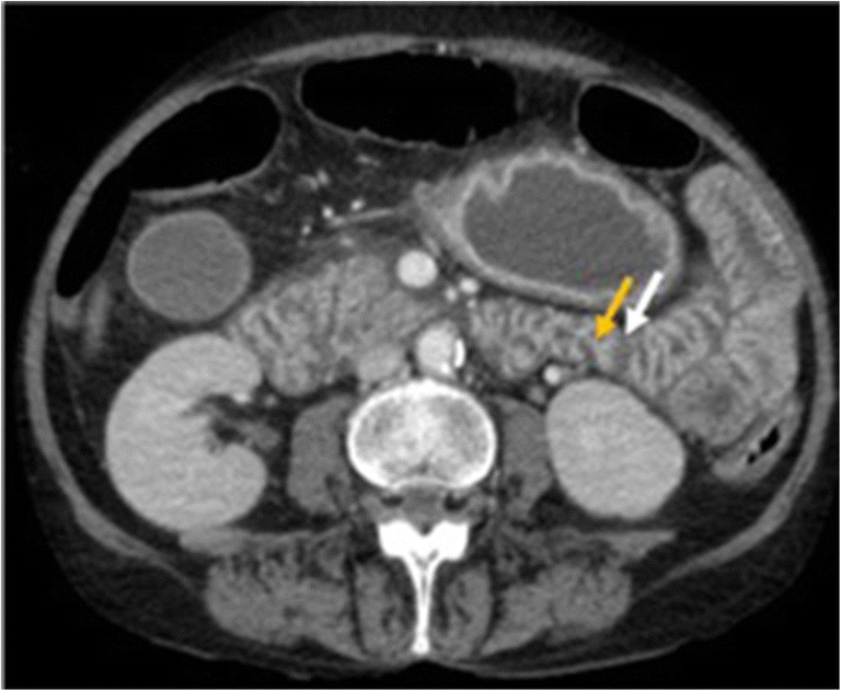
Acute mesenteric ischaemia (AMI) is an uncommon cause of acute hospital admission with high mortality rates (50-90%) that requires early diagnosis and treatment. With the increase in average life expectancy, AMI represents one of the most threatening abdominal conditions in elderly patients. Untreated, AMI will cause mesenteric infarction, intestinal necrosis, an overwhelming inflammatory response and death. Early intervention can reverse this process leading to a full recovery, but the diagnosis of AMI is difficult. The failure to recognise AMI before intestinal necrosis has developed is responsible for the high mortality of the disease. Unfortunately, common CT findings in bowel ischaemia are not specific. Therefore, it is often a combination of nonspecific clinical, laboratory and radiological findings that helps most in the correct interpretation of CT findings. The purpose of this article is to provide an overview of the anatomy, physiology of mesenteric perfusion and discussions of causes, pathogenesis and CT findings in various types of acute bowel ischaemia. Familiarity with various imaging features of mesenteric injury is essential to make a timely diagnosis that will lead to improved patient outcomes. TEACHING POINTS: • AMI is a potentially life-threatening disorder whose prognosis depends on early recognition, accurate diagnosis and timely intervention. • Arterial inflow occlusion due to thrombosis or embolisation is the most common cause of AMI. • Four aetiological types of AMI have been associated with different characteristics and risk factors (EAMI, TAMI, VAMI and NOMI). • Physical examination and laboratory findings are not sensitive or specific for diagnosing AMI; therefore, MDCT is still the first-line imaging method in suspected AMI. • Although a number of scoring systems for prognosis have been proposed, these have not been validated in large-scale studies.
Keywords: Computed tomography; Ischaemia/diagnosis; Mesenteric ischaemia; Mesenteric vascular occlusion/diagnosis; Pneumatosis intestinal.
Figures


Similar articles
-
Insights into acute mesenteric ischaemia: an up-to-date, evidence-based review from a mesenteric stroke centre unit.Br J Radiol. 2023 Nov;96(1151):20230232. doi: 10.1259/bjr.20230232. Epub 2023 Jul 26. Br J Radiol. 2023. PMID: 37493183 Free PMC article. Review.
-
ESTES guidelines: acute mesenteric ischaemia.Eur J Trauma Emerg Surg. 2016 Apr;42(2):253-70. doi: 10.1007/s00068-016-0634-0. Eur J Trauma Emerg Surg. 2016. PMID: 26820988 Free PMC article.
-
Review article: diagnosis and management of mesenteric ischaemia with an emphasis on pharmacotherapy.Aliment Pharmacol Ther. 2005 Feb 1;21(3):201-15. doi: 10.1111/j.1365-2036.2005.02269.x. Aliment Pharmacol Ther. 2005. PMID: 15691294 Review.
-
Management of acute mesenteric ischaemia: Results of a worldwide survey.Clin Nutr ESPEN. 2023 Apr;54:194-205. doi: 10.1016/j.clnesp.2022.12.022. Epub 2023 Jan 5. Clin Nutr ESPEN. 2023. PMID: 36963863
-
Biomarkers In Prediction of Acute Mesenteric Ischaemia: a prospective multicentre study (BIPAMI study): a study protocol.BMC Surg. 2024 Jul 3;24(1):201. doi: 10.1186/s12893-024-02491-3. BMC Surg. 2024. PMID: 38961419 Free PMC article.
Cited by
-
Comprehensive review of acute small bowel ischemia: CT imaging findings, pearls, and pitfalls.Emerg Radiol. 2022 Jun;29(3):531-544. doi: 10.1007/s10140-022-02028-2. Epub 2022 Feb 5. Emerg Radiol. 2022. PMID: 35122558 Review.
-
Acute superior mesenteric artery occlusion associated with COVID-19 pneumonia: a case report.Surg Case Rep. 2022 Jan 10;8(1):6. doi: 10.1186/s40792-022-01360-6. Surg Case Rep. 2022. PMID: 35001200 Free PMC article.
-
Our early experience with mesenteric ischemia in COVID-19 positive patients.Ann Vasc Surg. 2021 May;73:129-132. doi: 10.1016/j.avsg.2021.01.064. Epub 2021 Jan 27. Ann Vasc Surg. 2021. PMID: 33508450 Free PMC article.
-
Findings of Abdominal Imaging in Patients with COVID-19 - Part 1: Hollow Organs.Middle East J Dig Dis. 2022 Jul;14(3):278-286. doi: 10.34172/mejdd.2022.284. Epub 2022 Jul 30. Middle East J Dig Dis. 2022. PMID: 36619269 Free PMC article. Review.
-
Diagnostic potential of plasma biomarkers and exhaled volatile organic compounds in predicting the different stages of acute mesenteric ischaemia: protocol for a multicentre prospective observational study (TACTIC study).BMJ Open. 2023 Aug 29;13(8):e072875. doi: 10.1136/bmjopen-2023-072875. BMJ Open. 2023. PMID: 37643848 Free PMC article.
References
Publication types
LinkOut - more resources
Full Text Sources
Other Literature Sources
