

Ultrasound elastography: compression elastography and shear-wave elastography in the assessment of tendon injury
- PMID: 30120723
- PMCID: PMC6206379
- DOI: 10.1007/s13244-018-0642-1
Ultrasound elastography: compression elastography and shear-wave elastography in the assessment of tendon injury
Abstract
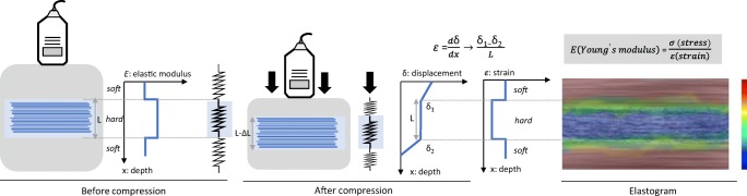
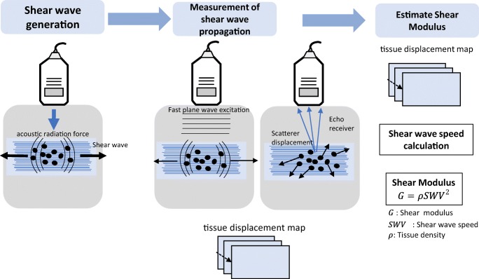
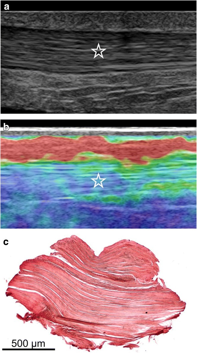
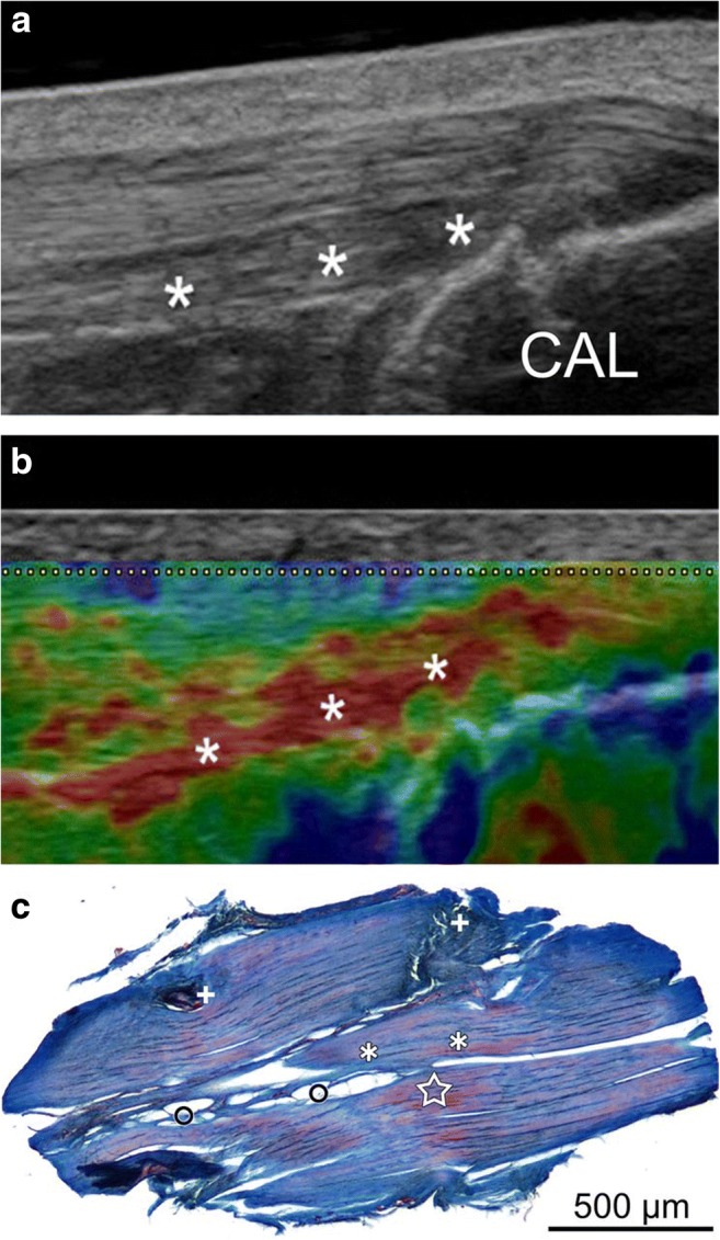
Ultrasound elastography (USE) is a recent technology that has experienced major developments in the past two decades. The assessment of the main mechanical properties of tissues can be made with this technology by characterisation of their response to stress. This article reviews the two major techniques used in musculoskeletal elastography, compression elastography (CE) and shear-wave elastography (SWE), and evaluates the studies published on major electronic databases that use both techniques in the context of tendon pathology. CE accounts for more studies than SWE. The mechanical properties of tendons, particularly their stiffness, may be altered in the presence of tendon injury. CE and SWE have already been used for the assessment of Achilles tendons, patellar tendon, quadriceps tendon, epicondylar tendons and rotator cuff tendons and muscles. Achilles tendinopathy is the most studied tendon injury with USE, including the postoperative period after surgical repair of Achilles rupture tendon. In relation to conventional ultrasound (US), USE potentially increases the sensitivity and diagnostic accuracy in tendinopathy, and can detect pathological changes before they are visible in conventional US imaging. Several technical limitations are recognised, and standardisation is necessary to ensure repeatability and comparability of the results when using these techniques. Still, USE is a promising technique under development and may be used not only to promote an early diagnosis, but also to identify the risk of injury and to support the evaluation of rehabilitation interventions. KEY POINTS: • USE is used for the assessment of the mechanical properties of tissues, including the tendons. • USE increases diagnostic performance when coupled to conventional US imaging modalities. • USE will be useful in early diagnosis, tracking outcomes and monitoring treatments of tendon injury. • Technical issues and lack of standardisation limits USE use in the assessment of tendon injury.
Keywords: Elastography; Sonoelastography; Tendinopathy; Tendon injury; Ultrasound.
Figures
Similar articles
-
Evaluation of Postoperative Changes in Patellar and Quadriceps Tendons after Total Knee Arthroplasty-A Comprehensive Analysis by Shear Wave Elastography, Power Doppler and B-mode Ultrasound.Acad Radiol. 2020 Jun;27(6):e148-e157. doi: 10.1016/j.acra.2019.08.015. Epub 2019 Sep 14. Acad Radiol. 2020. PMID: 31526688
-
Quantifying Mechanical Properties of the Patellar and Achilles Tendons Using Ultrasound Shear Wave Elastography: A Pilot Study.Diagnostics (Basel). 2025 Apr 1;15(7):879. doi: 10.3390/diagnostics15070879. Diagnostics (Basel). 2025. PMID: 40218229 Free PMC article.
-
"Soft, hard, or just right?" Applications and limitations of axial-strain sonoelastography and shear-wave elastography in the assessment of tendon injuries.Skeletal Radiol. 2014 Jan;43(1):1-12. doi: 10.1007/s00256-013-1695-3. Epub 2013 Aug 8. Skeletal Radiol. 2014. PMID: 23925561 Review.
-
Quantitative shear wave elastography compared to standard ultrasound (qualitative B-mode grayscale sonography and quantitative power Doppler) for evaluation of achillotendinopathy in treatment-naïve individuals: A cross-sectional study.Adv Clin Exp Med. 2022 Aug;31(8):847-854. doi: 10.17219/acem/147878. Adv Clin Exp Med. 2022. PMID: 35593220
-
The Use of Shear-Wave Ultrasound Elastography in the Diagnosis and Monitoring of Musculoskeletal Injuries.Crit Rev Biomed Eng. 2024;52(2):15-26. doi: 10.1615/CritRevBiomedEng.2023049807. Crit Rev Biomed Eng. 2024. PMID: 38305275 Review.
Cited by
-
2D shear wave elastography for the assessment of quadriceps entheses-a methodological study.Skeletal Radiol. 2024 Mar;53(3):455-463. doi: 10.1007/s00256-023-04425-1. Epub 2023 Aug 18. Skeletal Radiol. 2024. PMID: 37594519
-
Global trends in research of achilles tendon injury/rupture: A bibliometric analysis, 2000-2021.Front Surg. 2023 Mar 27;10:1051429. doi: 10.3389/fsurg.2023.1051429. eCollection 2023. Front Surg. 2023. PMID: 37051567 Free PMC article.
-
Elastography in the assessment of the Achilles tendon: a systematic review of measurement properties.J Foot Ankle Res. 2023 Apr 27;16(1):23. doi: 10.1186/s13047-023-00623-1. J Foot Ankle Res. 2023. PMID: 37101290 Free PMC article.
-
Ultrasonographic Evaluation of Thickness and Stiffness of Achilles Tendon and Plantar Fascia in Type 2 Diabetics Patients: A Cross-sectional Observation Study.J Med Ultrasound. 2023 Feb 13;31(4):282-286. doi: 10.4103/jmu.jmu_109_22. eCollection 2023 Oct-Dec. J Med Ultrasound. 2023. PMID: 38264597 Free PMC article.
-
In Vivo Effects of Joint Movement on Nerve Mechanical Properties Assessed with Shear-Wave Elastography: A Systematic Review and Meta-Analysis.Diagnostics (Basel). 2024 Feb 5;14(3):343. doi: 10.3390/diagnostics14030343. Diagnostics (Basel). 2024. PMID: 38337859 Free PMC article. Review.
References
-
- Ophir J, Céspedes I, Ponnekanti H, Yazdi Y, Li X. Elastography: a quantitative method for imaging the elasticity of biological tissues. Ultrason Imaging. 1991;13(2):111–134. - PubMed
Publication types
LinkOut - more resources
Full Text Sources
Other Literature Sources
Medical
Miscellaneous
