. 2018 Nov;19(11):840-853.
doi: 10.1111/tra.12610. Epub 2018 Sep 12.

T-complex protein 1-ring complex enhances retrograde axonal transport by modulating tau phosphorylation

Affiliations

T-complex protein 1-ring complex enhances retrograde axonal transport by modulating tau phosphorylation

Xu-Qiao Chen et al. Traffic. 2018 Nov.

Abstract

The cytosolic chaperonin T-complex protein (TCP) 1-ring complex (TRiC) has been shown to exert neuroprotective effects on axonal transport through clearance of mutant Huntingtin (mHTT) in Huntington's disease. However, it is presently unknown if TRiC also has any effect on axonal transport in wild-type neurons. Here, we examined how TRiC impacted the retrograde axonal transport of brain-derived neurotrophic factor (BDNF). We found that expression of a single TRiC subunit significantly enhanced axonal transport of BDNF, leading to an increase in instantaneous velocity with a concomitant decrease in pauses for retrograde BDNF transport. The transport enhancing effect by TRiC was dependent on endogenous tau expression because no effect was seen in neurons from tau knockout mice. We showed that TRiC regulated the level of cyclin-dependent kinase 5 (CDK5)/p35 positively, contributing to TRiC-mediated tau phosphorylation (ptau). Expression of a single TRiC subunit increased the level of ptau while downregulation of the TRiC complex decreased ptau. We further demonstrated that TRiC-mediated increase in ptau induced detachment of tau from microtubules. Our study has thus revealed that TRiC-mediated increase in tau phosphorylation impacts retrograde axonal transport.

Keywords: TRiC/CCT chaperonin; axonal transport; hyperphosphorylation; tau.


Conflict of interest statement

Conflict of interest

The authors declare no conflict of interest.

Figures

Figure 1.
Figure 1.. CCT5 promotes retrograde axonal transport of BDNF in rat E18 cortical neurons.
(A) Primary cultured rat cortical neurons at DIV5 were infected with hCCT5-Avi lentivirus at a MOI of 10 for 4 days followed by immunostaining with an anti-tau antibody. Representative images were shown. (B) Experimental design. (C) Representative kymographs of axonally transported QD-BDNF in cultured rat cortical neurons infected with control or CCT5-Avi lentivirus. Scale bar, 10 μm. The data for retrograde instantaneous velocity, percentage of pause events, average pause duration, average velocity of axonally transported QD-BDNF were quantitated and presented in D-G. Results are shown as mean ± SEM from three independent experiments with 60–100 QD-BDNF puncta recorded. The distribution of stationary versus non-stationary of QD-BDNF puncta is presented in H. *p<0.05, **p<0.01, unpaired student t-test.
Figure 2.
Figure 2.. CCT5-mediated enhancement of retrograde axonal transport of QD-BDNF is negated in tau knockout (tau−/−) mouse E18 cortical neurons.
(A) Experimental design. Cultured tau−/− mouse cortical neurons were infected with control-, tauWT- or tauP301L-lentivirus for 4 days. (B) The expression level of tauWT and tauP301L in tau−/− cortical neurons was detected by immunoblotting. (C) Representative kymographs of QD-BDNF retrograde transport in cultured tau−/− mouse cortical neurons infected with control or CCT5-Avi, and either tauWT or tauP301L lentivirus in microfluidic chamber. Scale bar, 10 μm. The data for retrograde instantaneous velocity, percentage of pause events, average pause duration, average velocity of axonally transported QD-BDNF were quantitated and presented in D-G. Results are shown as mean ± SEM from three independent experiments with 50–70 QD-BDNF puncta recorded. The distribution of stationary versus non-stationary of QD-BDNF puncta in each condition is presented in H, *p<0.05, **p<0.01, and ***p<0.001, unpaired student t-test.
Figure 3.
Figure 3.. CCT complex regulates the level of CDK5/p35 in a tau-dependent manner.
(A, B) The impact of CCT subunit(s) on the level of CDK5 and p35 in primary rat cortical neurons was analyzed. N=4–5; *p<0.05, **p<0.01, ns, not significant, paired student t-test. (C, D) The effect of CCT5 overexpression or knockdown of CCT complex on p25 protein level in primary neurons was analyzed. N=3; *p<0.05, paired student t-test. (E, F) As in A, the effects of CCT subunit(s) on the level and activity of GSK3β in primary neurons were analyzed. The activity of GSK3β was tested by two phosphor-specific antibodies to either Ser9 or Tyr216 of GSK3β. Asterisk indicates unspecific band. N=3. (G) Cultured tau−/− cortical neurons were infected with CCT5-Avi or control lentivirus and tauWT lentivirus or not for 4 days followed by detection of p35/CDK5 by Western blotting. N=3; *p<0.05, paired student t-test.
Figure 4.
Figure 4.. CCT complex modulates the phosphorylation level of endogenous tau in primary rat E18 cortical neurons.
(A) Rat cortical neurons infected with hCCT5-Avi or control lentivirus for 4 days were immunoblotted for ptau (AT8), tau and Avi. (B) The effects of lentivirus-mediated introduction of CCT3 and CCT6 on tau phosphorylation were analyzed. (C) Cultured rat cortical neurons were infected with control or rCCT2 shRNA-expressing lentivirus for analysis of AT8 levels as well as tau and CCT2. (D, E) The effect of CCT5 overexpression or knockdown of CCT complex on PHF signal in primary neurons was analyzed. (F) Rat E18 cultured cortical neurons that were infected with control or CCT5-Avi lentivirus for 4 days were treated with 20 μM roscovitine for 8 h. Neurons were harvested and analyzed by immunoblotting with indicated antibodies. (G) Rat cultured cortical neurons infected with or without CCT5-Avi or control lentivirus for 4 days were treated with 20 mM LiCl for 2 h or not before lysis for immunoblotting. N=3–5; *p<0.05, **p<0.01, paired student t-test in A, C, D, E, F. N=4; *p<0.05, ***p<0.001, one way-ANOVA test followed by Newman-Keuls multiple comparison test in G.
Figure 5.
Figure 5.. Upregulation of CCT5 reduces tau binding to microtubules and prevents tau-induced microtubule bundling.
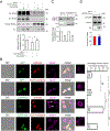
(A) Rat E18 cultured cortical neurons that were infected with control or CCT5-Avi lentivirus for 4 days were treated with vehicle or 20 μM roscovitine for 8 h followed by microtubule-binding assay as A. The supernatant (S) and pellet (P) fractions were analyzed by SDS–PAGE followed by immunoblotting with tau antibody to visualize tau and DM1A antibody to detect tubulin. The ratios of tau in P versus S fractions were quantitated. N=3; *p<0.05, one way-ANOVA test followed by Bonferroni multiple comparison test. (B) mCherry or CCT5-mCherry was co-transfected with either EGFP or EGFP-tau into CHO cells. Microtubules were stained with the DM1A antibody against α-tubulin and presented in magenta color. Arrows indicate cells containing tau-induced microtubule bundling. ***p<0.001, ns, not significant, one way-ANOVA test followed by Bonferroni multiple comparison test. (C) Lentivirus-infected primary neurons were lysed with hypotonic buffer. Equal volumes of supernatant (S) and pellet (P) proteins were separated by SDS-PAGE and immunoblotted with antibodies against α-tubulin (DM1A) and Avi. N=3. (D) Rat cortical neurons infected with hCCT5-Avi or control lentivirus for 4 days were analyzed for tubulin acetylation. N=3, n.s., not significant, paired student t-test.
Figure 6.
Figure 6.. CCT5 mediates promotion of retrograde axonal transport of BDNF likely through CDK5 pathway in rat E18 cortical neurons.
(A) Experimental design. (B) Representative kymographs of axonally transported QD-BDNF in cultured rat cortical neurons infected with control or CCT5-Avi lentivirus. These neurons were treated with 20 μM roscovitine (Ros) for 4 h in microfluidic chamber. Scale bar, 10 μm. The data for retrograde instantaneous velocity, percentage of pause events, average pause duration, average velocity of axonally transported QD-BDNF were quantitated and presented in C-F. Results are shown as mean ± SEM from three independent experiments with 50–60 QD-BDNF puncta recorded. The distribution of stationary versus non-stationary of QD-BDNF puncta is presented in G.

Similar articles

Cited by

References

    1. Lopez T, Dalton K, Frydman J. The Mechanism and Function of Group II Chaperonins. Journal of molecular biology 2015;427(18):2919–2930. - PMC - PubMed
    1. Yebenes H, Mesa P, Munoz IG, Montoya G, Valpuesta JM. Chaperonins: two rings for folding. Trends in biochemical sciences 2011;36(8):424–432. - PubMed
    1. Hartl FU, Bracher A, Hayer-Hartl M. Molecular chaperones in protein folding and proteostasis. Nature 2011;475(7356):324–332. - PubMed
    1. Spiess C, Meyer AS, Reissmann S, Frydman J. Mechanism of the eukaryotic chaperonin: protein folding in the chamber of secrets. Trends in cell biology 2004;14(11):598–604. - PMC - PubMed
    1. Tam S, Geller R, Spiess C, Frydman J. The chaperonin TRiC controls polyglutamine aggregation and toxicity through subunit-specific interactions. Nature cell biology 2006;8(10):1155–1162. - PMC - PubMed

Publication types

Substances