

Vamorolone treatment improves skeletal muscle outcome in a critical illness myopathy rat model
- PMID: 30120816
- PMCID: PMC8424699
- DOI: 10.1111/apha.13172
Vamorolone treatment improves skeletal muscle outcome in a critical illness myopathy rat model
Abstract
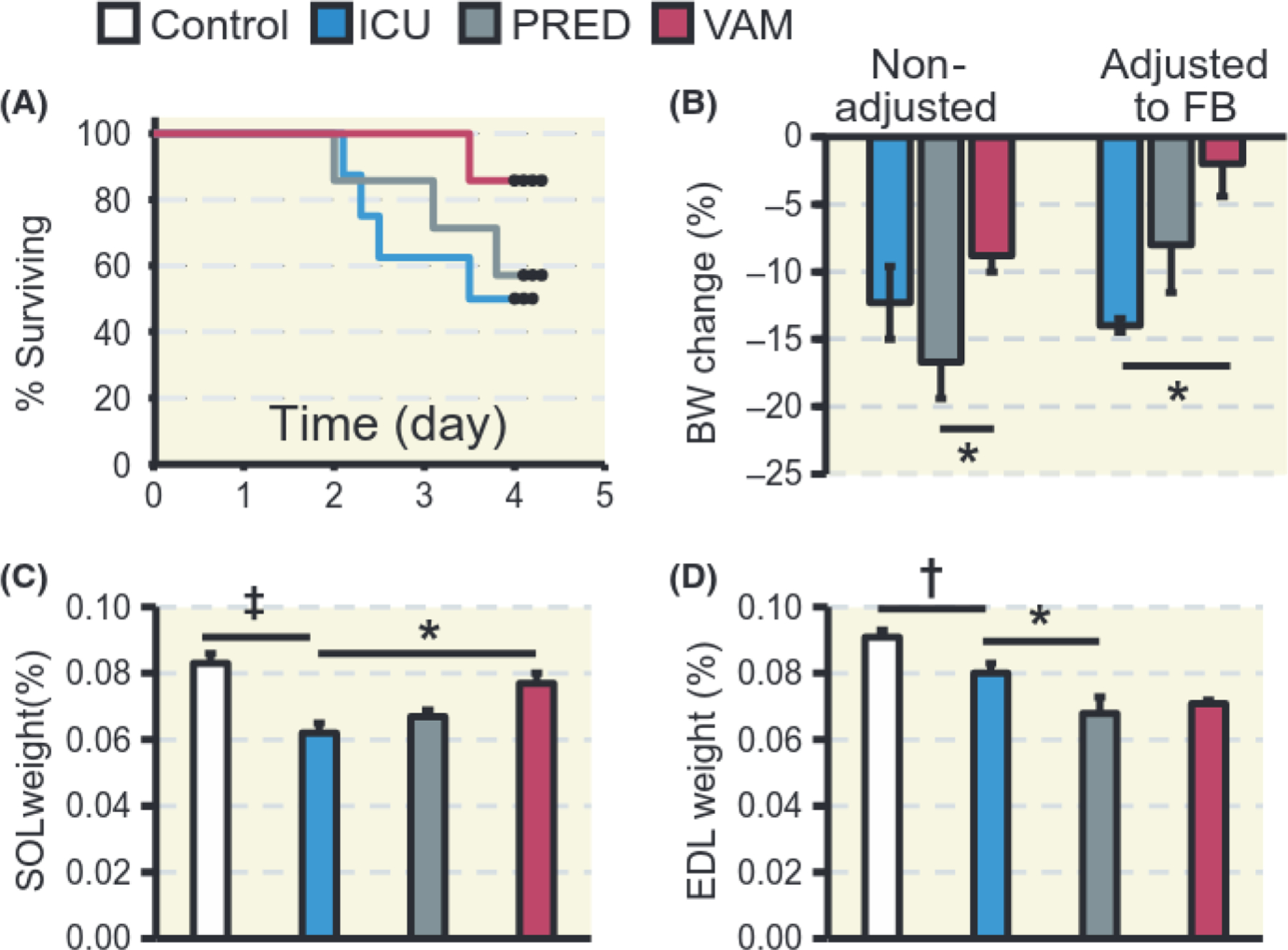
Aim: Critical illness myopathy (CIM) is a consequence of modern critical care, leading to skeletal muscle atrophy/paralysis with negative consequences for mortality/morbidity and health care costs. Glucocorticoids (GCs) have been proposed to trigger CIM. Here, we compare outcomes of two GCs, the commonly used prednisolone and the newly developed dissociative vamorolone in response to the intensive care unit (ICU) condition for 5 days, ie, sedation, immobilization, and mechanical ventilation.
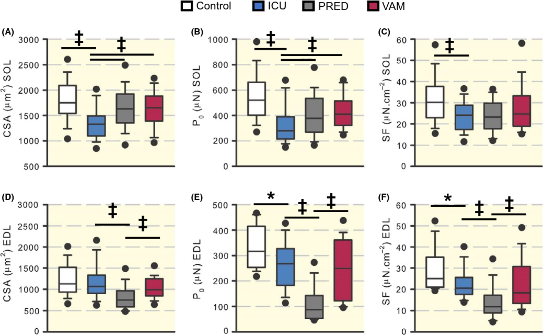
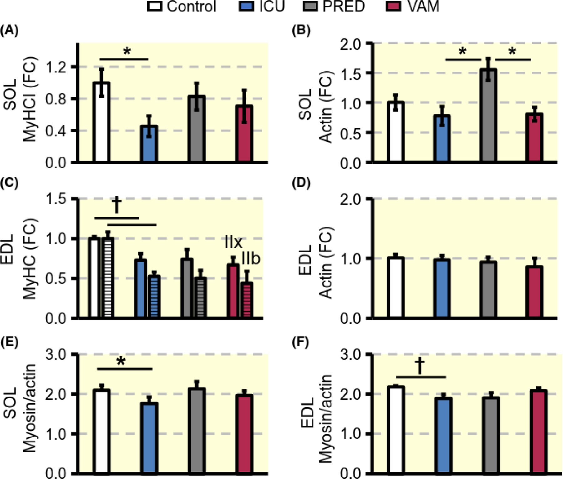
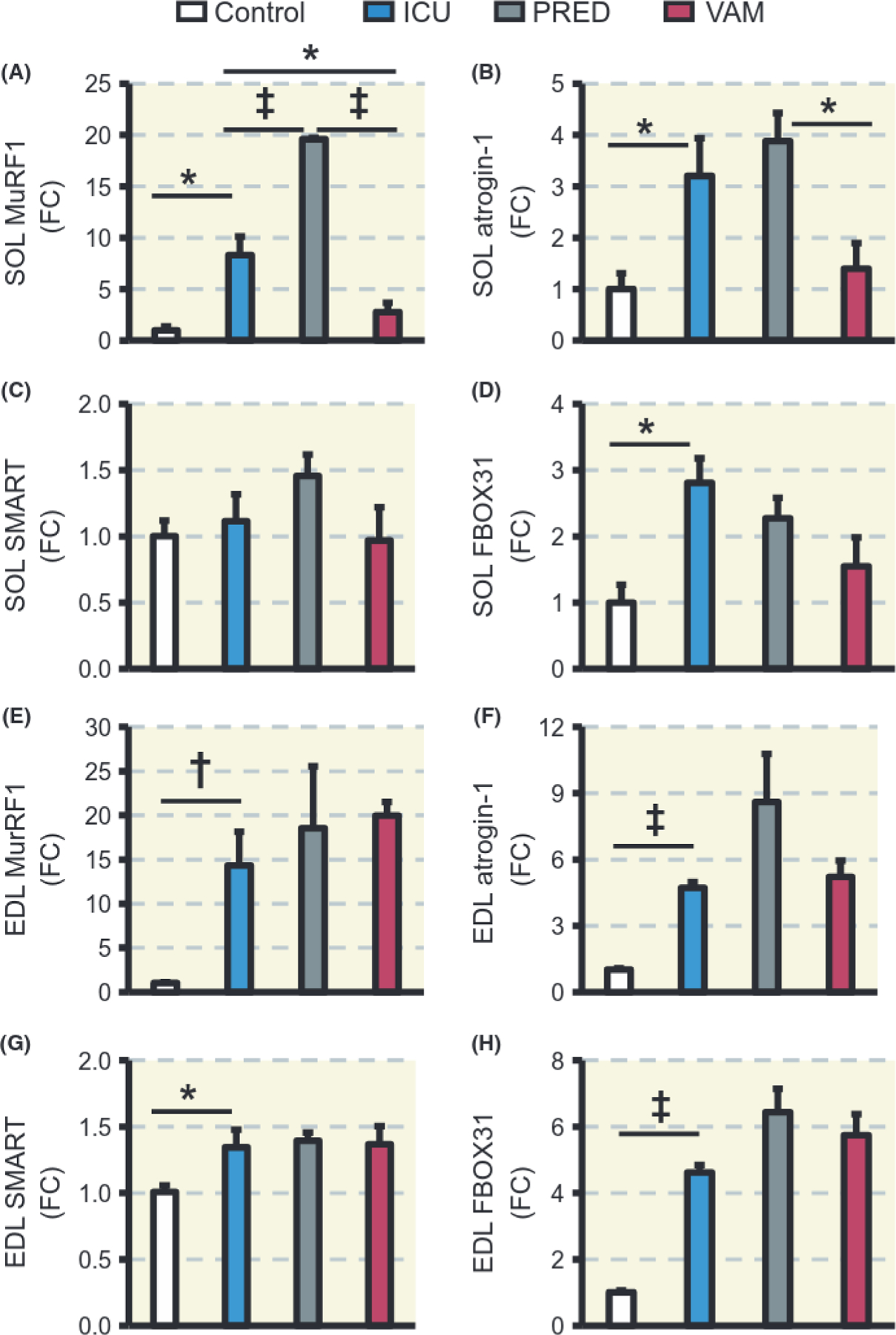
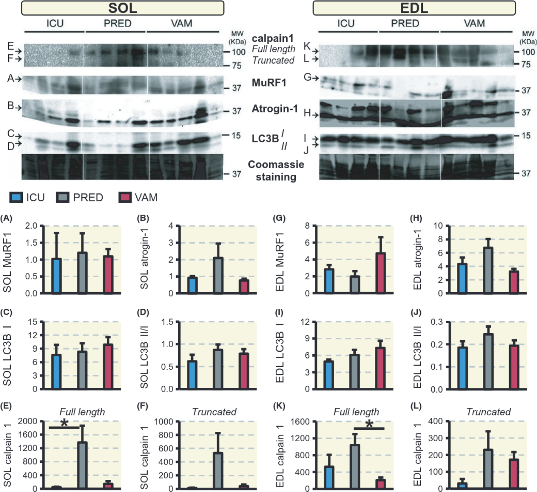
Methods: Rats were divided into a 0-day sham-operated control group, and three groups exposed to 5 days ICU condition during treatment with prednisolone (PRED) or vamorolone (VAM) or none of these GCs (ICU-group). Survival, body and muscle weights, cytokine concentrations, regulation of muscle contraction in single fast- and slow-twitch muscle fibres, myofibrillar protein expression and protein degradation pathways were studied.
Results: Critical illness myopathy geno- and pheno-types were confirmed in the ICU group. However, VAM and PRED groups showed reduced atrophy/weakness than the ICU group, and muscle specific differences with more severe negative effects on fast-twitch muscle fibres in the PRED than the other groups.
Conclusion: These results show that vamorolone provides a GC intervention superior to typical GCs in improving CIM outcomes. Further, the findings do not support the notion that moderate-dose GC treatment represents a factor triggering CIM.
Keywords: Glucocorticoids; ICU; muscle wasting; prednisolone; vamorolone.
© 2018 Scandinavian Physiological Society. Published by John Wiley & Sons Ltd.
Conflict of interest statement
CONFLICT OF INTEREST
The authors declare no conflict of interest.
Figures
Comment in
-
Critical illness myopathy: Glucocorticoids revisited?Acta Physiol (Oxf). 2019 Feb;225(2):e13205. doi: 10.1111/apha.13205. Epub 2018 Nov 2. Acta Physiol (Oxf). 2019. PMID: 30338655 No abstract available.
References
-
- Larsson L, Li XP, Edstrom L, et al.Acute quadriplegia and loss of muscle myosin in patients treated with nondepolarizing neuromuscular blocking agents and corticasteroids: mechanisms at the cellular and molecular levels. Crit Care Med 2000;28(1):34–45. - PubMed
-
- De Letter M, van Doom PA, Savelkoul HFJ, et al.Critical illness polyneuropathy and myopathy (CIPNM): evidence for local immune activation by cytokine-expression in the muscle tissue. J Neuroinununol 2000;106(1–2):206–213. - PubMed
-
- Henry J, Kaminski RLR. 1994. Endocrine myopathies (hyper-and hyponinction of adrenal, thyroid, pituitary, and parathyroid and iatrogenic corticosteroid myopathy) Chapter 66. In: Engel AG, Franzini-Armstrong C, (eds) Myology, pp. 1726–1753. McGraw-Hill, New York, NY, USA.
-
- Perrot S, Le Jeunne C. Steroid-induced myopathy. Presse Med 2012;41(4):422–426. - PubMed
-
- Tang BMP, Craig IC, Eslick GD, Seppelt I, McLean AS. Use of corticosteroids in acute lung injury and acute respiratory distress syndrome: a systematic review and meta-analysis. Crit Care Med 2009;37(5):1594–1603. - PubMed
Publication types
MeSH terms
Substances
Grants and funding
LinkOut - more resources
Full Text Sources
Other Literature Sources
Medical
Miscellaneous
