. 2018 Aug;24(3):1-9.
doi: 10.1117/1.JBO.24.3.031003.

Visualization of skin microvascular dysfunction of type 1 diabetic mice using in vivo skin optical clearing method

Affiliations

Visualization of skin microvascular dysfunction of type 1 diabetic mice using in vivo skin optical clearing method

Wei Feng et al. J Biomed Opt. 2018 Aug.

Abstract

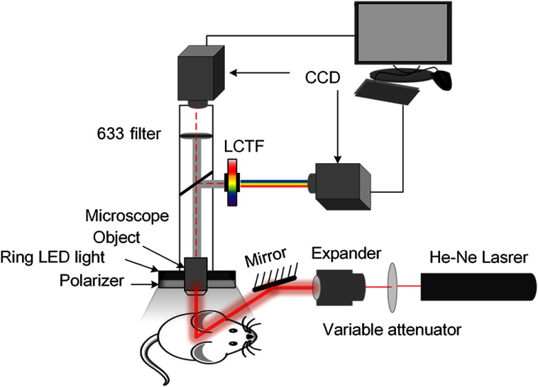
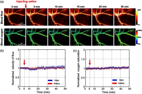
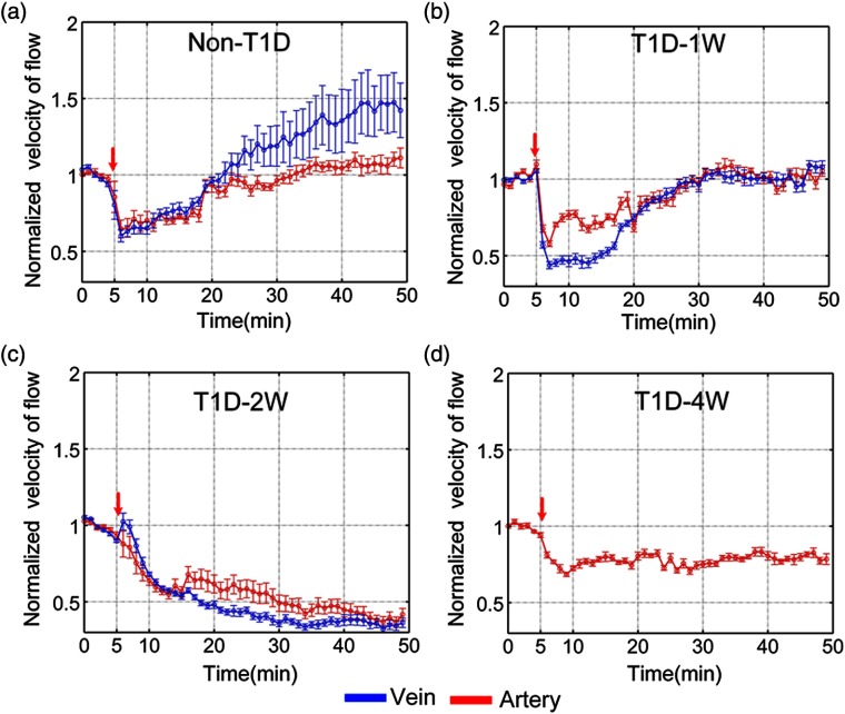
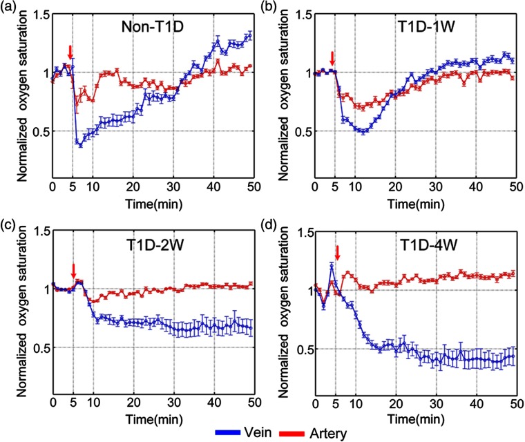
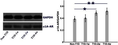
To realize visualization of the skin microvascular dysfunction of type 1 diabetic mice, we combined laser speckle contrast imaging and hyperspectral imaging to simultaneously monitor the noradrenaline (NE)-induced responses of vascular blood flow and blood oxygen with the development of diabetes through optical clearing skin window. The main results showed that venous and arterious blood flow decreased without recovery after injection of NE; furthermore, the decrease of arterious blood oxygen induced by NE greatly weakened, especially for 2- and 4-week diabetic mice. This change in vasoconstricting effect of NE was related to the expression of α1-adrenergic receptor. This study demonstrated that skin microvascular function was a potential research biomarker for early warning in the occurrence and development of diabetes. The in vivo skin optical clearing method provides a feasible solution to realize visualization of cutaneous microvessels for monitoring microvascular reactivity under pathological conditions. In addition, visual monitoring of skin microvascular function response has guiding significance for early diagnosis of diabetes and clinical research.

Keywords: diabetes; hyperspectral imaging; laser speckle imaging; microcirculation; skin optical clearing; vascular dysfunction.


Figures

Fig. 1
Fig. 1
Schematic of the imaging system.
Fig. 2
Fig. 2
Monitoring vascular blood flow and blood oxygen through optical clearing skin window. (a) Typical skin vascular blood flow and blood oxygen saturation maps before and after injection of saline. The time-lapse changes of (b) the corresponding blood flow and (c) oxygen saturation in artery (red line) and vein (blue line) before and after injection of saline. The red arrows refer to time of injection (n=8, mean ± standard deviation).
Fig. 3
Fig. 3
Dynamic monitoring of NE-induced skin vascular blood flow response at different stages of T1D through optical clearing skin window.
Fig. 4
Fig. 4
The time-lapse blood flow dynamic responses in artery (red line) and vein (blue line) at different stages of T1D: (a) non-T1D, (b) T1D-1w, (c) T1D-2w, and (d) T1D-4w. The red arrows refer to the time of injection (n=8 for each group, mean ± standard error).
Fig. 5
Fig. 5
Dynamic monitoring of NE-induced corresponding skin vascular blood oxygen response at different stages of T1D through optical clearing skin window.
Fig. 6
Fig. 6
The time-lapse blood oxygen dynamic responses in artery (red line) and vein (blue line) at different stages of T1D: (a) non-T1D, (b) T1D-1w, (c) T1D-2w, and (d) T1D-4w. The red arrows refer to the time of injection (n=8 for each group, mean ± standard error).
Fig. 7
Fig. 7
The expression level of α1-AR at different stages of T1D assessed by western blot. ★ p<0.05 and ★★ p<0.01 compared with non-T1D (n=9 for each group, mean ± standard error).

Similar articles

Cited by

References

    1. Cameron N. E., Cotter M. A., “The relationship of vascular changes to metabolic factors in diabetes mellitus and their role in the development of peripheral nerve complications,” Diabetes/Metab. Rev. 10(3), 189–224 (1994).DMREEG10.1002/dmr.5610100302 - DOI - PubMed
    1. Roosterman D., et al. , “Neuronal control of skin function: the skin as a neuroimmunoendocrine organ,” Physiol. Rev. 86(4), 1309–1379 (2006).PHREA710.1152/physrev.00026.2005 - DOI - PubMed
    1. Duff M., et al. , “Cutaneous manifestation of diabetes mellitus,” Clin. Diabetes 33(1), 40–48 (2015).10.2337/diaclin.33.1.40 - DOI - PMC - PubMed
    1. Quondamatteo F., “Skin and diabetes mellitus: what do we know?” Cell Tissue Res. 355(1), 1–21 (2014).10.1007/s00441-013-1751-2 - DOI - PubMed
    1. Goncalves N. P., et al. , “Schwann cell interactions with axons and microvessels in diabetic neuropathy,” Nat. Rev. Neurol. 13(3), 135–147 (2017).10.1038/nrneurol.2016.201 - DOI - PMC - PubMed

Publication types