

The neural basis of motivational influences on cognitive control
- PMID: 30120846
- PMCID: PMC6866502
- DOI: 10.1002/hbm.24348
The neural basis of motivational influences on cognitive control
Abstract
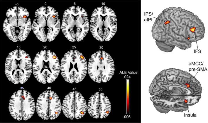
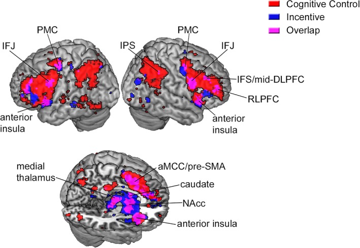
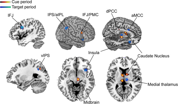
Cognitive control mechanisms support the deliberate regulation of thought and behavior based on current goals. Recent work suggests that motivational incentives improve cognitive control and has begun to elucidate critical neural substrates. We conducted a quantitative meta-analysis of neuroimaging studies of motivated cognitive control using activation likelihood estimation (ALE) and Neurosynth to delineate the brain regions that are consistently activated across studies. The analysis included studies that investigated changes in brain activation during cognitive control tasks when reward incentives were present versus absent. The ALE analysis revealed consistent recruitment in regions associated with the frontoparietal control network including the inferior frontal sulcus and intraparietal sulcus, as well as regions associated with the salience network including the anterior insula and anterior mid-cingulate cortex. As a complementary analysis, we performed a large-scale exploratory meta-analysis using Neurosynth to identify regions that are recruited in studies using of the terms cognitive control and incentive. This analysis replicated the ALE results and also identified the rostrolateral prefrontal cortex, caudate nucleus, nucleus accumbens, medial thalamus, inferior frontal junction, premotor cortex, and hippocampus. Finally, we separately compared recruitment during cue and target periods, which tap into proactive engagement of rule-outcome associations, and the mobilization of appropriate viscero-motor states to execute a response, respectively. We found that largely distinct sets of brain regions are recruited during cue and target periods. Altogether, these findings suggest that flexible interactions between frontoparietal, salience, and dopaminergic midbrain-striatal networks may allow control demands to be precisely tailored based on expected value.
Keywords: cognitive control; control network; frontoparietal; reward.
© 2018 Wiley Periodicals, Inc.
Conflict of interest statement
The authors declared no conflict of interest.
Figures
Similar articles
-
The wandering brain: meta-analysis of functional neuroimaging studies of mind-wandering and related spontaneous thought processes.Neuroimage. 2015 May 1;111:611-21. doi: 10.1016/j.neuroimage.2015.02.039. Epub 2015 Feb 25. Neuroimage. 2015. PMID: 25725466 Review.
-
Neural correlates of interference resolution in the multi-source interference task: a meta-analysis of functional neuroimaging studies.Behav Brain Funct. 2018 Apr 10;14(1):8. doi: 10.1186/s12993-018-0140-0. Behav Brain Funct. 2018. PMID: 29636070 Free PMC article.
-
Motivation by potential gains and losses affects control processes via different mechanisms in the attentional network.Neuroimage. 2015 May 1;111:549-61. doi: 10.1016/j.neuroimage.2015.02.047. Epub 2015 Feb 27. Neuroimage. 2015. PMID: 25731995
-
Motivated cognitive control: reward incentives modulate preparatory neural activity during task-switching.J Neurosci. 2010 Aug 4;30(31):10294-305. doi: 10.1523/JNEUROSCI.2052-10.2010. J Neurosci. 2010. Retraction in: J Neurosci. 2013 May 15;33(20):8922. doi: 10.1523/JNEUROSCI.1696-13.2013. PMID: 20685974 Free PMC article. Retracted.
-
Common and distinct networks underlying reward valence and processing stages: a meta-analysis of functional neuroimaging studies.Neurosci Biobehav Rev. 2011 Apr;35(5):1219-36. doi: 10.1016/j.neubiorev.2010.12.012. Epub 2010 Dec 24. Neurosci Biobehav Rev. 2011. PMID: 21185861 Free PMC article. Review.
Cited by
-
Learning when effort matters: neural dynamics underlying updating and adaptation to changes in performance efficacy.Cereb Cortex. 2023 Feb 20;33(5):2395-2411. doi: 10.1093/cercor/bhac215. Cereb Cortex. 2023. PMID: 35695774 Free PMC article.
-
Dissociable influences of reward and punishment on adaptive cognitive control.PLoS Comput Biol. 2021 Dec 28;17(12):e1009737. doi: 10.1371/journal.pcbi.1009737. eCollection 2021 Dec. PLoS Comput Biol. 2021. PMID: 34962931 Free PMC article.
-
Interactions of Motivation and Cognitive Control.Curr Opin Behav Sci. 2018 Feb;19:83-90. doi: 10.1016/j.cobeha.2017.11.009. Epub 2017 Nov 24. Curr Opin Behav Sci. 2018. PMID: 30035206 Free PMC article.
-
Age-Related Structural and Functional Changes of the Hippocampus and the Relationship with Inhibitory Control.Brain Sci. 2020 Dec 19;10(12):1013. doi: 10.3390/brainsci10121013. Brain Sci. 2020. PMID: 33352718 Free PMC article.
-
Control adjustment costs limit goal flexibility: Empirical evidence and a computational account.bioRxiv [Preprint]. 2025 Feb 3:2023.08.22.554296. doi: 10.1101/2023.08.22.554296. bioRxiv. 2025. Update in: Psychol Rev. 2025 Jul 28. doi: 10.1037/rev0000576. PMID: 37662382 Free PMC article. Updated. Preprint.
References
-
- Aron, A. R. , Robbins, T. W. , & Poldrack, R. A. (2004). Inhibition and the right inferior frontal cortex. Trends in Cognitive Sciences, 8(4), 170–177. - PubMed
-
- Asaad, W. F. , Rainer, G. , & Miller, E. K. (2000). Task‐specific neural activity in the primate prefrontal cortex. Journal of Neurophysiology, 84(1), 451–459. - PubMed
Publication types
MeSH terms
LinkOut - more resources
Full Text Sources
Other Literature Sources
Research Materials
Miscellaneous
