. 2019 Jan;139(1):146-156.
doi: 10.1016/j.jid.2018.07.025. Epub 2018 Aug 16.

A Critical Role of the IL-1β-IL-1R Signaling Pathway in Skin Inflammation and Psoriasis Pathogenesis

Affiliations

A Critical Role of the IL-1β-IL-1R Signaling Pathway in Skin Inflammation and Psoriasis Pathogenesis

Yihua Cai et al. J Invest Dermatol. 2019 Jan.

Abstract

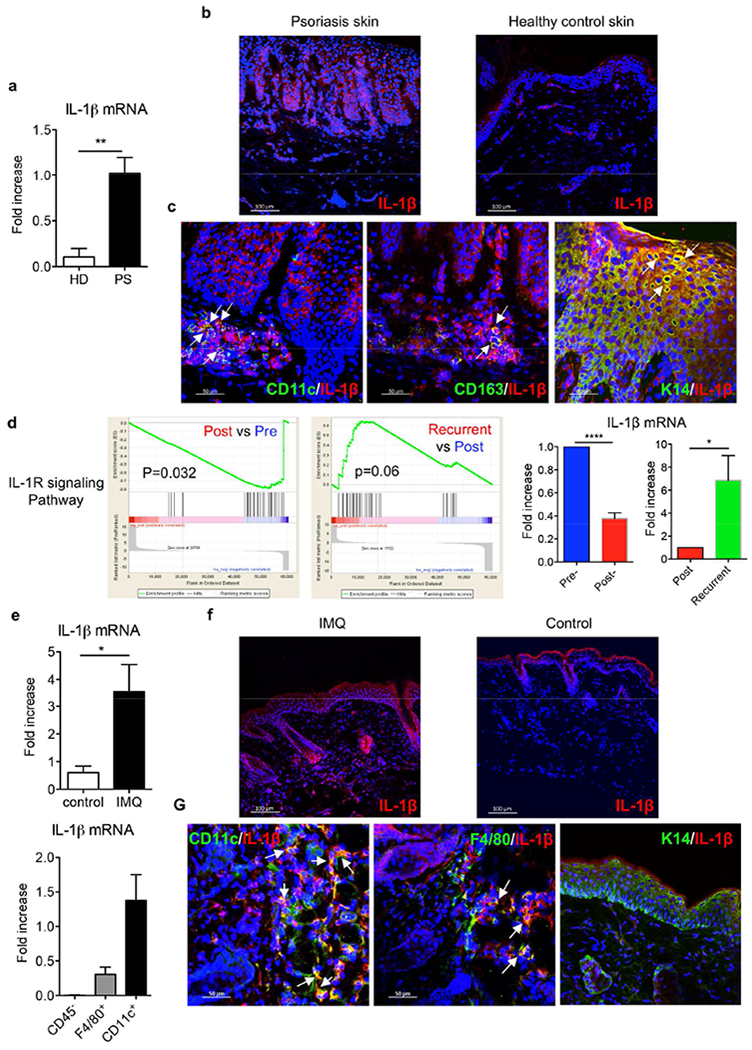
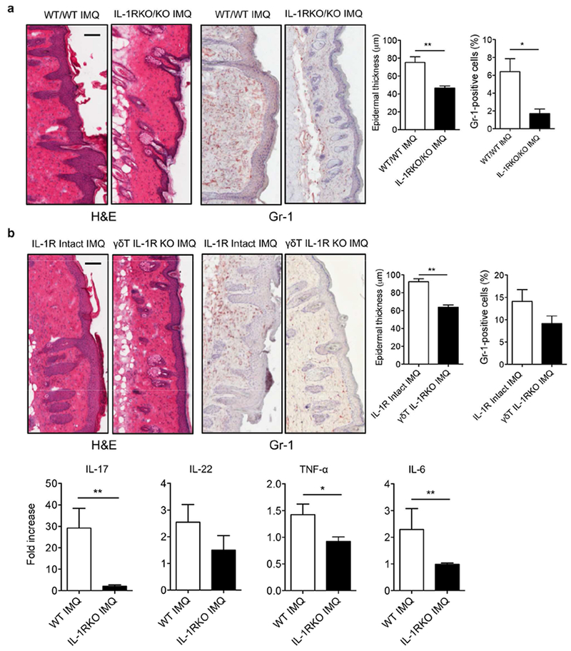
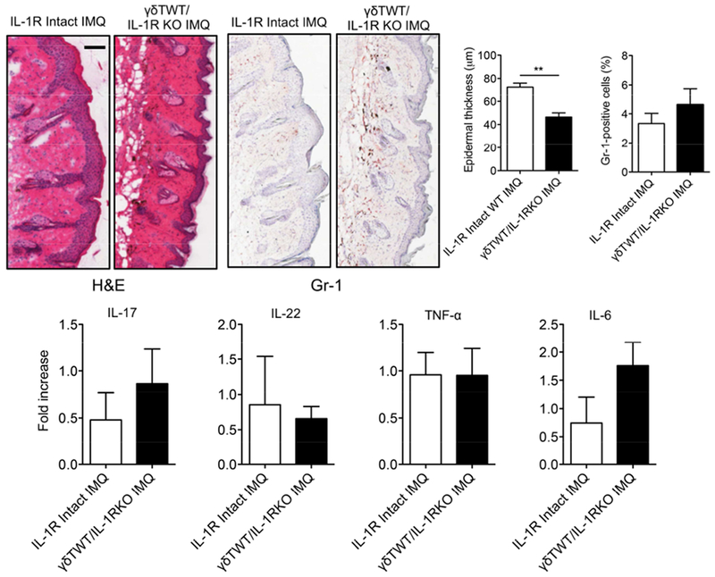
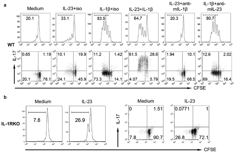
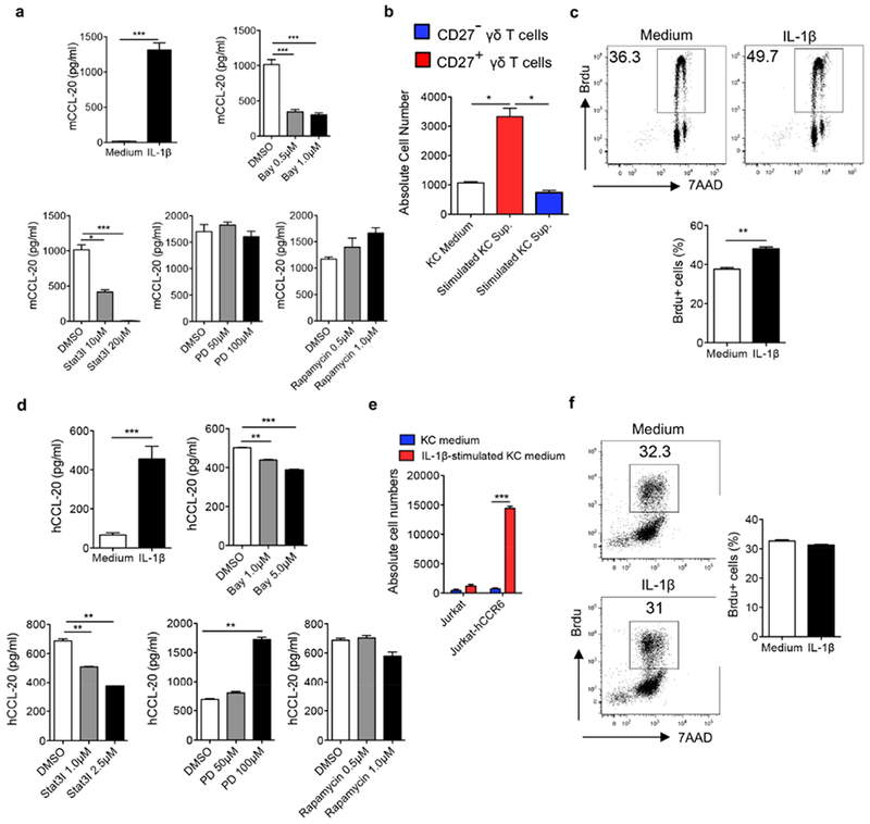
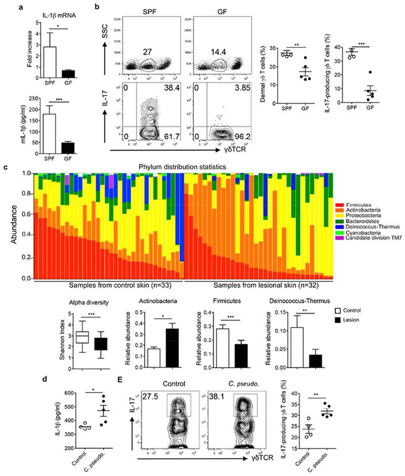
The IL-1 signaling pathway has been shown to play a critical role in the pathogenesis of chronic, autoinflammatory skin diseases such as psoriasis. However, the exact cellular and molecular mechanisms have not been fully understood. Here, we show that IL-1β is significantly elevated in psoriatic lesional skin and imiquimod-treated mouse skin. In addition, IL-1R signaling appears to correlate with psoriasis disease progression and treatment response. IL-1 signaling in both dermal γδ T cells and other cells such as keratinocytes is essential to an IMQ-induced skin inflammation. IL-1β induces dermal γδ T cell proliferation and IL-17 production in mice. In addition, IL-1β stimulates keratinocytes to secrete chemokines that preferentially chemoattract peripheral CD27- CCR6+IL-17 capable of producing γδ T cells (γδT17). Further studies showed that endogenous IL-1β secretion is regulated by skin commensals to maintain dermal γδT17 homeostasis in mice. Mouse skin associated with Corynebacterium species, bacteria enriched in human psoriatic lesional skin, has increased IL-1β and dermal γδT17 cell expansion. Thus, the IL-1β-IL-1R signaling pathway may contribute to skin inflammation and psoriasis pathogenesis via the direct regulation of dermal IL-17-producing cells and stimulation of keratinocytes for amplifying inflammatory cascade.


Conflict of interest statement

Conflict of Interest: YZ declared a conflict of interest and all other authors declared no conflict of interests in this study.

Figures

Figure 1.
Figure 1.. IL-1β expression was significantly increased in both murine and human psoriatic lesions.
(a) IL-1β mRNA levels from skin biopsies collected from psoriasis patients (PS, n=5) and healthy donors (HD, n=4). (b) Frozen sections were stained with IL-1β mAb (red) and DAPI (blue). Scale bar =100 μm. (c) Frozen sections from psoriatic lesional skin were stained with IL-1β mAb (red) and human CD11c mAb (green) or human CD163 mAb (green) or keratin 14 mAb (green), and DAPI (blue). Scale bar =50 μm. (d) Gene set enrichment analysis identifies significant transcriptional downregulation of IL-1R signaling pathway in psoriasis patients initially effectively treated with glucocorticoid while upregulation in recurrent patients after stopping treatment (NCBI GEO with the accession number GSE114729). The mRNA expression of IL-1β by real-time PCR analysis was calculated using Pre- or Post-treatment level as base level. (e) C57BL/6 mice received daily topical application with IMQ or vehicle control for 3-5 days. IL-1β mRNA levels from IMQ-treated or vehicle control mouse skin were measured. CD11c+ cells, F4/80+ cells and CD45 cells were sorted from 3 days of IMQ-treated mouse skin and IL-1β mRNA level was measured. (f) Skin frozen sections from IMQ-or vehicle control-treated mice were stained with IL-1β mAb (red) and DAPI (blue). Scale bar =100 μm. (g) Skin frozen sections from IMQ-treated mice were stained with IL-1β mAb (red) and mouse CD11 c mAb (green), or mouse F4/80 mAb (green), or keratin 14 mAb (green), and DAPI (blue). Scale bar =50 μm. *p< 0.05, **p< 0.01, ***p< 0.001.
Figure 2.
Figure 2.. IL-1R signaling in γδ T cells is essential to induce skin inflammation.
(a) Reconstituted WT or IL-1R KO chimeric mice (n=3) were treated daily for 5 days with IMQ or vehicle control. Representative H&E-stained sections and frozen sections stained with Gr-1 are shown. Gr-1 positive cells are brown. Skin tissues were also stained with CD45 and Gr-1 assessed by flow cytometry. Epidermal thickness and percentage of CD45+Gr-1+ cells were measured. Scale bar =100 μm. Data are representative of two independent experiments with similar results. (b) Reconstituted mice (n=3) with WT γδ T cells or IL-1R deficiency in γδ T cells were treated daily for 5 days with IMQ or vehicle control. Representative H&E-stained sections and frozen sections stained with Gr-1 are shown. Gr-1 positive cells are brown. Skin tissues were also stained with CD45 and Gr-1 assessed by flow cytometry. Epidermal thickness and percentage of CD45+Gr-1+ cells were measured. Scale bar =100 μm. Data are representative of two independent experiments with similar results. The mRNA levels of IL-17, IL-22, TNF-α and IL-6 were measured by real-time PCR analysis. *p< 0.05, **p< 0.01.
Figure 3.
Figure 3.. IL-1R signaling in other cells is important to induce full-fledged skin inflammation.
Reconstituted mice (n=3) with WT γδ T cells and IL-1R intact or deficiency in other cells were treated daily for 5 days with IMQ or vehicle control. Representative H&E-stained sections and frozen sections stained with Gr-1 are shown. Gr-1 positive cells are brown. Skin tissues were also stained with CD45 and Gr-1 assessed by flow cytometry. Epidermal thickness and percentage of CD45+Gr-1+ cells were measured at day 5. Scale bar =100 μm. Data are representative of two independent experiments with similar results. The mRNA levels of IL-17, IL-22, TNF-α and IL-6 were measured by qPCR analysis. *p < 0.05.
Figure 4.
Figure 4.. IL-1R signaling is essential in dermal γδ T cell proliferation and IL-17 production.
(a) Whole skin cell suspensions from C57BL/6 WT mice were labeled with CFSE and then stimulated with IL-23, IL-1β, IL-23 plus IL-1β for 3 days. CFSE dilution and intracellular IL-17 production by dermal γδ T cells were determined by flow cytometry. Flow plots gated on CD3int γδTCRint cells are representative of at least three independent experiments with similar results. (b) Whole skin cell suspensions from IL-1R KO mice labeled with CFSE were stimulated with IL-23 for 3 days. CFSE dilution and intracellular IL-17 production by dermal γδ T cells were determined by flow cytometry. Flow plots gated on CD3int γδTCRint cells are representative of at least three independent experiments with similar results.
Figure 5.
Figure 5.. IL-1β stimulates keratinocytes (KCs) to secrete chemokines, which are capable of chemoattracting γδT17 cells.
(a) Primary mouse KCs were stimulated with IL-1β in the absence or presence of signaling pathway inhibitors at indicated concentrations. CCL20 level was measured by ELISA. (b) Sorted CD27/CD27+ γδ T cells were added on the top of insert and incubated with supernatants from mouse KCs stimulated with or without IL-1β. Cells were harvested and quantified by flow cytometry. (c) Primary mouse KCs were stimulated with IL-1β for 24 hours. Brdu was added in the culture for the last 2.5 hours. Cells were stained with anti-Brdu Ab and 7-AAD and analyzed by flow cytometry. Representative dot plots and summarized Brdu+ cells are shown. (d) Primary human KCs were stimulated with IL-1β in the absence or presence of signaling pathway inhibitors. Human CCL20 level was measured by ELISA. (e) Cultured Jurkat or Jurkat-hCCR6 cells were added on the top of insert and incubated with supernatants from human KCs stimulated with or without IL-1β. Cells were harvested and quantified by flow cytometry. (f) Primary human KCs were cultured and stimulated with IL-1β for 24 hours. Brdu was added in the culture for the last 2.5 hours. Cells were stained with anti-Brdu Ab and 7-AAD and analyzed by flow cytometry. Representative dot plots and summarized Brdu+ cells are shown. *p< 0.05, **p< 0.01, ***p< 0.001.
Figure 6.
Figure 6.. Skin commensals or altered skin microbiota contribute to IL-1β-mediated dermal γδT17 expansion.
(a) The mRNA and protein levels of IL-1β in C57BL/6 SPF mice versus GF mice. (b) The frequency of dermal γδ T cells and γδT17 cells in C57BL/6 SPF and GF mice are shown. Flow plots gated on CD3+ cells (top) or CD3intγδTCRint cells (bottom) are representative of two independent experiments with similar results. (c) Skin swabs from psoriasis patients and healthy individuals were sequenced for skin microbiota analysis. Bacterial abundance at the phylum level, the Shannon index, and relative abundances of Actinobacteria, Firmicutes, and Deinococcus-Thermus between lesional skin and healthy control skin are shown. (d) The IL-1β protein levels from C57BL/6 mice associated with or without Corynebacterium pseudodiptheriticum were measured by ELISA. (e). Representative dot plots and summarized dermal γδT17 cells in mice associated with or without Corynebacterium pseudodiptheriticum are shown. Flow plots gated on CD3intγδTCRint cells are representative of two independent experiments with similar results. *p< 0.05, **p< 0.01, ***p< 0.001.

Similar articles

Cited by

References

    1. Alekseyenko AV, Perez-Perez GI, De Souza A, Strober B, Gao Z, Bihan M, et al. Community differentiation of the cutaneous microbiota in psoriasis. Microbiome 2013;1(1):31. - PMC - PubMed
    1. Caccamo N, La Mendola C, Orlando V, Meraviglia S, Todaro M, Stassi G, et al. Differentiation, phenotype, and function of interleukin-17-producing human Vgamma9Vdelta2 T cells. Blood 2011;118(1): 129–38. - PubMed
    1. Cai Y, Shen X, Ding C, Qi C, Li K, Li X, et al. Pivotal Role of Dermal IL-17-Producing gammadelta T Cells in Skin Inflammation. Immunity 2011;35(4):596–610. - PMC - PubMed
    1. Cai Y, Xue F, Fleming C, Yang J, Ding C, Ma Y, et al. Differential developmental requirement and peripheral regulation for dermal Vgamma4 and Vgamma6T17 cells in health and inflammation. Nature communications 2014;5:3986. - PMC - PubMed
    1. Cho KA, Suh JW, Lee KH, Kang JL, Woo SY. IL-17 and IL-22 enhance skin inflammation by stimulating the secretion of IL-1beta by keratinocytes via the ROS-NLRP3-caspase-1 pathway. International immunology 2012;24(3):147–58. - PubMed

Publication types

MeSH terms