. 2018 Aug 18;11(1):69.
doi: 10.1186/s13048-018-0439-3.

Characterization of stem cell and cancer stem cell populations in ovary and ovarian tumors

Affiliations

Characterization of stem cell and cancer stem cell populations in ovary and ovarian tumors

Seema C Parte et al. J Ovarian Res. .

Abstract

Background: Ovarian cancer is a complicated malady associated with cancer stem cells (CSCs) contributing to 238,700 estimated new cases and 151,900 deaths per year, worldwide. CSCs comprise a tiny fraction of tumor-bulk responsible for cancer recurrence and eventual mortality. CSCs or tumor initiating cells are responsible for self-renewal, differentiation and proliferative potential, tumor initiation capability, its progression, drug resistance and metastatic spread. Although several biomarkers are implicated in these processes, their distribution within the ovary and association with single cell type has neither been established nor demonstrated across ovarian tumor developmental stages. Therefore, precise identification, thorough characterization and effective targeted destruction of dormant and highly proliferating potent CSC populations is an immediate need.

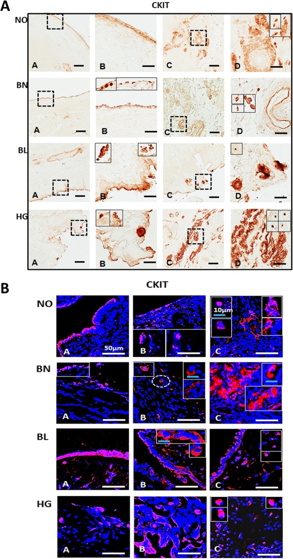
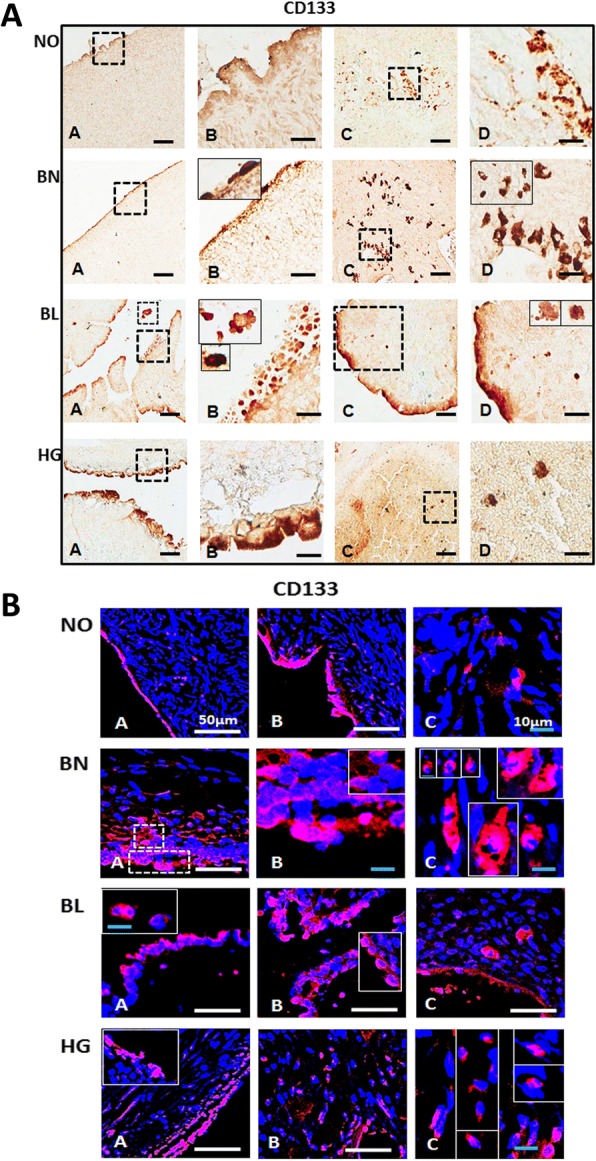
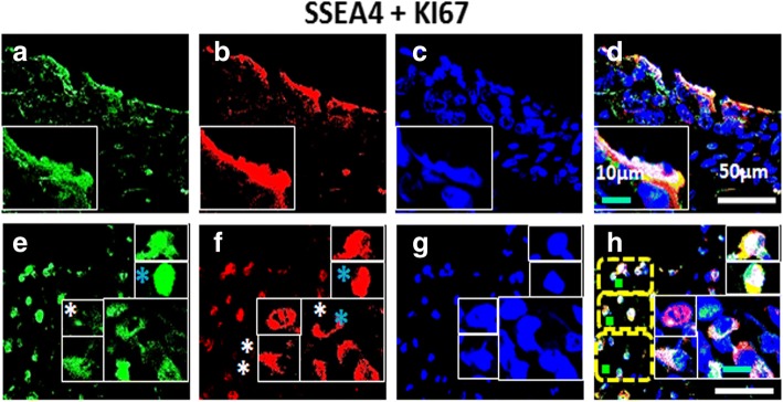
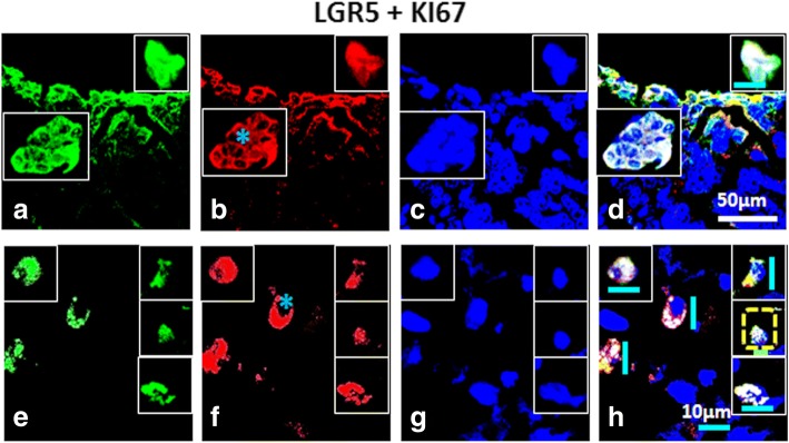
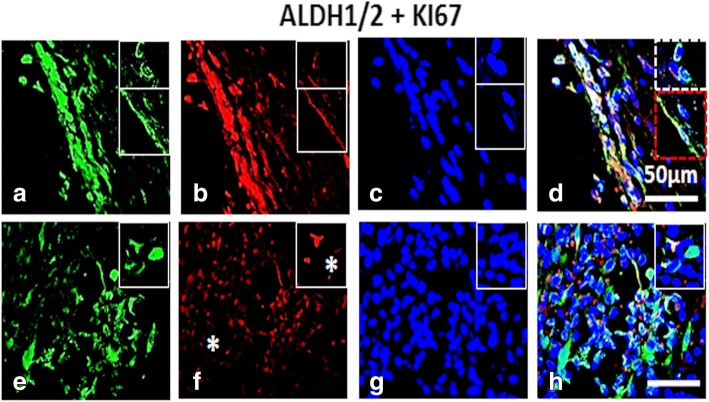
Results: In view of this, distribution of various CSC (ALDH1/2, C-KIT, CD133, CD24 and CD44) and cell proliferation (KI67) specific markers in the ovarian surface epithelium (OSE) and cortex regions in normal ovary, and benign, borderline and high grade metastatic ovarian tumors by immuno-histochemistry and confocal microscopy was studied. We further confirmed their expression by RT-PCR analysis. Co-expression analysis of stem cell (OCT4, SSEA4) and CSC (ALDH1/2, CD44 and LGR5) markers with proliferation marker (KI67) in HG tumors revealed dual positive proliferating stem and CSCs, few non-proliferating stem/CSC (SSEA4+/KI67- and ALDH1/2+/KI67-) and only KI67+ cells in cortex, signifying dynamic populations and interesting cellular hierarchy in cortex region. Smaller spherical (≤ 5 μm) and larger spindle/elliptical shaped (~ 10 μm) cell populations with high nucleo-cytoplasmic ratio were detected across all samples (including normal ovaries) but with variable distribution and characteristic stage-wise marker expression across different tumor stages.

Conclusions: Diverse stem and CSC populations expressing characteristic markers revealing distinct phenotypes (spherical ≤5 μm and spindle/elliptical ~ 10 μm) were distributed within different tumor stages studied signifying dynamic and probable functional hierarchy within these cell types. Involvement of extra-ovarian sites of origin of stem and CSCs requires rigorous evaluation. Quantitative analysis of potent CSC populations, their mechanisms and pathways for self-renewal, chemo-resistance, metastatic spread etc. with respect to various markers studied, will provide better insights and targets for developing effective therapeutics to prevent metastasis and eventually help improve patient mortality.

Keywords: Cancer stem cells; High grade metastatic ovarian cancer; Ki67; Ovarian cancer; Ovarian cancer stem cells; Ovarian stem cells; Ovarian tumor.


Conflict of interest statement

Ethics approval and consent to participate

Ovarian normal and tumor tissues were obtained from James Graham Brown Cancer Center by adhering to standards and protocols approved by the Institutional Review Board of University of Louisville. A written informed consent from the patient was obtained before surgery by the Brown Cancer Center, and tissues were acquired at the Clinical and Translational Research building for further processing and study.

Consent for publication

Concerned permission from University of Louisville was obtained prior to submission of manuscript.

Competing interests

The Authors declare that they have no competing interest.

Publisher’s Note

Springer Nature remains neutral with regard to jurisdictional claims in published maps and institutional affiliations.

Figures

Fig. 1
Fig. 1
Immunolocalization of CD117/C-KIT in normal ovarian and tumor tissue sections: [A] Immunohistochemical detection of Anti-C-KIT polyclonal antibody in OSE (A, B) and ovarian cortex (C, D). Region between dotted boxes in A, C is magnified in B, D respectively. Diffused, faint signals were localized to OSE layer of NO and BN, BL and HG ovarian tumor tissue. C-KIT+ cells appear as single isolated or as clusters in cortex region across all tissue sections. More C-KIT+ immuno-stained cells per field were noticed in BL and HG than NO and BN. Insets denote individual spherical and elongated/spindle shaped cells at higher magnification from other fields of focus. Scale bar = 100 μm (A, C) and 25 μm (B, D) respectively. [B] Immunofluorescence staining of CD117/C-KIT in OSE layer (A, B) and cortex (C) both reveal specific CD117+ cells consistently in NO, BN as well as BL and HG ovarian cancer tissue, where these CD117+ cell numbers are higher in BL OSE and HG cortex per field of focus, compared to NO and BN. White scale bar = 50 μm; blue scale bar = 10 μm. Secondary antibody was conjugated with Alexa fluor 568 and sections were counterstained with nucleus specific dye DAPI
Fig. 2
Fig. 2
Immunostaining for CD133 in normal ovarian and tumor tissue sections: [A] Mouse monoclonal anti-CD133 antibody was localized in both OSE (A, B) and ovarian cortex (C, D) by immunohistochemistry. Region between dotted boxes in A, C is magnified in B, D respectively. Polar staining of CD133 is obvious in OSE layer especially in NO, BL and HG ovaries. BL ovaries exhibit multi-layered OSE. Cortex comprised of CD133+ cells arranged in clusters with elongated/spindle shaped morphology in NO and BN ovaries. BL ovarian cortex harbours single spherical cell clusters distributed throughout. HG comprised more of large CD133+ cells in OSE and few clusters in the cortex per field focussed. Insets include magnified images of cells from different fields. Scale bar = 100 μm (A, C) and 25 μm (B, D) respectively. [B] Immunofluorescence staining of CD133 in OSE layer (A, B) as well as cortex (C) reveals specific CD133+ cells with relatively higher cell numbers in BL and HG. Area within dotted lines in BN OSE (A) are magnified in (B) while elliptical/spindle shaped CD133+ cells in cortex from various fields were represented in the composite image in (C) of BN and HG. Large CD133+ cells in cortex were also observed. White scale bar = 50 μm; blue scale bar = 10 μm. Secondary antibody employed was conjugated with Alexa fluor 568 and tissue sections were counterstained with nucleus specific dye DAPI
Fig. 3
Fig. 3
Immunolocalization of surface marker CD44 in normal ovarian and tumor tissue sections: [A] Monoclonal anti-CD44 antibody was localized to the OSE (A, B) and ovarian cortex (C, D) with few CD44+ cells visible within OSE layer in NO, BN, BL ovaries per field as compared to HG. Region between dotted boxes in A, C is magnified in B, D respectively. NO and BN ovaries appear to harbour more CD44+ cells in cortex as compared to BL ovaries. Some regions within the ovarian cortex possess more CD44+ cells adjacent to OSE layer in HG. Typically spindle/elongated shaped CD44+ cells were present in NO, BN, BL and HG cortex with round spherical cells located moreover in OSE layer. Cortex region of HG possessed both spindle and spherical shaped CD44+ cells. Insets in D of NO, BN, HG ovaries depict representative individual cell morphology and distribution density and localization within the cortex. Scale bar = 100 μm (A, C) and 25 μm (B, D) respectively. [B] Immunofluorescence staining of CD44 in OSE layer (A) and cortex (B, C) where C and insets in BN, BL and HG (B, C) represent images captured at higher magnification. BN (B) typically represents large round fluffy cells within cortex similar to those observed in HG cortex (C). CD44+ spindle shaped cells are found throughout all the tissue samples including NO. Single large CD44+ cells and multi-nucleated clusters in BN, BL (white arrows) and HG cortex were present. White scale bar = 50 μm; blue scale bar = 10 μm. Secondary antibody conjugated with Alexa fluor 568 was employed and sections were counterstained with nuclear dye DAPI
Fig. 4
Fig. 4
Dual labelling of High Grade (HG) ovarian tumor with OCT4 and KI67: Panel comprises of (a-d) OSE layer and (e-h) cortex [C] of which (a, e) OCT4, Alexa fluor 488, (b, f) KI67, Alexa fluor 568, (c, g) DAPI and (d, h) were merge composites. Both OSE layer (a-d) and cortex (e-f) regions of HG ovary revealed OCT4+/KI67+ cells. Cortex comprised of cells expressing nuclear OCT4+/KI67+ and cytoplasmic KI67+ (blue asterisk). Few OCT4+/KI67 cells (white asterisk) were also observed in the cortex. Tiny spherical (VSELs-like) stem cells were indicated by yellow dotted square. Blue asterisk in (f) denotes cytoplasmic KI67. White scale bar = 50 μm, blue scale bar = 10 μm, green scale bar = 5 μm
Fig. 5
Fig. 5
Co-localization of SSEA4 and KI67 in High Grade (HG) ovarian tumor: Panel comprises of (a-d) OSE layer and (e-h) cortex [C] of which (a, e) SSEA4, Alexa fluor 488, (b, f) KI67, Alexa fluor 568, (c, g) DAPI and (d, h) were merge composites. OSE layer prominently showed SSEA4+ cells that were KI67+ (nuclear and cytoplasmic). In some fields SSEA4/KI67+ cells were also observed in cortex. Cytoplasmic SSEA4+/KI67+ were also observed in some fields of focus. Insets denote representative cells from same field of focus compiled in one panel. Tiny spherical (VSELs-like) stem cells were indicated by yellow dotted square. Blue asterisk denotes cytoplasmic KI67. White scale bar = 50 μm, blue scale bar = 10 μm, green scale bar=5μm (H)
Fig. 6
Fig. 6
Dual labelling of High Grade (HG) ovarian tumor with CD44 and KI67: Panel comprises of (a-d) OSE layer and (e-h) cortex [C] of which (a, e) CD44, Alexa fluor 488, (b, f) KI67, Alexa fluor 568, (c, g) DAPI and (d, h) were merge composites. CD44+ cells situated just below OSE layer expressed KI67. Some KI67+ cells were prominently visible in cortex. Nuclear as well as cytoplasmic KI67+ cells were visible in cortex. White asterisk = CD44 cells, Blue asterisk denotes cytoplasmic KI67. Tiny spherical (VSELs-like) stem cells were indicated by yellow dotted square. White scale bar = 50 μm, blue scale bar = 10 μm, yellow scale bar = 10 μm (D) and green scale bar = 5 μm (D, H)
Fig. 7
Fig. 7
Co-expression of High Grade (HG) ovarian tumor for LGR5 and KI67: Panel comprises of (a-d) OSE layer and (e-h) cortex [C] of which (a, e) LGR5, Alexa fluor 488, (b, f) KI67, Alexa fluor 568, (c, g) DAPI and D, H) were merge composites. OSE layer and few clusters in some fields expressed both i.e. LGR5+/KI67+. Membrane bound LGR5+ and cytoplasmic KI67+ were observed within the cortex. Inset reveals cells from different fields captured at high magnification. Tiny spherical (VSELs-like) stem cells were indicated by yellow dotted square. Blue asterisk denotes cytoplasmic KI67. White scale bar = 50 μm, blue scale bar = 10 μm, green scale bar = 5 μm (H)
Fig. 8
Fig. 8
Dual labelling of High Grade (HG) ovarian tumor with ALDH1/2 and KI67: Panel comprises of (a-d) OSE layer and (e-h) cortex [C] of which (a, e) ALDH1/2, Alexa fluor 488 (b, f) KI67, Alexa fluor 568 (c, g) DAPI and (d, h) were merge composites. OSE layer revealed presence of ALDH1/2+/KI67+ cells. Some spindle shaped ALDH1/2+ (inset) were KI67+. Many cells observed within the cortex were ALDH1/2+/KI67. White dotted square denotes disseminated OSE cells. Red dotted square denotes spindle shaped ALDH1/2+ OSE cells. KI67 cells = white asterisk. White scale bar = 50 μm
Fig. 9
Fig. 9
RT-PCR and mRNA transcript analysis for cancer stem cell and proliferation markers in normal ovarian and tumor tissue: Expression pattern of RT-PCR amplicons for cancer stem cell related genes (Aldh, Ckit, Cd133, Cd24, Cd44) and proliferation marker (Ki67) and housekeeping gene (Gapdh) were detected in various normal human ovarian and tumor tissue samples. 1 = NO, 2 = BN, 3 = BL, 4 = HG; A, B, C, D = set 1–4 of patient samples. NO = normal ovarian tissue, BN = benign, BL = borderline, HG = high grade ovarian tumor

References

    1. Chen WC, Qiu JT, Lai CH, et al. Outcomes and prognoses of patients with ovarian cancer using bevacizumab: 6-year experience in a tertiary care hospital of northern Taiwan. PLoS One. 2017;12(5):e0175703. doi: 10.1371/journal.pone.0175703. - DOI - PMC - PubMed
    1. Bapat SA, Mali AM, Koppikar CB, Kurrey NK. Stem and progenitor-like cells contribute to the aggressive behavior of human epithelial ovarian Cancer. Cancer Res. 2005;65(8):3025–3029. doi: 10.1158/0008-5472.CAN-04-3931. - DOI - PubMed
    1. Latifi A, Luwor RB, Bilandzic M, Nazaretian S, Stenvers K, Pyman J, Zhu H, Thompson EW, Quinn MA, Findlay JK, Ahmed N. Isolation and characterization of tumor cells from the ascites of ovarian Cancer patients: molecular phenotype of Chemoresistant ovarian tumors. PLoS One. 2012;7(10):e46858. doi: 10.1371/journal.pone.0046858. - DOI - PMC - PubMed
    1. Kakar SS, Ratajczak MZ, Powell KS, Moghadamfalahi M, Miller DM, Batra SK, Singh SK. Withaferin a alone and in combination with cisplatin suppresses growth and metastasis of ovarian Cancer by targeting putative Cancer stem cells. PLoS One. 2014;9(9):e107596. doi: 10.1371/journal.pone.0107596. - DOI - PMC - PubMed
    1. Foster R, Buckanovich RJ, Rueda BR. Ovarian cancer stem cells: working towards the root of stemness. Cancer Letts. 2013;338:147–157. doi: 10.1016/j.canlet.2012.10.023. - DOI - PubMed

MeSH terms