. 2019 Mar:92:155-194.
doi: 10.1016/j.simyco.2018.06.003. Epub 2018 Jul 5.

Phylogeny and genetic diversity of the banana Fusarium wilt pathogen Fusarium oxysporum f. sp. cubense in the Indonesian centre of origin

Affiliations

Phylogeny and genetic diversity of the banana Fusarium wilt pathogen Fusarium oxysporum f. sp. cubense in the Indonesian centre of origin

N Maryani et al. Stud Mycol. 2019 Mar.

Abstract

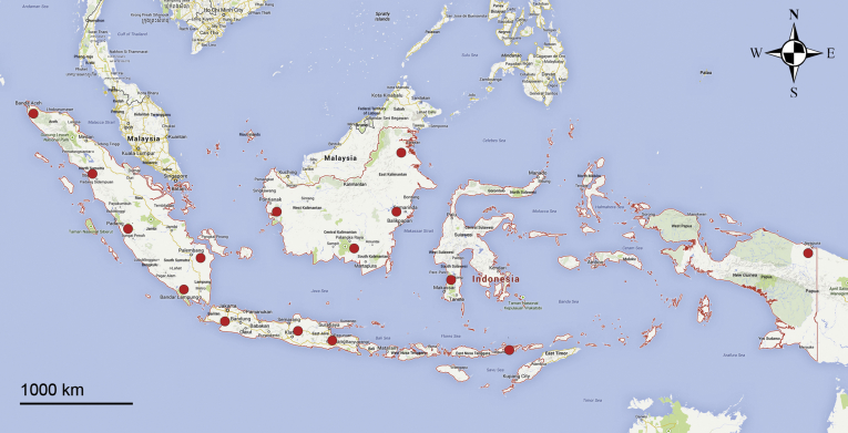
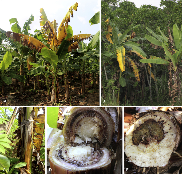
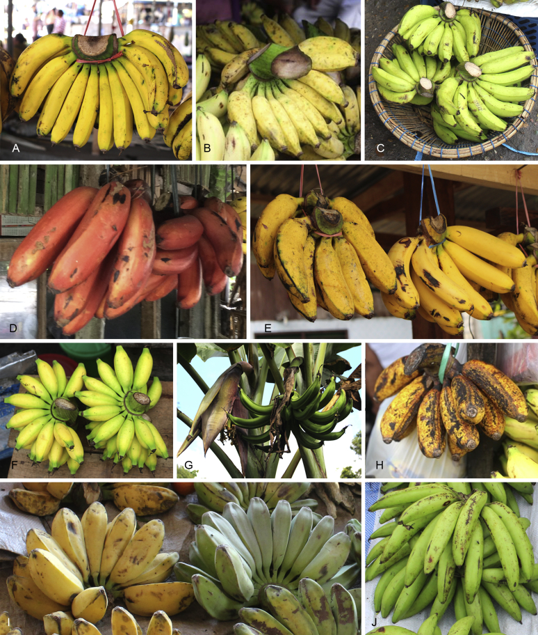
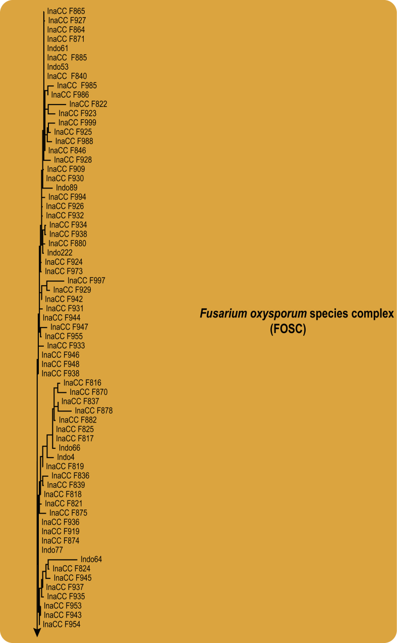
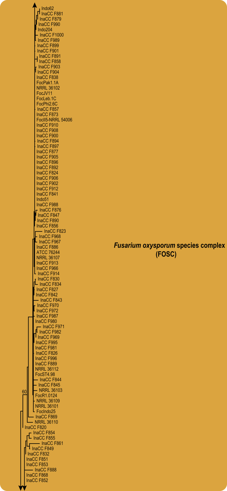
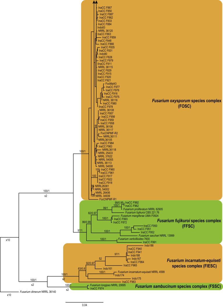
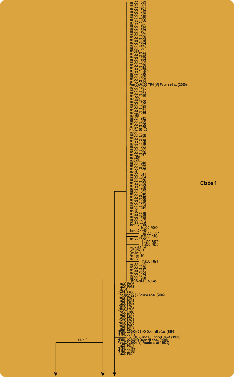
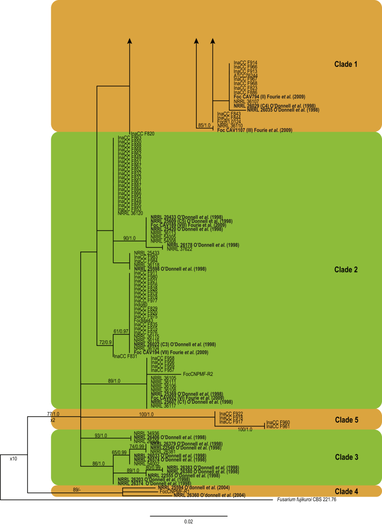
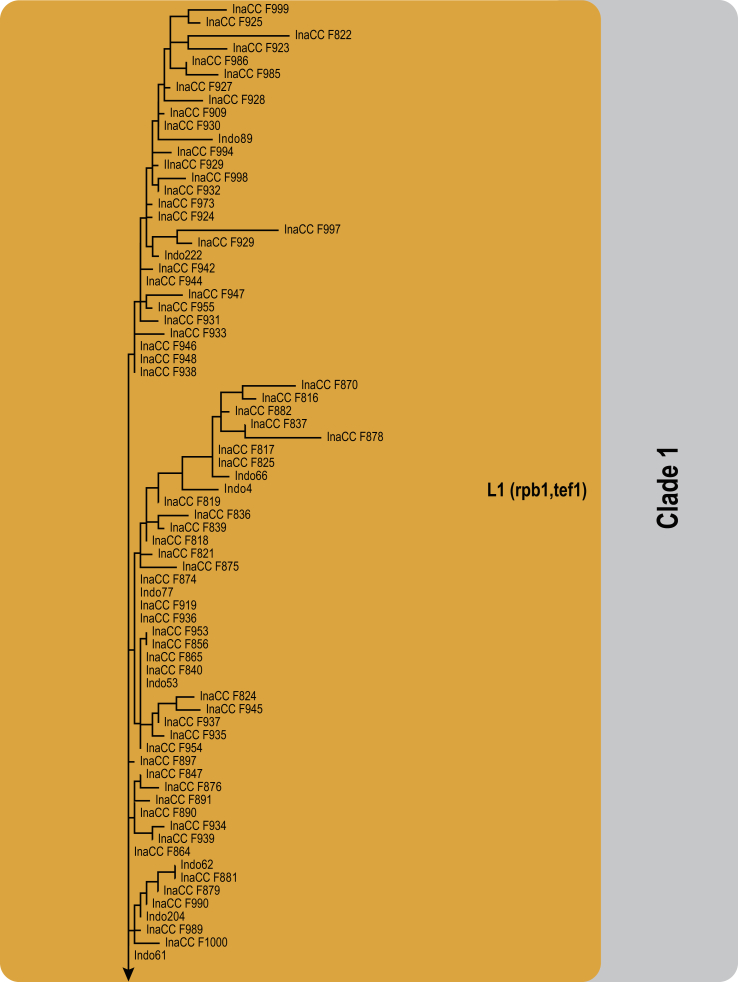
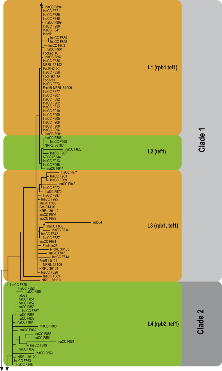
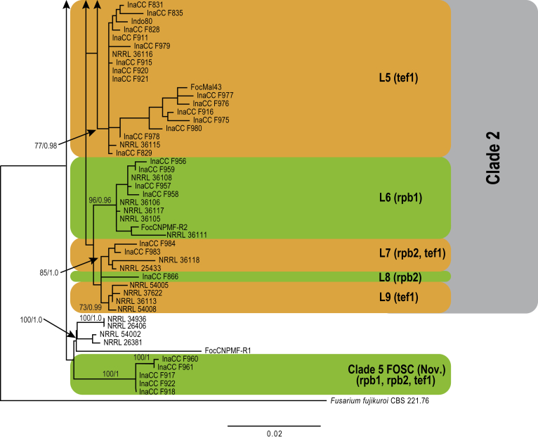
Fusarium oxysporum f. sp. cubense (Foc), the causal agent of Fusarium wilt or Panama disease on banana, is one of the major constraints in banana production worldwide. Indonesia is the centre of origin for wild and cultivated bananas, which likely co-evolved with Foc. This study explored the widest possible genetic diversity of Foc by sampling across Indonesia at 34 geographically and environmentally different locations in 15 provinces at six islands. This resulted in a comprehensive collection of ∼200 isolates from 40 different local banana varieties. Isolates were identified and assessed using sequence analysis of the translation elongation factor-1alpha (tef1), the RNA polymerase II largest subunit (rpb1), and the RNA polymerase II second largest subunit (rpb2). Phylogenetic analyses of these genes allowed the identification of 180 isolates of Fusarium oxysporum f. sp. cubense (Foc), and 20 isolates of the Fusarium fujikuroi species complex (FFSC), the Fusarium incarnatum-equiseti species complex (FIESC), and the Fusarium sambucinum species complex (FSSC). Further analyses, incorporating a worldwide collection of Foc strains, revealed nine independent genetic lineages for Foc, and one novel clade in the Fusarium oxysporum species complex (FOSC). Selected isolates from each lineage were tested on the banana varieties Gros Michel and Cavendish to characterise their pathogenicity profiles. More than 65 % of the isolates were diagnosed as Tropical Race 4 (Foc-TR4) due to their pathogenicity to Cavendish banana, which supports the hypothesis that Foc-TR4 is of Indonesian origin. Nine independent genetic lineages for Foc are formally described in this study. This biodiversity has not been studied since the initial description of Foc in 1919. This study provides a detailed overview of the complexity of Fusarium wilt on banana and its diversity and distribution across Indonesia.

Keywords: 11 New taxa; F. duoseptatum N. Maryani, L. Lombard, Kema & Crous; F. grosmichelii N. Maryani, L. Lombard, Kema & Crous; F. hexaseptatum N. Maryani, L. Lombard, Kema & Crous; F. kalimantanense N. Maryani, L. Lombard, Kema & Crous; F. odoratissimum N. Maryani, L. Lombard, Kema & Crous; F. phialophorum N. Maryani, L. Lombard, Kema & Crous; F. purpurascens N. Maryani, L. Lombard, Kema & Crous; F. sangayamense N. Maryani, L. Lombard, Kema & Crous; F. tardichlamydosporum N. Maryani, L. Lombard, Kema & Crous; F. tardicrescens N. Maryani, L. Lombard, Kema & Crous; Fusarium cugenangense N. Maryani, L. Lombard, Kema & Crous; Morphology; New species; Panama disease; Pathogenicity; Tropical Race 4.


Figures

Fig. 1
Fig. 1
Map of sampling collection in 2014–2015 in the island of Java, Sumatra, Kalimantan, Sulawesi, Papua, and Flores.
Fig. 2
Fig. 2
Symptoms of Fusarium wilt on banana. A. External wilting symptom on leaves in a monoculture plantation in Lampung, Sumatra. B. External wilting symptom in a backyard home plantation in Cianjur, West Java. C. Splitting of the pseudostem. D. Internal symptoms, discoloration of the pseudostem. E. Discoloration of the corm.
Fig. 3
Fig. 3
Local Indonesian banana varieties. A. Pisang Raja Bulu (AAB). B. Pisang Awak (ABB). C. Pisang Ambon Hijau (AAA). D. Pisang Udang (ABB). E. Left, Pisang Raja Manten (AAB), right, Pisang Barangan (AAA). F. Pisang Mas Lampung (AA). G. Pisang Tanduk (AAB). H. Pisang Susu (AAB). I. Pisang Kepok (ABB). J. Pisang Jarum (AA).
Fig. 4
Fig. 4
Maximum likelihood tree inferred from the combined rpb1, rpb2 and tef1 genes sequence data set of 244 isolates. The bootstrap support values (BP) and Bayesian posterior probabilities (PP) are given at nodes. Coloured blocks indicate the various Fusarium species complexes included. The tree is rooted to Fusarium dimerum (NRRL 36140).
Fig. 4
Fig. 4
Maximum likelihood tree inferred from the combined rpb1, rpb2 and tef1 genes sequence data set of 244 isolates. The bootstrap support values (BP) and Bayesian posterior probabilities (PP) are given at nodes. Coloured blocks indicate the various Fusarium species complexes included. The tree is rooted to Fusarium dimerum (NRRL 36140).
Fig. 5
Fig. 5
Maximum likelihood tree inferred from the tef1 gene sequence data set of 183 Indonesian isolates in the FOSC clade. Included are representatives of the studies by O'Donnell et al., 1998, O'Donnell et al., 2004 and Fourie et al. (2009), indicated in bold. The bootstrap support values >70 % (BS) and Bayesian posterior probabilities >0.95 (PP) are given at nodes. The tree is rooted to Fusarium fujikuroi (CBS 221.76).
Fig. 5
Fig. 5
Maximum likelihood tree inferred from the tef1 gene sequence data set of 183 Indonesian isolates in the FOSC clade. Included are representatives of the studies by O'Donnell et al., 1998, O'Donnell et al., 2004 and Fourie et al. (2009), indicated in bold. The bootstrap support values >70 % (BS) and Bayesian posterior probabilities >0.95 (PP) are given at nodes. The tree is rooted to Fusarium fujikuroi (CBS 221.76).
Fig. 6
Fig. 6
Maximum likelihood tree inferred from the combined rpb1, rpb2 and tef1 genes sequence data sets. The bootstrap support values >70 % (BS) and Bayesian posterior probabilities >0.95 (PP) are given at nodes. Foc lineages are numbered based on the consensus from single and combine gene data sets represented by the coloured blocks. The tree is rooted to Fusarium fujikuroi (CBS 221.76).
Fig. 6
Fig. 6
Maximum likelihood tree inferred from the combined rpb1, rpb2 and tef1 genes sequence data sets. The bootstrap support values >70 % (BS) and Bayesian posterior probabilities >0.95 (PP) are given at nodes. Foc lineages are numbered based on the consensus from single and combine gene data sets represented by the coloured blocks. The tree is rooted to Fusarium fujikuroi (CBS 221.76).
Fig. 7
Fig. 7
Fusarium odoratissimum (InaCC F817). A. Culture grown on PDA. B–C. Sporodochia on carnation leaves. D–E. Sporodochial branched conidiophores with monophialides. F. False head. G. Falcate-shaped macroconidia. H. Microconidia. I. Chlamydospores. J. Polyphialides. Scale bars D–J = 10 µm.
Fig. 8
Fig. 8
Fusarium odoratissimum (ex-type InaCC F822). A. Culture grown on PDA. B. Sporodochia on carnation leaves. C. Monophialides with initial conidia being formed. D. Falcate-shaped macroconida. E. Branched conidophores. F. Elliptical microconidia. G. Thick-walled chlamydospores. Scale bars C–G = 10 µm.
Fig. 9
Fig. 9
Fusarium purpurascens (ex-type InaCC F886). A. Culture grown on PDA. B–C. Sporodochia grown on carnation leaves. D. Falcate-shaped macroconidia. E. False heads. F. Microconidia. G. Monophialides. Scale bars D–G = 10 µm.
Fig. 10
Fig. 10
Fusarium phialophorum (ex-type InaCC F971) A. Culture grown on PDA. B–C. Sporodochia on carnation leaves. D. Aerial conidiophore on carnation leaves. E–F. Sporodochial phialides. G. Falcate-shaped macroconidia. H. Microconidia. I. False head. J. Lateral monophialides with long collaretes. K. Thick-walled chlamydospores. Scale bars E–K = 10 µm.
Fig. 11
Fig. 11
Fusarium grosmichelii (ex-type InaCC F833). A. Culture grown on PDA. B. Sporodochia on carnation leaves. C. Aerial conidiophores from stereo microscope. D. Falcate-shaped macroconidia. E. Microconidia. F. Chlamydospores. G–H. Sporodochial phialides. I. False heads. J. Polyphialides. K. Branched conidiophore. Scale bars D–F, H–K = 10 µm, G = 20 µm.
Fig. 12
Fig. 12
Fusarium duoseptatum (ex-type InaCC F916). A. Culture grown on PDA. B–C. Sporodochia on carnation leaves. D. Falcate-shaped macroconidia. E. Microconidia. F. Polyphialidic conidiogenous cells. G. False heads. H. Chlamydospores. Scale bars D–H = 10 µm.
Fig. 13
Fig. 13
Fusarium tardichlamydosporum (ex-type InaCC F958). A. Culture grown on PDA. B. Sporodochia on carnation leaves. C. Aerial conidiophore. D. Microconidia. E. Falcate-shaped macroconidia. F. Chlamydospores. G. Sporodochial phialides. H. False heads. Scale bars D–H = 10 µm.
Fig. 14
Fig. 14
Fusarium cugenangense (ex-type InaCC F984). A. Culture grown on PDA. B–C. Sporodochia on carnation leaves. D. Falcate-shaped macroconidia. E. Microconidia. F. Chlamydospores. G. False heads. H. Monophialidic conidiogenous cells. I–J. Branched conidiophores. Scale bars D–J = 10 µm.
Fig. 15
Fig. 15
Fusarium hexaseptatum (ex-type InaCC F866). A. Culture grown on PDA. B. Sporodochia on carnation leaves. C. Microconidia. D. Falcate-shaped macroconidia. E. Thick-walled chlamydospores. F. False heads. G. Monophialides and polyphialides. Scale bars C–G = 10 µm.
Fig. 16
Fig. 16
Fusarium tardicrescens (ex-tyoe CBS 102024). A. Culture grown on PDA. B. Sporodochia on carnation leaves. C. Falcate-shaped macroconidia. D. Microconidia. E. Thick-walled chlamydospores. F. Monophialides produce microconidia and macroconidia. G. False head. Scale bars C–G = 10 µm.
Fig. 17
Fig. 17
Fusarium kalimantanense (ex-type InaCC F917). A. Culture grown on PDA. B–C. Sporodochia on carnation leaves. D–E. Sporodochial phialides. F. Falcate-shaped macroconidia. G. Microconidia. H. Thick-walled chlamydospores. I. Monophialides producing macroconidia. J. Branched conidiophores. K. False heads. Scale bars D–K = 10 µm.
Fig. 18
Fig. 18
Fusarium sangayamense (ex-type InaCC F960). A. Culture grown on PDA. B–C. Sporodochia on carnation leaves. D. Aerial conidiophore. E–F. Sporodochial phialides. G. Falcate-shaped macroconidia. H. Microconidia. I. Short monophialides. J. Thick-walled chlamydospores. Scale bars D–J = 10 µm.
Fig. 19
Fig. 19
Pathogenicity assays. A. External wilting symptoms. B–C. Left panel Cavendish and right panel Gros Michel, corm symptom caused by Foc-Race1, Fusarium tardichlamydosporum. D–E. Left panel Cavendish and right panel Gros Michel, corm symptom caused by Foc-TR4, Fusarium odoratissimum.

References

    1. Baayen R.P., O'Donnell K., Bonants P.J.M. Gene genealogies and AFLP analyses in the Fusarium oxysporum complex identify monophyletic and nonmonophyletic formae speciales causing wilt and rot disease. Phytopathology. 2000;90:891–900. - PubMed
    1. Banke S., Peschon A., McDonald B.A. Phylogenetic analysis of globally distributed Mycosphaerella graminicola populations based on three DNA sequence loci. Fungal Genetics and Biology. 2004;41:226–238. - PubMed
    1. Bentley S., Pegg K.G., Dale J.L. Genetic variation among a world-wide collection of isolates of Fusarium oxysporum f. sp. cubense analysed by RAPD-PCR fingerprinting. Mycological Research. 1995;99:1378–1384.
    1. Bentley S., Pegg K.G., Moore N.Y. Genetic variation among vegetative compatibility groups of Fusarium oxysporum f. sp. cubense analyzed by DNA fingerprinting. Phytopathology. 1998;88:1283–1293. - PubMed
    1. Boehm E., Ploetz R., Kistler C.H. Statistical analysis of electrophoretic karyotype variation among vegetative compatibility groups of Fusarium oxysporum f. sp. cubense. MPMI-Molecular Plant Microbe Interactions. 1994;7:196–207.