

Differentiating Extensor Plantar Response in Pathological and Normal Population
- PMID: 30122841
- PMCID: PMC6073965
- DOI: 10.4103/aian.AIAN_254_17
Differentiating Extensor Plantar Response in Pathological and Normal Population
Abstract
Introduction: Approximately 5%-11% of neurologically normal population has extensor plantar response (EPR).
Method: This study is aimed to identify differentiating features of EPR between physiological and pathological population.
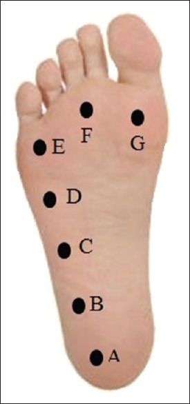
Results: A total of 43 patients with pyramidal lesions and 113 normal controls were recruited for this study. The pathological EPRs were more reproducible, with 89.4% having at least two positive Babinski responses and 91.5% having two positive Chaddock responses (vs. 14.3% and 4.8% in controls, P < 0.001). The pathological EPR was more sensitive to stimulation, in which 89.1% were elicited when the stimulation reached mid-lateral sole (vs. 11.9% in controls, P < 0.001). Most (93.6%) pathological cases had sustained big toe extension throughout stimulation (vs. 73.8% in controls, P < 0.001). As compared to those with brain lesion, the plantar responses in those with spinal lesion are less likely to have ankle dorsiflexion (5.3% vs. 25%, P < 0.05) more likely to have sustained extensor response with Babinski (94.7% vs. 71.4%, P < 0.05), Chaddock (89.5% vs. 64.3%, P < 0.05), and Schaefer (26.3% vs. 3.6%, P < 0.05) methods. A scoring system was computed using four variables, i.e., two consecutive positive Babinski or Chaddock responses, extensor response at mid-lateral sole, and sustained extension throughout stimulation. A score ≥3 is predictive of pathological origin, with sensitivity and specificity of 78.7% and 95.2%, respectively.
Conclusion: The pathological EPR is more reproducible, sensitive to stimulation, and sustainable compared to physiological extensor response.
Keywords: Babinski; Chaddock; Schaefer; physiological plantar response; plantar response; reflex.
Conflict of interest statement
There are no conflicts of interest.
Figures
Similar articles
-
[Reverse Chaddock sign].Brain Nerve. 2011 Aug;63(8):839-50. Brain Nerve. 2011. PMID: 21817175 Japanese.
-
History of the extensor plantar response: Babinski and Chaddock signs.Semin Neurol. 2002 Dec;22(4):391-8. doi: 10.1055/s-2002-36761. Semin Neurol. 2002. PMID: 12539060
-
[Clinical studies on chaddock reflex].Hokkaido Igaku Zasshi. 1982 Nov;57(6):741-51. Hokkaido Igaku Zasshi. 1982. PMID: 6985142 Japanese.
-
The Plantar Reflex.In: Walker HK, Hall WD, Hurst JW, editors. Clinical Methods: The History, Physical, and Laboratory Examinations. 3rd edition. Boston: Butterworths; 1990. Chapter 73. In: Walker HK, Hall WD, Hurst JW, editors. Clinical Methods: The History, Physical, and Laboratory Examinations. 3rd edition. Boston: Butterworths; 1990. Chapter 73. PMID: 21250238 Free Books & Documents. Review.
-
[What does the Babinski sign have to offer 100 years after its description?].Rev Neurol (Paris). 1998 Jan;154(1):22-7. Rev Neurol (Paris). 1998. PMID: 9773021 Review. French.
Cited by
-
Extensor Plantar Response: The Examination Technique Makes a Crucial Difference.Ann Indian Acad Neurol. 2019 Oct-Dec;22(4):513. doi: 10.4103/aian.AIAN_425_18. Epub 2019 Oct 25. Ann Indian Acad Neurol. 2019. PMID: 31736587 Free PMC article. No abstract available.
References
-
- Rehman HU. Babinski sign. Neurologist. 2002;8:316–8. - PubMed
-
- Van Gijn J. The Plantar Reflex: A Historical, Clinical and Electromyographic Study. Krips Repro, Meppel. 1977
-
- Morrow MJ, Mary MR. 'The babinski sign'. Br J Hosp Med. 2011;72:157–8. - PubMed
-
- Miller TM, Johnston SC. Should the babinski sign be part of the routine neurologic examination? Neurology. 2005;65:1165–8. - PubMed
-
- Isaza Jaramillo SP, Uribe Uribe CS, García Jimenez FA, Cornejo-Ochoa W, Alvarez Restrepo JF, Román GC, et al. Accuracy of the babinski sign in the identification of pyramidal tract dysfunction. J Neurol Sci. 2014;343:66–8. - PubMed
LinkOut - more resources
Full Text Sources
Other Literature Sources
Research Materials
Miscellaneous
