. 2018 Aug 1:12:2389-2402.
doi: 10.2147/DDDT.S154814. eCollection 2018.

Zafirlukast in combination with pseudohypericin attenuates spinal cord injury and motor function in experimental mice

Affiliations

Zafirlukast in combination with pseudohypericin attenuates spinal cord injury and motor function in experimental mice

Xiao-Gang Chen et al. Drug Des Devel Ther. .

Erratum in

Abstract

Background: Biosynthesis of leukotriene (LT) by arachidonic acid involves 5-lipoxygenase (5-LO) as an important precursor. Here, we evaluated the role of pseudohypericin (PHP) for its postulated 5-LO inhibitory activity along with a Cys-LT receptor antagonist zafirlukast (ZFL) against inflammatory response and tissue injury in mice.

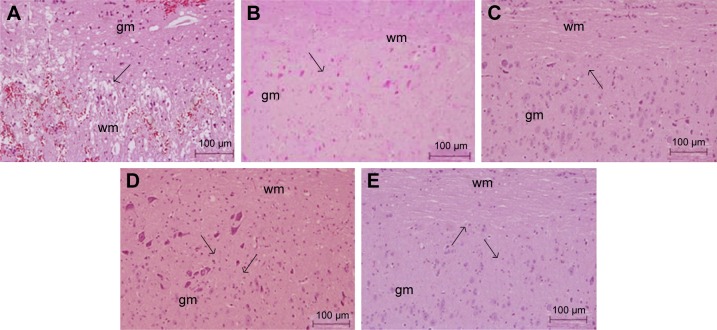
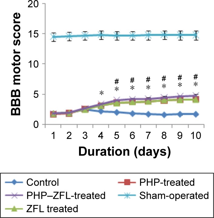
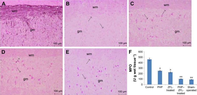
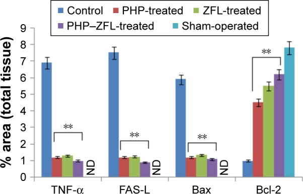
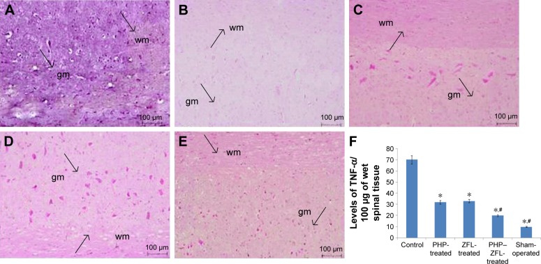
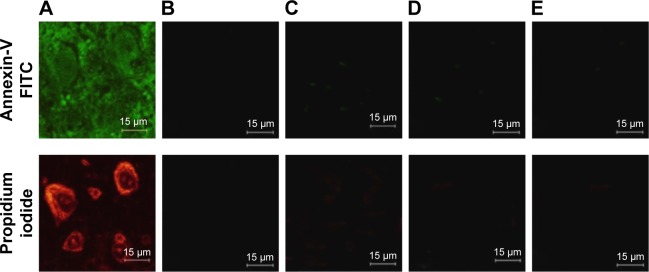
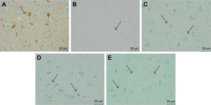
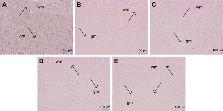
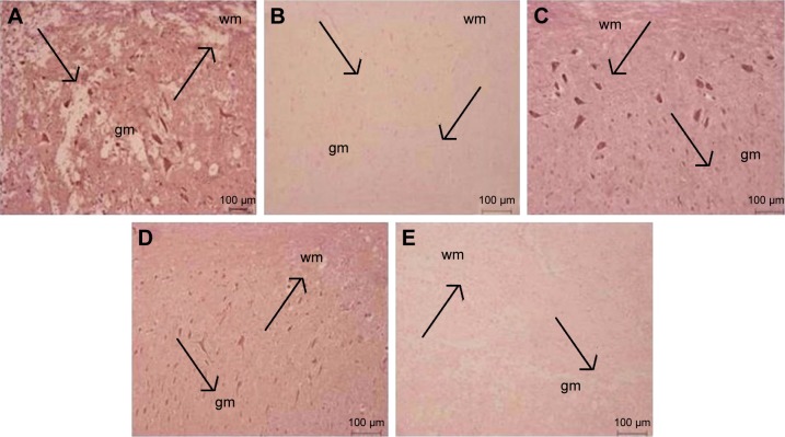
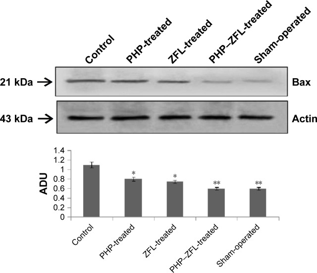
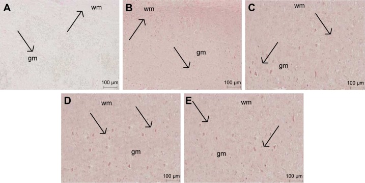
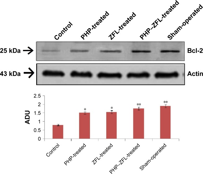
Materials and methods: The spinal injury was induced by two-level laminectomy of T6 and T7 vertebrae. The inflammation was assessed by histology, inflammatory mediators by enzyme-linked immunosorbent assay, apoptosis by Annexin-V, FAS staining, terminal deoxynucleoti-dyltransferase-mediated UTP end labeling (TUNEL) assay and expression of Bax and Bcl-2 by Western blot. Effect on motor recovery of hind limbs was evaluated for 10 days postinjury.

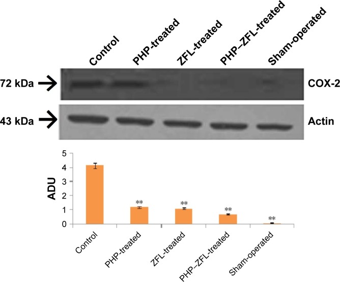
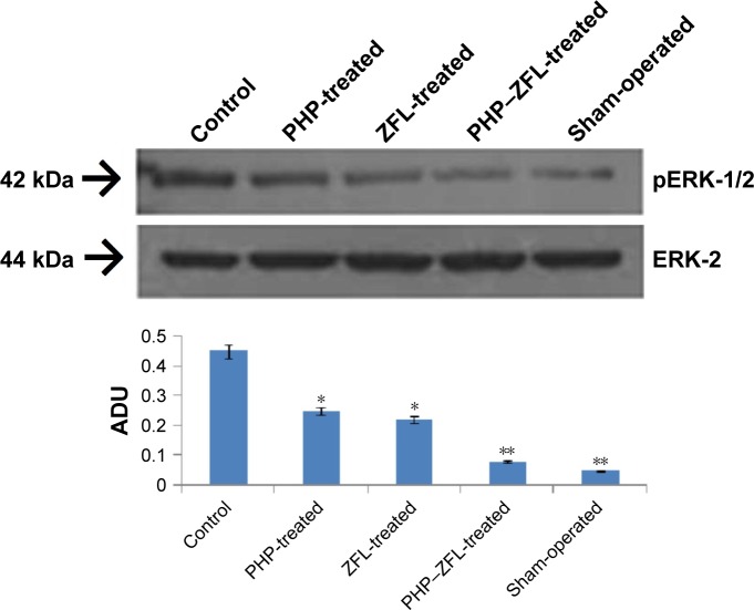
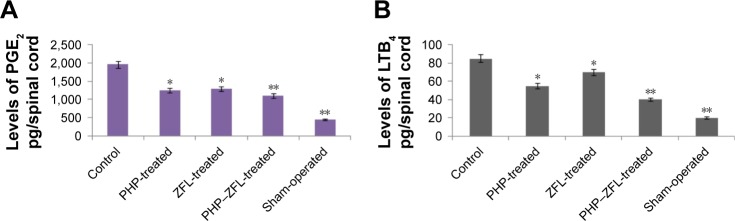
Results: The spinal injury resulted in tissue damage, apoptosis, edema, infiltration of neutrophils with increased expression of tumor necrosis factor-α (TNF-α) and cyclooxygenase-2 (COX-2). The spinal tissue showed elevated levels of prostaglandin E2 (PGE2), and LTB4 and increased phosphorylation of injured extracellular signal-regulated kinase-1/2 (ERK1/2). The PHP, ZFL and combination decreased inflammation, tissue injury and infiltration of neutrophils. Treatment also decreased the levels of PGE2, phosphorylation of extracellular signal-regulated kinase-1/2 (pERK 1/2), LT, TNF-α and COX-2 with a marked reduction in apoptosis and improved the motor function.

Conclusion: The present study confirmed 5-LO antagonist activity of PHP and established its neuroprotective role along with ZFL.

Keywords: 5-lipoxygenase; Cys-LT; mice; pseudohypericin; zafirlukast.


Conflict of interest statement

Disclosure The authors report no conflicts of interest in this work.

Figures

Figure 1
Figure 1
Effect of pseudohypericin and zafirlukast on histological changes on the spinal cord tissue 24 h after inducing spinal injury. Notes: (A) Vehicle-treated control group showing occurrence of edema and changes in white matter of perilesional zone. (B) Sham-vehicle-treated group mice showing no signs of changes. The severity of spinal cord injury decreased in tissues with reduction in edema and preservation of white matter observed in sections (C) pseudohypericin-and (D) zafirlukast-treated mice. The results were improved in (E) after combining pseudohypericin and zafirlukast. The produced figures are representatives of three experiments. The arrows show the changes in white matter suggesting spinal tissue injury. Abbreviations: wm, white matter; gm, gray matter.
Figure 2
Figure 2
Effect of PHP and ZFL on motor dysfunction after spinal cord injury. Notes: The findings were the outcome of BBB motor score, the mice were evaluated for motor activity daily for 10 days after inducing spinal injury. Highly significant (*P<0.001) improvement in motor score was seen in mice treated with PHP, ZFL and their combination compared to control-vehicle-treated group. The values of BBB motor score were significant in PHP–ZFL-treated mice compared to their individual treatment (#P<0.05). Abbreviations: PHP, pseudohypericin; ZFL, zafirlukast; BBB, Basso–Beattie– Bresnahan.
Figure 3
Figure 3
Immunohistochemical localization and activity of MPO in spinal tissue of mice treated with PHP and ZFL their combination. Notes: In vehicle-treated control group mice, the tissue stained positive for MPO and presented increased MPO activity (A and F). Sham-operated group mice, tissue sections showed no positive staining for MPO and also had normalized MPO activity (B and F). MPO staining in tissues sections from PHP (C), ZFL (D) and their combination (E) a significant reduction in positive staining for MPO activity was seen compared to vehicle-treated control group. The produced figures are representatives of three experiments. Data are mean ±%RSD (n=10), *P<0.01, **P<0.001 compared to control vehicle-treated mice. The arrows show the pink coloration that indicates the localization of MPO activity. Abbreviations: MPO, myeloperoxidase; PHP, pseudohypericin; ZFL, zafirlukast; wm, white matter; gm, gray matter; RSD, relative standard deviation.
Figure 4
Figure 4
Densitometry studies of immunocytochemistry images of spinal tissue sections for levels of TNF-α, Bax, FAS-L and Bcl-2. Notes: All the data are presented as % of total tissue area. **P<0.001 compared to control group. Abbreviations: TNF-α, tumor necrosis factor-α; ND, not detectable; PHP, pseudohypericin; ZFL, zafirlukast; FAS-L, FAS ligand.
Figure 5
Figure 5
Effect of PHP and ZFL on tissue levels and immunohistochemical localization of TNF-α. Notes: The control vehicle-treated group showed increased levels of TNF-α and positive staining in spinal tissues (A). (B) Sham-group did not exhibit staining. The treatment of PHP, ZFL and their combination significantly (*P<0.01) reduced levels of TNF-α compared to control (F), also reduced the positive staining in spinal tissues (C, D and E, respectively). The results were highly significant (#P<0.001) in mice treated with combination of PHP and ZFL and sham group compared to control. The arrows indicate the localization of TNF staining in spinal tissues. Abbreviations: PHP, pseudohypericin; ZFL, zafirlukast; TNF-α, tumor necrosis factor-α; wm, white matter; gm, gray matter.
Figure 6
Figure 6
Apoptosis studies after 24 h of spinal injury. Control group mice showed positive staining for Annexin-V FITC indicating the cells undergoing apoptosis. Notes: Late-stage apoptosis was also shown by positive staining for PI in control group mice (A). The sham-group mice did not stain positive for Annexin-V and PI (B). The mice treated with pseudohypericin (C), zafirlukast (D) and their combination (E) demonstrated significant downfall in Annexin-V and PI-positive cells, the results were highly significant with their combination. Abbreviation: PI, propidium iodide.
Figure 7
Figure 7
TUNEL assay of spinal cord tissue of mice subjected to spinal injury followed by treatment of pseudohypericin, zafirlukast and their combination. Notes: The tissue harvested after 24 h of injury and received treatment of vehicle only showed appearance of brown color cells suggesting apoptosis (A). In contrast the tissues of mice treated with pseudohypericin (C), zafirlukast (D) and their combination (E) demonstrated reduction in number of apoptotic cells, no apoptotic fragments were detected in sham-operated vehicle-treated mice spinal tissues (B). TUNEL-terminal deoxynucleotidyltransferase-mediated UTP end labeling assay. The arrows indicate the TUNEL positive cells. Abbreviation: TUNEL, terminal deoxynucleotidyltransferase-mediated UTP end labeling.
Figure 8
Figure 8
Results of immunohistological staining for FAS-L. Notes: The spinal tissues sections obtained after 24 h of spinal injury showed positive staining for FAS-L (A). The spinal tissue sections of mice after receiving treatment of pseudohypericin (C) and zafirlukast (D) showed decrease in number of cells positive for FAS-L. The positive stained FAS-L cells reduced more significantly in spinal tissues of mice receiving combined treatment of pseudohypericin and zafirlukast (E). The sham-operated mice tissues showed no staining for FAS-L (B). The arrows indicate the staining for FAS-L in spinal tissues. Abbreviations: FAS-L, FAS ligand; wm, white matter; gm, gray matter.
Figure 9
Figure 9
Effect of pseudohypericin and zafirlukast treatment on Bax staining in spinal tissues of mice. Notes: The degree of positive staining was higher in vehicle-treated control mice tissues (A). (B) Sham-group did not exhibit positive staining. The extent of Bax staining decreased in pseudohypericin (C)- and zafirlukast (D)-treated mice tissues compared to control. The results were even better and degree of positive staining for Bax decreased further in tissues of mice treated with combination of pseudohypericin and zafirlukast (E). The arrows indicate the Bax staining in spinal tissues. Abbreviations: wm, white matter; gm, gray matter.
Figure 10
Figure 10
Western blot and densitometry analysis were done on the tissue homogenates of spinal tissue collected 24 h after inducing injury. Notes: Expression of Bax levels was more prominent in tissues of vehicle-treated mice. The expression levels decreased significantly (*P<0.05) in PHP- and ZFL-treated mice, whereas the levels reduced more significantly (*P<0.01, **P<0.001) in mice treated with combination. All the data were normalized against actin levels as loading control. Abbreviations: PHP, pseudohypericin; ZFL, zafirlukast; ADU, arbitrary density units.
Figure 11
Figure 11
Evaluation of spinal tissues for Bcl-2 levels, after 24 h of injury. Note: The control vehicle-treated (A) mice showed reduction in positive staining for Bcl-2, whereas the positive staining for Bcl-2 increased in tissues of sham-group mice (B), treated with pseudohypericin (C), zafirlukast (D) and their combination (E). The arrows show positive staining for Bcl-2. Abbreviations: wm, white matter; gm, gray matter.
Figure 12
Figure 12
Western blot and densitometry analysis were done on the tissue homogenates of spinal tissue collected 24 h after inducing injury. Notes: Expression of Bcl-2 levels decreased in tissues of vehicle-treated mice. The expression levels increased significantly (*P<0.05) in PHP- and ZFL-treated mice, whereas the levels increased more significantly (**P<0.001) in mice treated with combination compared to control group. All the data were normalized against actin levels as loading control. Abbreviations: PHP, pseudohypericin; ZFL, zafirlukast; ADU, arbitrary density units.
Figure 13
Figure 13
Western blot and densitometry analysis were done on the tissue homogenates of spinal tissue collected 24 h after inducing injury. Notes: The spinal injury resulted in increased expression of COX-2 in tissues of vehicle-treated mice. The expression levels decreased more significantly (**P<0.01) in PHP, ZFL and their combination compared to control group. All the data were normalized against actin levels as loading control. Abbreviations: COX-2, cyclooxygenase-2; PHP, pseudohypericin; ZFL, zafirlukast; ADU, arbitrary density units.
Figure 14
Figure 14
Western blot and densitometry analysis were done on the tissue homogenates of spinal tissue collected 24 h after inducing injury. Notes: Extent of ERK-1/2 phosphorylation increased in tissues of vehicle-treated mice. The expression levels decreased significantly (*P<0.05) in PHP- and ZFL-treated mice, whereas the decrease in levels was more significant (**P<0.001) in mice treated with combination compared to control group. All the data were normalized against ERK-2 levels as loading control. Abbreviations: ERK-1/2, extracellular signal-regulated kinase-1/2; pERK-1/2, phosphorylation of ERK-1/2; PHP, pseudohypericin; ZFL, zafirlukast; ADU, arbitrary density units.
Figure 15
Figure 15
Effect of PHP and ZFL on levels of PGE2 (A) and LTB4 (B). Notes: The levels were measured in spinal tissues obtained 24 h after injury. The tissues levels of PGE2 and LTB4 were elevated on vehicle-treated group. A significant decrease in levels of PGE2 and LTB4 was observed in PHP- and ZFL-treated mice (*P<0.05) compared to control. The decrease in levels was highly significant (**P<001) in group treated with combination of PHP and ZFL. Abbreviations: PHP, pseudohypericin; ZFL, zafirlukast; PGE2, prostaglandin E2; LTB4, leukotriene B4.

Similar articles

Cited by

References

    1. Beattie MS. Inflammation and apoptosis: linked therapeutic targets in spinal cord injury. Trends Mol Med. 2004;10(12):580–583. - PubMed
    1. Gris D, Marsh DR, Oatway MA, et al. Transient blockade of the CD11d/ CD18 integrin reduces secondary damage after spinal cord injury, improving sensory, autonomic, and motor function. J Neurosci. 2004;24(16):4043–4051. - PMC - PubMed
    1. Carlson GD, Gorden C. Current developments in spinal cord injury research. Spine J. 2002;2(2):116–128. - PubMed
    1. Anderson AJ. Mechanisms and pathways of inflammatory responses in CNS trauma: spinal cord injury. J Spinal Cord Med. 2002;25(2):70–79. - PubMed
    1. Jung SB, Song CH, Yang CS, et al. Role of the phosphatidylinositol 3-kinase and mitogen-activated protein kinase pathways in the secretion of tumour necrosis factor-alpha and interleukin-10 by the PPD antigen of Mycobacterium tuberculosis. J Clin Immunol. 2005;25(5):482–490. - PubMed

MeSH terms