

Astragaloside IV improves renal function and fibrosis via inhibition of miR-21-induced podocyte dedifferentiation and mesangial cell activation in diabetic mice
- PMID: 30122901
- PMCID: PMC6084069
- DOI: 10.2147/DDDT.S170840
Astragaloside IV improves renal function and fibrosis via inhibition of miR-21-induced podocyte dedifferentiation and mesangial cell activation in diabetic mice
Abstract
Background: Podocyte dedifferentiation and mesangial cell (MC) activation play an important role in many glomerular diseases associated with fibrosis. MicroRNA-21 (miR-21) is closely linked to renal fibrosis, but it is unknown whether and how miR-21 promotes podocyte dedifferentiation and MC activation and whether astragaloside IV (AS-IV) improves renal function and fibrosis through the regulation of miR-21.
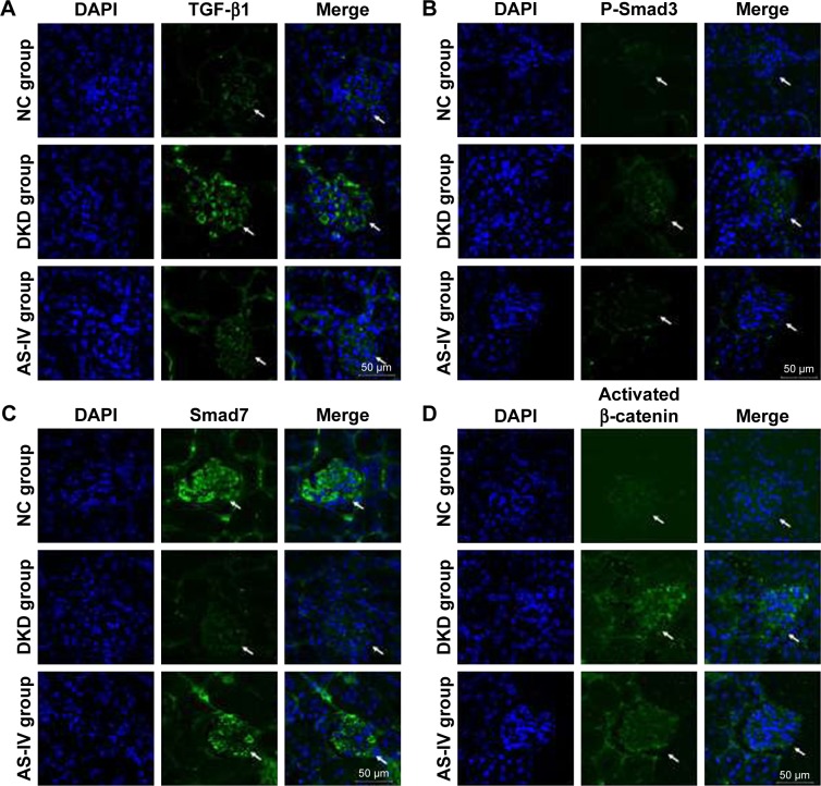
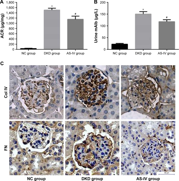
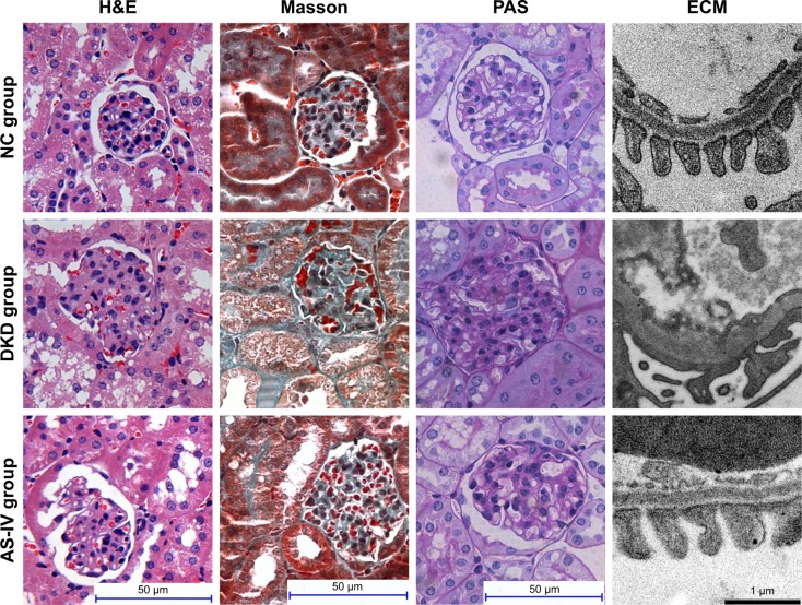
Materials and methods: Cultured MCs, primary mouse podocytes, and diabetic KK-Ay mice were treated with AS-IV. Cell transfection, Western blot, real-time PCR, immunofluorescence assay, immunohistochemical assay, and electronic microscopy were used to detect the markers of podocyte dedifferentiation and MC activation and to observe the renal morphology.
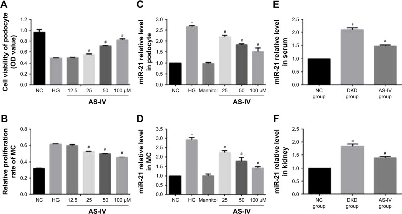
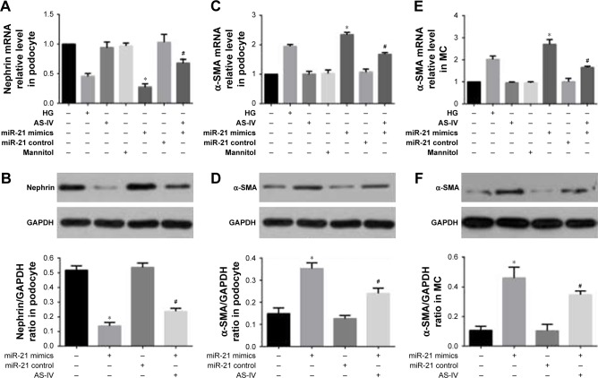
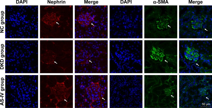
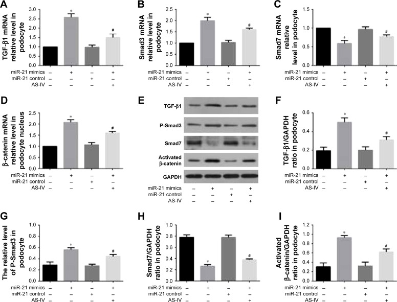
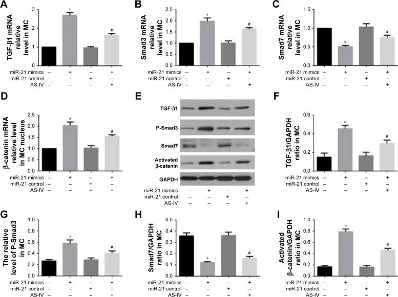
Results: Our data showed that miR-21 expression was increased and that AS-IV decreased miR-21 levels in cells, serum, and kidney. Overexpressed miR-21 promoted podocyte dedifferentiation and MC activation, and treatment with AS-IV reversed this effect. Furthermore, the overexpression of miR-21 activated the β-catenin pathway and the transforming growth factor (TGF)-β1/Smads pathway in the process of podocyte dedifferentiation and MC activation, which was abolished by AS-IV treatment. In addition, both the Wnt/β-catenin pathway inhibitor XAV-939 and the TGF-β1/Smads pathway inhibitor SB431542 reversed the effect of AS-IV. Furthermore, AS-IV improved renal function and fibrosis in diabetic KK-Ay mice.
Conclusion: Our results indicated that AS-IV ameliorates renal function and renal fibrosis by inhibiting miR-21 overexpression-induced podocyte dedifferentiation and MC activation in diabetic kidney disease. These findings pave way for future studies investigating AS-IV as a potential therapeutic agent in the management of glomerular diseases.
Keywords: TGF-β1/Smads pathway; astragaloside IV; mesangial cell activation; miR-21; podocyte dedifferentiation; renal fibrosis; β-catenin pathway.
Conflict of interest statement
Disclosure The authors report no conflicts of interest in this work.
Figures

Similar articles
-
Astragaloside IV represses high glucose-induced mesangial cells activation by enhancing autophagy via SIRT1 deacetylation of NF-κB p65 subunit.Drug Des Devel Ther. 2018 Sep 12;12:2971-2980. doi: 10.2147/DDDT.S174058. eCollection 2018. Drug Des Devel Ther. 2018. PMID: 30254426 Free PMC article.
-
Astragaloside IV ameliorates diabetic nephropathy involving protection of podocytes in streptozotocin induced diabetic rats.Eur J Pharmacol. 2014 Aug 5;736:86-94. doi: 10.1016/j.ejphar.2014.04.037. Epub 2014 May 6. Eur J Pharmacol. 2014. PMID: 24809932
-
Astragaloside IV protects against podocyte apoptosis by inhibiting oxidative stress via activating PPARγ-Klotho-FoxO1 axis in diabetic nephropathy.Life Sci. 2021 Mar 15;269:119068. doi: 10.1016/j.lfs.2021.119068. Epub 2021 Jan 18. Life Sci. 2021. PMID: 33476631
-
[Pathomechanisms of podocyte injury in diabetic nephropathy and interventional effects of Chinese herbal medicine].Zhongguo Zhong Yao Za Zhi. 2016 Jul;41(13):2416-2421. doi: 10.4268/cjcmm20161308. Zhongguo Zhong Yao Za Zhi. 2016. PMID: 28905562 Review. Chinese.
-
Glomerular mesangial cell and podocyte injuries in diabetic nephropathy.Nephrology (Carlton). 2018 Oct;23 Suppl 4:32-37. doi: 10.1111/nep.13451. Nephrology (Carlton). 2018. PMID: 30298646 Review.
Cited by
-
Sirtuins and Renal Oxidative Stress.Antioxidants (Basel). 2021 Jul 27;10(8):1198. doi: 10.3390/antiox10081198. Antioxidants (Basel). 2021. PMID: 34439446 Free PMC article. Review.
-
Integrating network pharmacology and experimental validation to explore the pharmacological mechanism of Astragaloside IV in alleviating urotensin II-mediated renal tubular epithelial cell injury.PLoS One. 2024 Dec 20;19(12):e0310210. doi: 10.1371/journal.pone.0310210. eCollection 2024. PLoS One. 2024. PMID: 39705287 Free PMC article.
-
A systematic review of astragaloside IV effects on animal models of diabetes mellitus and its complications.Heliyon. 2024 Feb 22;10(5):e26863. doi: 10.1016/j.heliyon.2024.e26863. eCollection 2024 Mar 15. Heliyon. 2024. PMID: 38439832 Free PMC article. Review.
-
Remission Induced by Shichimotsukokato in an Older Adult With Nephrotic Syndrome Secondary to Diabetic Kidney Disease: A Case Report.Cureus. 2025 Mar 24;17(3):e81115. doi: 10.7759/cureus.81115. eCollection 2025 Mar. Cureus. 2025. PMID: 40276452 Free PMC article.
-
Recent Advances in Traditional Chinese Medicine for Treatment of Podocyte Injury.Front Pharmacol. 2022 Feb 25;13:816025. doi: 10.3389/fphar.2022.816025. eCollection 2022. Front Pharmacol. 2022. PMID: 35281899 Free PMC article. Review.
References
-
- Durvasula RV, Shankland SJ. Podocyte injury and targeting therapy: an update. Curr Opin Nephrol Hypertens. 2006;15(1):1–7. - PubMed
-
- Gruden G, Perin PC, Camussi G. Insight on the pathogenesis of diabetic nephropathy from the study of podocyte and mesangial cell biology. Curr Diabetes Rev. 2005;1(1):27–40. - PubMed
MeSH terms
Substances
LinkOut - more resources
Full Text Sources
Other Literature Sources
Medical
Research Materials
