

A GPC3-specific aptamer-mediated magnetic resonance probe for hepatocellular carcinoma
- PMID: 30122918
- PMCID: PMC6078089
- DOI: 10.2147/IJN.S168268
A GPC3-specific aptamer-mediated magnetic resonance probe for hepatocellular carcinoma
Abstract
Purpose: To construct and test a hepatocellular carcinoma (HCC)-targeted magnetic resonance probe based on a glypican-3 (GPC3)-specific aptamer (AP613-1) with ultrasmall superpara-magnetic iron oxide (USPIO).
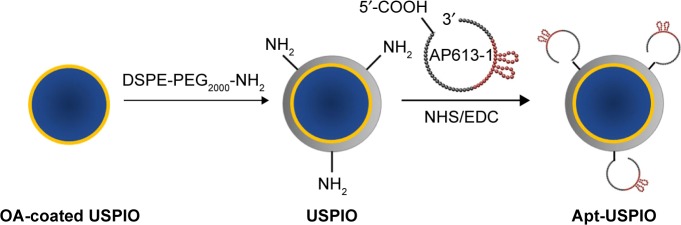
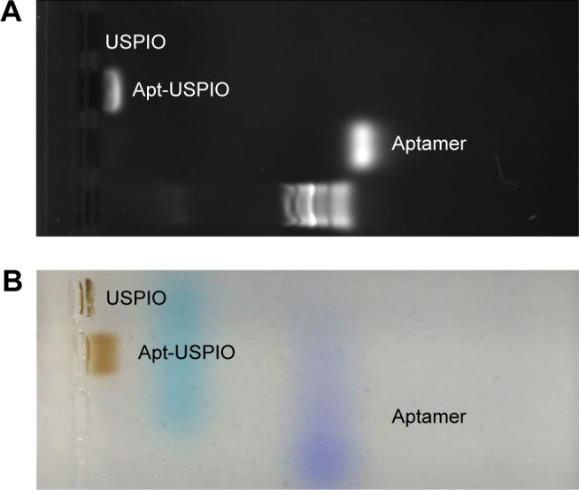
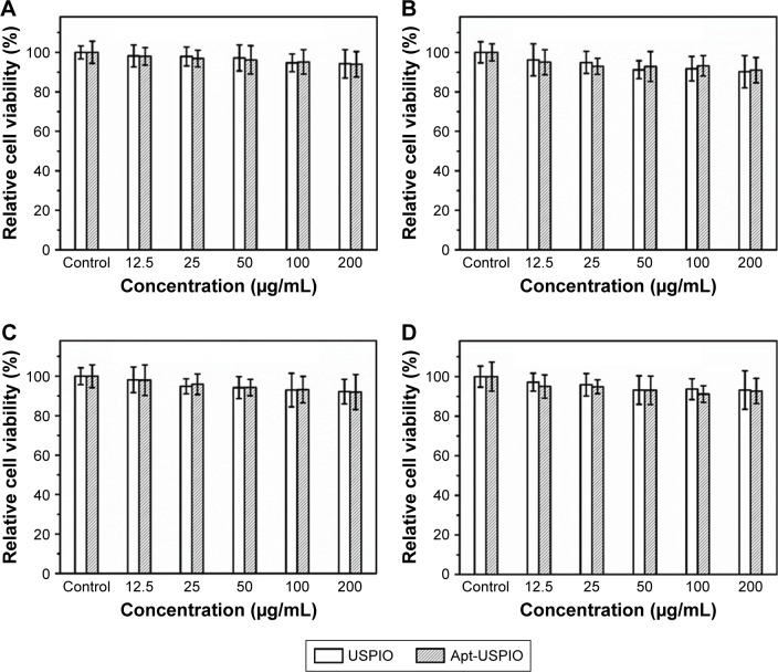
Methods: Oleic acid-coated USPIO nanoparticles were modified with amino polyethylene glycol on the surface. Amino groups of the USPIO nanoparticles were reacted with the carboxyl group of 5' carboxyl-modified AP613-1, forming an aptamer-mediated USPIO (Apt-USPIO) probe. The material characterization of this probe including transmission electron microscopy (TEM), zeta potential, dynamic laser scattering, and magnetic behavior was carried out. The targeting efficiency and magnetic resonance imaging (MRI) performance of Apt-USPIO were evaluated both in vitro and in vivo with USPIO alone as a control. The cytotoxicity and bio-compatibility of Apt-USPIO and USPIO were analyzed by cell counting kit-8 tests in vitro and animal experiments in vivo.
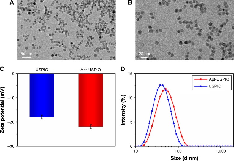
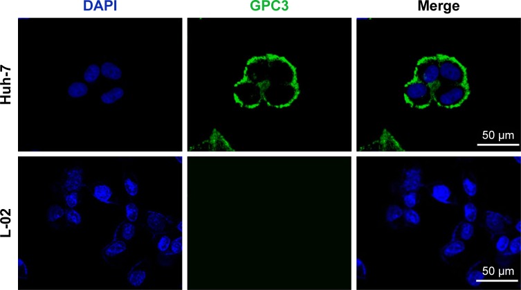
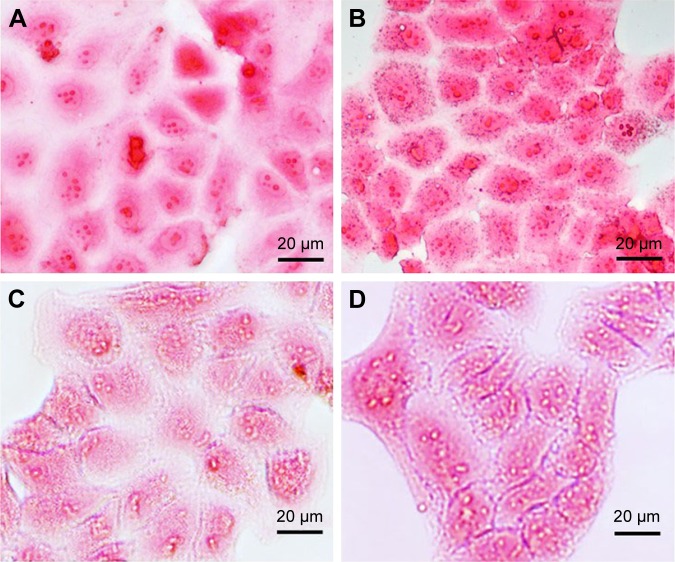
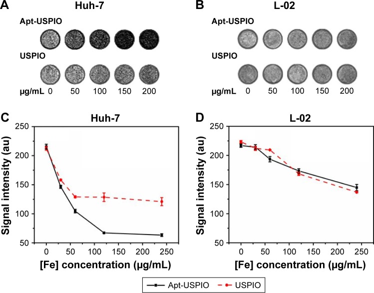
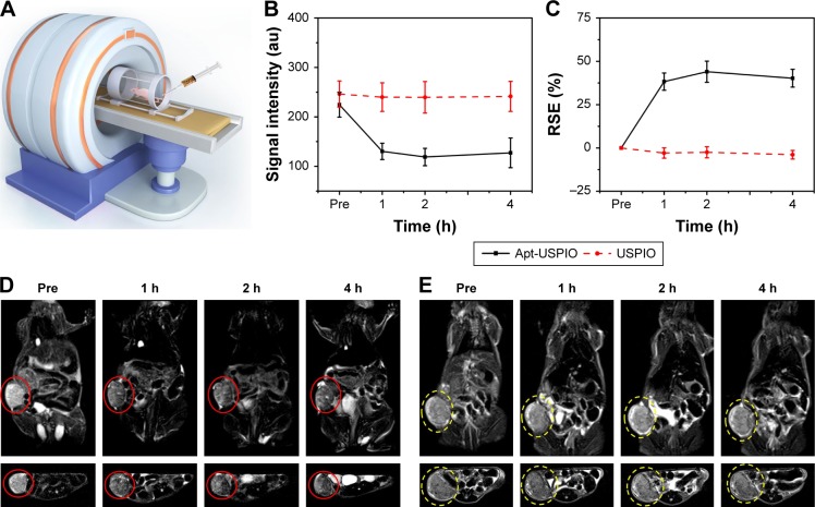
Results: TEM imaging revealed that the Apt-USPIO nanoparticles were spherical in shape and well dispersed. Specific uptake of Apt-USPIO in Huh-7 cells could be observed using the Prussian blue staining test; however, no uptake of USPIO could be found. In vitro phantom T2-weighted MRI showed a significant decrease of the signal intensity in Apt-USPIO-incubated Huh-7 cells compared to USPIO-incubated Huh-7 cells. In vivo T2-weighted MRI showed significantly negative enhancement in the Huh-7 tumors enhanced with Apt-USPIO, whereas no enhancement was found with USPIO alone. Excellent biocompatibility of Apt-USPIO and USPIO was also demonstrated.
Conclusion: In this study, a molecular MRI probe which was highly specific to GPC3 on HCC was successfully prepared. Our results validated the targeted imaging effect of this Apt-USPIO probe in vivo for GPC3-expressing HCCs in xenograft mice.
Keywords: MRI; aptamer; carcinoma; glypican-3; hepatocellular; tumor-targeted imaging; ultrasmall superparamagnetic iron oxide.
Conflict of interest statement
Disclosure The authors report no conflicts of interest in this work.
Figures

Similar articles
-
Development and in vitro study of a bi-specific magnetic resonance imaging molecular probe for hepatocellular carcinoma.World J Gastroenterol. 2019 Jun 28;25(24):3030-3043. doi: 10.3748/wjg.v25.i24.3030. World J Gastroenterol. 2019. PMID: 31293339 Free PMC article.
-
Detecting GPC3-Expressing Hepatocellular Carcinoma with L5 Peptide-Guided Pretargeting Approach: In Vitro and In Vivo MR Imaging Experiments.Contrast Media Mol Imaging. 2018 Sep 10;2018:9169072. doi: 10.1155/2018/9169072. eCollection 2018. Contrast Media Mol Imaging. 2018. PMID: 30275801 Free PMC article.
-
Preparation of magnetic resonance probes using one-pot method for detection of hepatocellular carcinoma.World J Gastroenterol. 2015 Apr 14;21(14):4275-83. doi: 10.3748/wjg.v21.i14.4275. World J Gastroenterol. 2015. PMID: 25892879 Free PMC article.
-
Preparation and in vitro studies of MRI-specific superparamagnetic iron oxide antiGPC3 probe for hepatocellular carcinoma.Int J Nanomedicine. 2012;7:4593-611. doi: 10.2147/IJN.S32196. Epub 2012 Aug 22. Int J Nanomedicine. 2012. PMID: 22956868 Free PMC article.
-
Glypican-3: A promising biomarker for hepatocellular carcinoma diagnosis and treatment.Med Res Rev. 2018 Mar;38(2):741-767. doi: 10.1002/med.21455. Epub 2017 Jun 16. Med Res Rev. 2018. PMID: 28621802 Review.
Cited by
-
Aptamers used for molecular imaging and theranostics - recent developments.Theranostics. 2022 May 13;12(9):4010-4050. doi: 10.7150/thno.72949. eCollection 2022. Theranostics. 2022. PMID: 35673581 Free PMC article. Review.
-
Engineered aptamers for molecular imaging.Chem Sci. 2023 Nov 21;14(48):14039-14061. doi: 10.1039/d3sc03989g. eCollection 2023 Dec 13. Chem Sci. 2023. PMID: 38098720 Free PMC article. Review.
-
Magnetic Nanoparticles as MRI Contrast Agents.Top Curr Chem (Cham). 2020 May 7;378(3):40. doi: 10.1007/s41061-020-00302-w. Top Curr Chem (Cham). 2020. PMID: 32382832 Free PMC article. Review.
-
Imaging Characteristics of USPIO Nanoparticles (<5 nm) as MR Contrast Agent In Vitro and in the Liver of Rats.Contrast Media Mol Imaging. 2019 Jul 21;2019:3687537. doi: 10.1155/2019/3687537. eCollection 2019. Contrast Media Mol Imaging. 2019. PMID: 31427909 Free PMC article.
-
Aptamers as Versatile Ligands for Biomedical and Pharmaceutical Applications.Int J Nanomedicine. 2020 Feb 17;15:1059-1071. doi: 10.2147/IJN.S237544. eCollection 2020. Int J Nanomedicine. 2020. PMID: 32110008 Free PMC article. Review.
References
-
- Torre LA, Bray F, Siegel RL, Ferlay J, Lortet-Tieulent J, Jemal A. Global cancer statistics, 2012. CA Cancer J Clin. 2015;65(2):87–108. - PubMed
-
- Venook AP, Papandreou C, Furuse J, de Guevara LL. The incidence and epidemiology of hepatocellular carcinoma: a global and regional perspective. Oncologist. 2010;15(Suppl 4):5–13. - PubMed
-
- Semelka RC, Helmberger TK. Contrast agents for MR imaging of the liver. Radiology. 2001;218(1):27–38. - PubMed
-
- Cruite I, Schroeder M, Merkle EM, Sirlin CB. Gadoxetate disodium-enhanced MRI of the liver: part 2, protocol optimization and lesion appearance in the cirrhotic liver. AJR Am J Roentgenol. 2010;195(1):29–41. - PubMed
MeSH terms
Substances
LinkOut - more resources
Full Text Sources
Other Literature Sources
Medical
