. 2018 Aug 1:15:28.
doi: 10.1186/s12983-018-0274-9. eCollection 2018.

Neuromuscular transmitter candidates of a centipede (Lithobius forficatus, Chilopoda)

Affiliations

Neuromuscular transmitter candidates of a centipede (Lithobius forficatus, Chilopoda)

Hendrik Langeloh et al. Front Zool. .

Abstract

Background: The neuromuscular junction is the chemical synapse where motor neurons communicate with skeletal muscle fibers. Whereas vertebrates and many invertebrates use acetylcholine as transmitter at the neuromuscular junction, in those arthropods examined up to now, glutamate and GABA are used instead. With respect to taxon sampling in a phylogenetic context, there is, however, only a limited amount of data available, focusing mainly on crustaceans and hexapods, and neglecting other, arthropod groups. Here we investigate the neurotransmitter equipment of neuromuscular synapses of a myriapod, Lithobius forficatus, using immunofluorescence and histochemical staining methods.

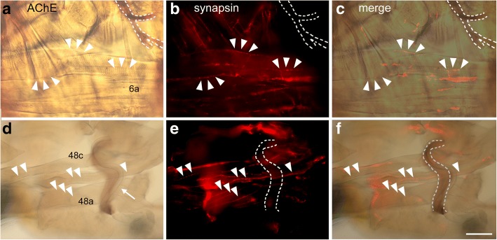
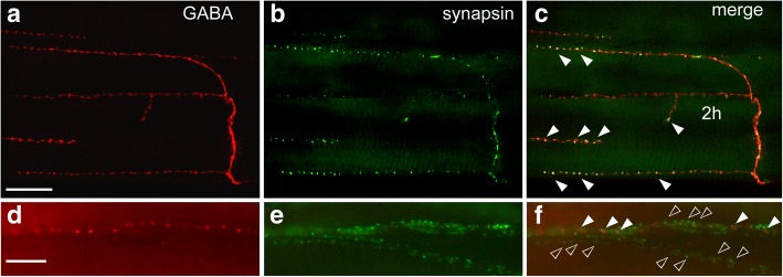
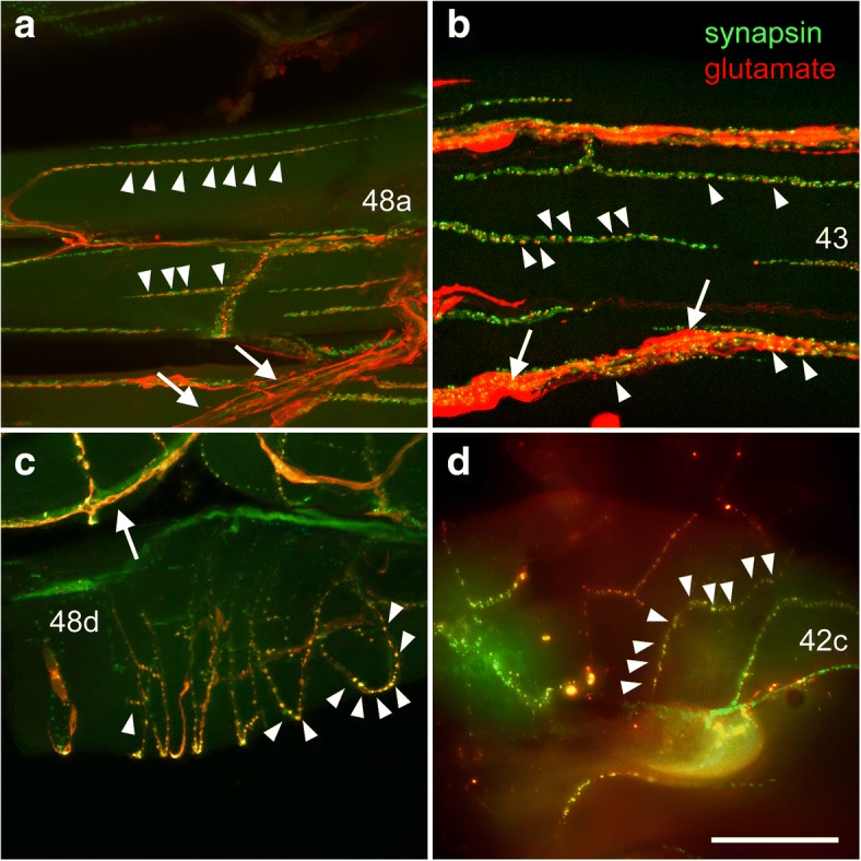
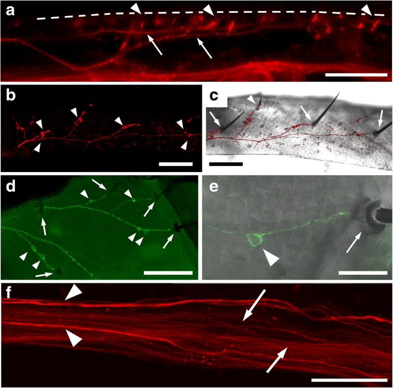
Results: Glutamate and GABA could be found colocalised with synapsin in synaptic boutons of body wall and leg muscles of Lithobius forficatus. Acetylcholinesterase activity as a marker for cholinergic synapses was found abundantly in the central nervous system and also in some peripheral nerves, but not at neuromuscular junctions. Furthermore, a large number of leg sensory neurons displayed GABA-immunofluorescence and was also labeled with an antiserum against the GABA-synthesizing enzyme, glutamate decarboxylase.

Conclusions: Our data indicate that glutamate and GABA are neurotransmitters at Lithobius forficatus neuromuscular junctions, whereas acetylcholine is very unlikely to play a role here. This is in line with the concept of glutamate as excitatory and GABA as the main inhibitory neuromuscular transmitters in euarthropods. Furthermore, we have, to our knowledge for the first time, localized GABA in euarthropod leg sensory neurons, indicating the possibility that neurotransmitter panel in arthropod sensory systems may be far more extensive than hitherto assumed.

Keywords: Acetylcholine; GABA; Glutamate; Neuromuscular junction; Synapse.


Conflict of interest statement

Not applicable. Not applicable. The authors declare that they have no competing interests. Springer Nature remains neutral with regard to jurisdictional claims in published maps and institutional affiliations.

Figures

Fig. 1
Fig. 1
Schematic diagrams of the muscles, where neuromuscular junctions were analyzed (adapted and simplified from Rilling, 1960). a Right half of 2 ½ tergites with dorsal body wall muscles. b left half of a single sternite with ventral body wall muscles. c posterior ts view of a right walking leg in three different planes (anterior, medial, posterior) with leg flexor muscles. Muscles analyzed in this study are shaded in grey and numbers according to Rilling (1960) are indicated. a, b: anterior is to the top, c dorsal is to the top. All scale bars 500 μm
Fig. 2
Fig. 2
Horizontal vibratome sections of ventral nerve cord ganglia. a Acetylcholine esterase staining (brown). b Merge of acetylcholine esterase staining (brown, same section as in a) and synapsin-IR (red). c Glutamate-IR (red) and nuclear labeling (DAPI, blue). Arrowheads point to clusters of glutamate-IR cell bodies, arrow points to midline cell bodies. d Left hemiganglion labeled with GABA antibody (red) and DAPI (blue). e right hemi ganglion labeled with glutamate decarboxylase antibody (GAD, red) and DAPI (blue). Anterior is to the left in all panels. Scale bars 200 μm, scale bar in b also applies for a, scale bar in e also applies for d
Fig. 3
Fig. 3
Localisation of acelylcholine esterase staining (brown) and synapsin-IR(red). a-c Dorsal body wall muscle 6a and peripheral nerve stained for acetylcholine esterase (a) and immunolabeled for synapsin (b), c is a merge of a and b. Peripheral nerve is outlined in white, selected synaptic boutons from B are marked with arrowheads in all panels. d-f: Tibia of a walking leg stained for acetylcholine esterase (d) and immunolabeled for synapsin (b), f is a merge of d and e. Peripheral nerve 4 is outlined in white, selected synaptic boutons from e are marked with arrowheads in all panels. Arrow points to a trachea accompanying the leg nerve. Note absence of colocalization of AChE and synapsin. Muscle numbers are indicated. Anterior is to the left in a-c, distal is to the left in d-f. Scale bar 100 μm
Fig. 4
Fig. 4
Colocalisation of synapsin- with glutamate-IR at body wall neuromuscular junctions. a-c: Confocal images (maximum projection of 18 images at 1 μm intervals) of dorsal body wall muscles 6a (intersegmental muscles of a segment with long tergite) labeled with antibodies against glutamate (a) and synapsin (b) displaying numerous co-labeled boutons (c) Arrows point to two motor nerves innervating the muscle. d-f: Confocal images (maximum projection of 28 images at 1 μm intervals) of ventral body wall muscles 2 h and 2 g labeled with antibodies against glutamate (d) and synapsin (e) displaying numerous co-labeled boutons (f, arrowheads).). g-j: Confocal images (maximum projection of 11 images at 1.6 μm intervals) of a ventral body wall muscle labeled with antibodies against glutamate (g) and synapsin (h). Presynaptic sites appear as aggregates of several small dots, most of which are co-labeled by both antibodies (j, arrowheads). Anterior is to the left in all panels. Scale bar: a-c 100 μm, d-f 50 μm, g-j 20 μm
Fig. 5
Fig. 5
Colocalisation of synapsin- with GABA-IR at body wall neuromuscular junctions. a-c: Confocal images (maximum projection of 18 images at 1 μm intervals) of ventral body wall muscle 2 h labeled with antibodies against GABA (a) and synapsin (b) displaying numerous co-labeled boutons (c). d-f: Fluorescence images of lateral body wall muscle 34 (a dorsoventral muscle not depicted on Fig. 1). Presynaptic sites appear as aggregates of several small synapsin-IR dots, some of which are co-labeled against GABA (f, white arrowheads), whereas most of them are not labeled for GABA (hollow arrowheads). Anterior is to the left in a-c, dorsal is to the left in d-f. Scale bar: a-c 100 μm, d-f 10 μm
Fig. 6
Fig. 6
Colocalisation of synapsin- (green) with glutamate-IR (red) at leg neuromuscular junctions. a Merged confocal images (maximum projection of 33 images at 1 μm intervals) of muscle 48a in the femur, displaying numerous co-labeled boutons (arrowheads). A motor nerve containing several fibers labeled for glutamate is indicated by arrows. b Merged confocal images (maximum projection of 25 images at 1 μm intervals) of muscle 43 in the trochanter 2, displaying numerous co-labeled boutons (arrowheads) and strong glutamate-immunoreactivity surrounding synapsin-IR motor endings, indicating presumptive glia cells (arrows). c Merged confocal images (maximum projection of 28 images at 1 μm intervals) of muscle 48d (claw flexor) in the femur displaying numerous co-labeled boutons (arrowheads). Presumptive glial cells surrounding synapsin-IR motor endings are strongly labeled for glutamate (arrow). d Merged fluorescent images of muscle 42c in the trochanter 2 displaying numerous co-labeled boutons (arrowheads). Numbers refer to muscle numbers. Distal is to the right in all panels. Scale bars: a 100 μm, b-d 50 μm
Fig. 7
Fig. 7
Colocalisation of synapsin- (green) with GABA-IR (red) at leg neuromuscular junctions. a Merged confocal images (maximum projections of 35 images at 1 μm intervals) of muscle 45a. b Merged confocal images (maximum projections of 35 images at 1 μm intervals) of muscle 42 displaying numerous co-labeled boutons (arrowheads) and junctions only labeled against synapsin (arrows). c Merged confocal images (maximum projection of 30 images at 1 μm intervals) of muscle 45 displaying numerous co-labeled boutons and junctions only labeled against synapsin (arrows). Numbers refer to muscle numbers. Distal is to the right in all panels. Scale bars: a 100 μm, b, c 50 μm
Fig. 8
Fig. 8
GABA and GAD in sensory neurons of walking legs. a Fluorescence image of GABA-IR in the tibia of a walking leg. The position of the cuticle is indicated by a dashed line. Arrowheads point to individual sensory neurons extending a dendrite through the cuticle. Arrows point to sensory nerve branches collecting the axons of sensory neurons. b Confocal image (maximum projection of 17 images at 3 μm intervals) of GAD-IRin sensory neurons (arrowheads) in the tibia. c Combination of the same confocal image as in b with a transillumination photomicrograph of the cuticle, displaying three large (arrows) and one smaller (arrowhead) sensilla innervated by dendrites of GAD-IR sensory neurons and corresponding sensory nerve branches (red). d Confocal image (maximum projection of 19 images at 1.8 μm intervals) of GAD-IR (green) in the distal part of the femur. One or two sensory neurons (arrowheads) extend dendrites into each of the sensilla (arrows) that can be seen as mdark outlines against the deliberately strong green background fluorescence. e Combination of a confocal image (maximum projection of 13 images at 2 μm intervals) of a GAD-IR sensory neuron (arrowhead) extending a dendrite into the socket of a sensillum (arrow). f Confocal image (maximum projection of 23 images at 1 μm intervals) of GABA-IR in the leg nerve (n4) in the region of the trochanter. Arrowheads point to large GABA-IRaxons (presumptive motor neuron axons), arrows point to small GABA-IR axons (presumptive afferents). Distal is to the right in all panels. Scale bars: a 200 μm, b-d 100 μm, e 20 μm, f 50 μm

Similar articles

Cited by

References

    1. Otsuka M, Kravitz EA, Potter DD. Physiological and chemical architecture of a lobster ganglion with particular reference to gamma-aminobutyrate and glutamate. J Neurophysiol. 1967;30:725–752. doi: 10.1152/jn.1967.30.4.725. - DOI - PubMed
    1. Usherwood PN, Machili P, Leaf G. L-glutamate at insect excitatory nerve-muscle synapses. Nature. 1968;219:1169–1172. doi: 10.1038/2191169a0. - DOI - PubMed
    1. Jan LY, Jan YN. L-glutamate as an excitatory transmitter at the Drosophila larval neuromuscular junction. J Physiol. 1976;262:215–236. doi: 10.1113/jphysiol.1976.sp011593. - DOI - PMC - PubMed
    1. Bicker G, Schäfer S, Ottersen OP, Storm-Mathisen J. Glutamate-like immunoreactivity in identified neuronal populations of insect nervous systems. J Neurosci. 1988;8:2108–2122. doi: 10.1523/JNEUROSCI.08-06-02108.1988. - DOI - PMC - PubMed
    1. Johansen J, Halpern ME, Johansen KM, Keshishian H. Stereotypic morphology of glutamatergic synapses on identified muscle cells of Drosophila larvae. J Neurosci. 1989;9:710–725. doi: 10.1523/JNEUROSCI.09-02-00710.1989. - DOI - PMC - PubMed

LinkOut - more resources