. 2018 Aug 3:9:26.
doi: 10.1186/s13100-018-0131-6. eCollection 2018.

Length variations within the Merle retrotransposon of canine PMEL: correlating genotype with phenotype

Affiliations

Length variations within the Merle retrotransposon of canine PMEL: correlating genotype with phenotype

Sarah C Murphy et al. Mob DNA. .

Abstract

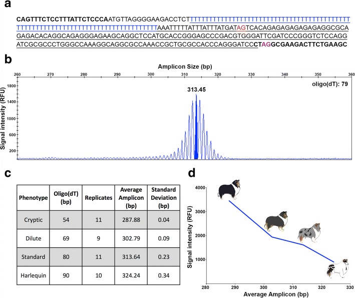
Background: The antisense insertion of a canine short interspersed element (SINEC_Cf) in the pigmentation gene PMEL (or SILV) causes a coat pattern phenotype in dogs termed merle. Merle is a semi-dominant trait characterized by patches of full pigmentation on a diluted background. The oligo(dT) tract of the Merle retrotransposon is long and uninterrupted and is prone to dramatic truncation. Phenotypically wild-type individuals carrying shorter oligo(dT) lengths of the Merle allele have been previously described and termed cryptic merles. Two additional coat patterns, dilute merle (uniform, steely-grey coat) and harlequin merle (white background with black patches), also appear in breeds segregating the Merle allele.

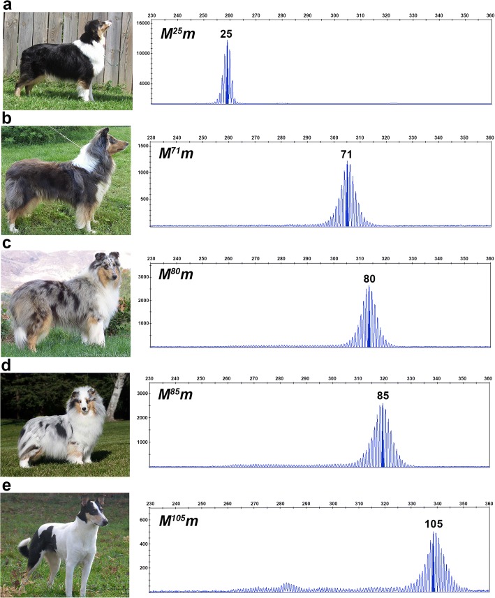
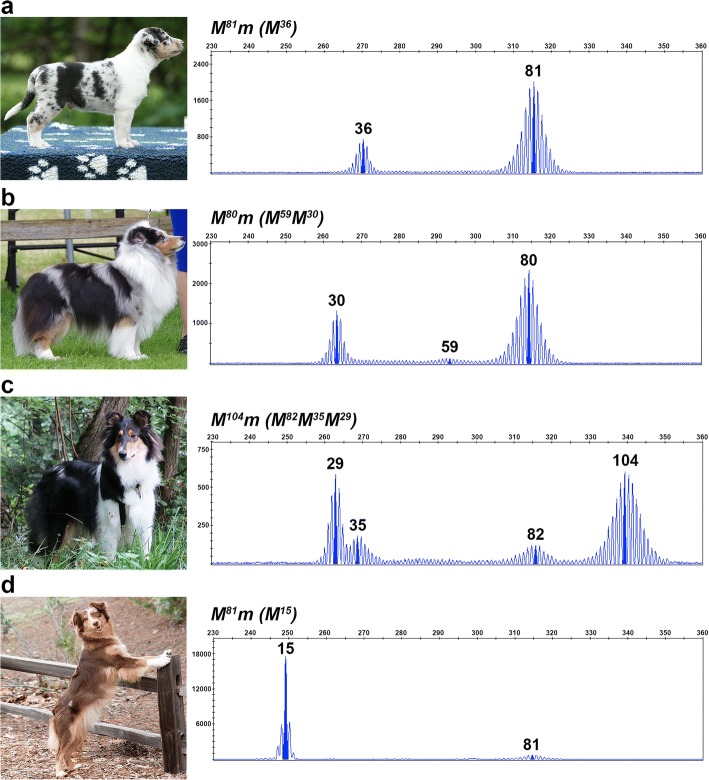
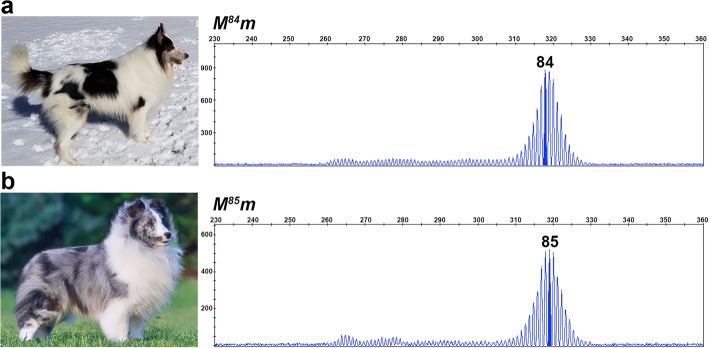
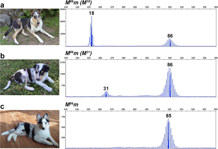
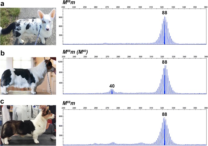
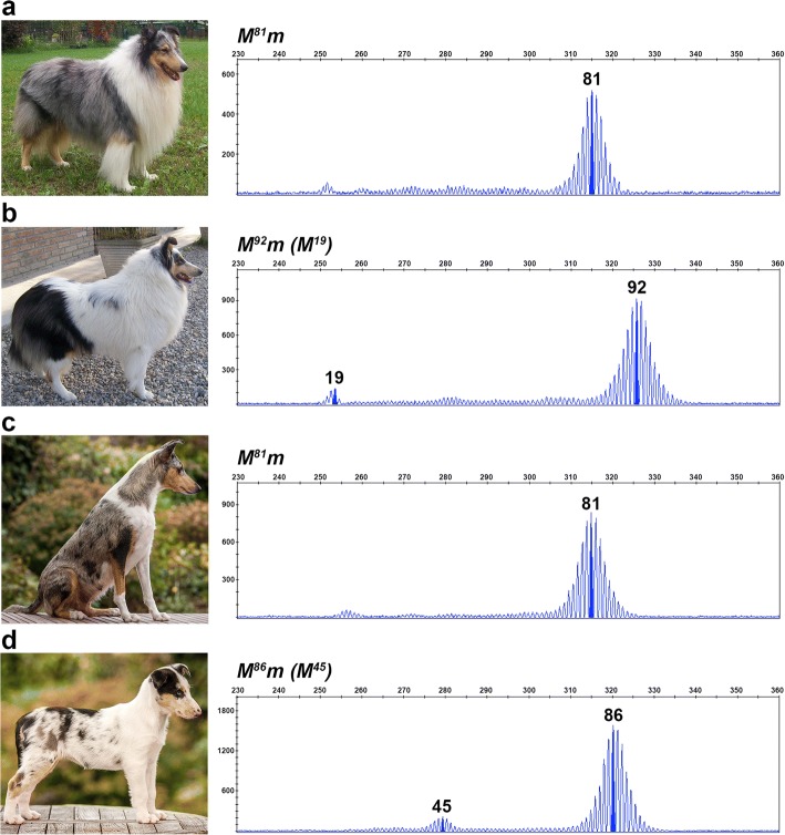
Results: Sequencing of all PMEL exons in a dilute and a harlequin merle reveals that variation exists solely within the oligo(dT) tract of the SINEC_Cf insertion. In fragment analyses from 259 dogs heterozygous for Merle, we observed a spectrum of oligo(dT) lengths spanning 25 to 105 base pairs (bp), with ranges that correspond to the four varieties of the merle phenotype: cryptic (25-55 bp), dilute (66-74 bp), standard (78-86 bp), and harlequin (81-105 bp). Somatic contractions of the oligo(dT) were observed in 43% of standard and 51% of harlequin merle dogs. A small proportion (4.6%) of the study cohort inherited de novo contractions or expansions of the Merle allele that resulted in dilute or harlequin coat patterns, respectively.

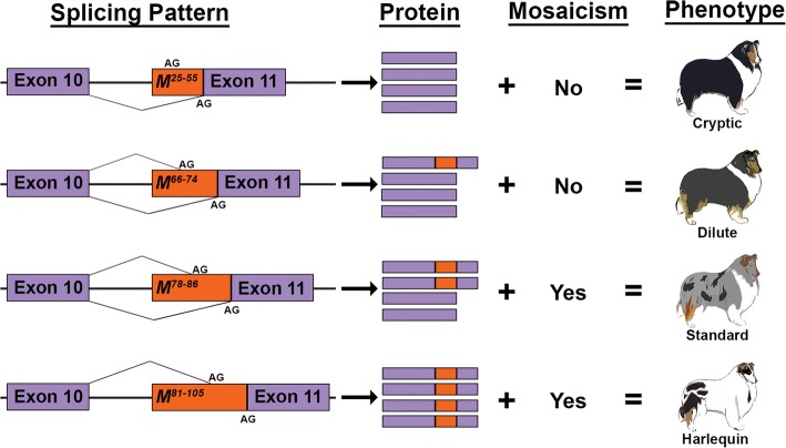
Conclusions: The phenotypic consequence of the Merle SINE insertion directly depends upon oligo(dT) length. In transcription, we propose that the use of an alternative splice site increases with oligo(dT) length, resulting in insufficient PMEL and a pigment dilution spectrum, from dark grey to complete hypopigmentation. We further propose that during replication, contractions and expansions increase in frequency with oligo(dT) length, causing coat variegation (somatic events in melanocytes) and the spontaneous appearance of varieties of the merle phenotype (germline events).

Keywords: Alternative splicing; Coat pattern; Dilution; Dog; Exonization; Mononucleotide repeat; Pigmentation; SILV; SINE; Slippage.


Conflict of interest statement

All samples were obtained with informed owner consent according to protocols approved by the Clemson University Institutional Review Board (IBC2008–17) and IACUC (2012–039). Not applicable. The authors declare that they have no competing interests. Springer Nature remains neutral with regard to jurisdictional claims in published maps and institutional affiliations.

Figures

Fig. 1
Fig. 1
Evaluation of an assay for determining the length of the Merle SINE oligo(dT). (a) Sequence of the PCR product is shown, with primer sequences in bold. The retrotransposon is underlined, and the oligo(dT) is in blue. Non-oligo(dT) sequences total 234 bp. The wild-type splice site is in purple, while the alternative splice site is in red. (b) A chromatogram from fragment analysis depicts amplicon size in base pairs (x-axis) and signal intensity in relative fluorescent units (RFU) (y-axis). For determination of oligo(dT) length, 234 is subtracted from the size of the amplicon peak with the highest RFU (highlighted), rounded to the nearest whole number. (c) Standard deviation of amplicon size is given for technical replicates from one dog representing each of four phenotypes. (d) Average signal intensity from the technical replicates is plotted against average amplicon size
Fig. 2
Fig. 2
Oligo(dT) lengths correspond to the merle phenotypic spectrum. Photographs and fragment analysis chromatograms are shown for dogs representing each of the four merle varieties: cryptic (a), dilute (b), standard (c), and harlequin (e), as well as one dog (d) that displays characteristics of both standard and harlequin merles. Amplicon size in base pairs is shown on the x-axis, and the y-axis measures signal intensity (RFU). The most abundant Merle amplicon peak is highlighted for each dog with corresponding oligo(dT) length written above. Genotypes are reported in the top left of the chromatogram, with M representing the Merle allele depicted in the chromatogram and m signifying the wild-type allele, confirmed through gel electrophoresis (not pictured). The cryptic and harlequin merle dogs pictured respectively possess the shortest and longest oligo(dT) lengths identified in the study
Fig. 3
Fig. 3
Somatic contractions of the oligo(dT) reflect the proportion of full pigmentation in merle coats. Photographs and chromatograms depicting fragment analysis data from blood and buccal cells are given for two standard (a and b) and two predominantly solid merle dogs (c and d). Amplicon size in base pairs is shown on the x-axis, and the y-axis measures signal intensity (RFU). In these four dogs, the amplicon with the longest oligo(dT) represents the inherited allele, while smaller amplicons stem from somatic contractions. Genotypes are listed above each chromatogram, with M representing the inherited Merle allele, m indicating the wild-type allele (not pictured), and (M) denoting the contracted Merle amplicons
Fig. 4
Fig. 4
Oligo(dT) lengths of 81–86 bp are observed in standard and harlequin merle dogs. Photographs and chromatograms from fragment analysis data are shown for a harlequin (a) and a standard merle (b). Amplicon size in base pairs is shown on the x-axis, and the y-axis measures signal intensity (RFU). Genotypes are given for each dog above their chromatogram with M denoting their inherited Merle allele, and m representing their non-pictured, wild-type allele
Fig. 5
Fig. 5
Inheritance of Merle oligo(dT) lengths. Fragment analysis chromatograms and photographs are shown for a predominantly solid dam (a) and her harlequin merle progeny (b and c). Amplicon size in base pairs is shown on the x-axis, and the y-axis measures signal intensity (RFU). Genotypes are given for each dog above their chromatogram with M denoting the inherited Merle allele, m representing their non-pictured, wild-type allele, and (M) signifying somatic contractions of the inherited Merle allele
Fig. 6
Fig. 6
Somatic oligo(dT) contractions and resulting merle phenotypes can vary between individuals. Photographs and fragment analysis chromatograms are shown for three littermates. Amplicon size in base pairs is shown on the x-axis, and the y-axis measures signal intensity (RFU). All three dogs share an identical inherited Merle allele in the harlequin range, but possess either zero (a), one (b), or two (c) contracted Merle amplicons that correlate with the degree of pigmentation in their coats. Genotypes are listed above each chromatogram, with M representing the inherited Merle allele, m indicating the wild-type allele (not pictured), and (M) denoting the contracted Merle amplicons
Fig. 7
Fig. 7
Germline de novo expansion of the Merle oligo(dT) tract. Photographs and fragment analysis chromatograms are shown for a standard merle sire (a) and his harlequin merle daughter (b), as well as a standard merle dam (c) and her harlequin merle daughter (d). Amplicon size in base pairs is shown on the x-axis, and the y-axis measures signal intensity (RFU). Genotypes are given above the chromatogram for each dog with M denoting the inherited Merle allele, m representing the wild-type allele (not pictured), and (M) signifying somatic contractions of the longer de novo Merle allele
Fig. 8
Fig. 8
Proposed mechanism for merle phenotypic variation. Suggested patterns of splicing are shown for cryptic, dilute, standard, and harlequin merles. The Merle SINE is depicted in orange with oligo(dT) length ranges superscripted. The original and alternative (within SINE) splice acceptor sites are denoted by “AG.” The proportion of wild-type (solid purple) to aberrant (purple and orange) PMEL protein illustrates the proposed frequency of alternative splicing, as it corresponds to oligo(dT) length. Mosaicism reflects whether somatic oligo(dT) contractions were observed in each phenotypic group herein. Together, the rates of alternative splicing and somatic oligo(dT) contractions confer the background coat color intensity and variegation, respectively

References

    1. Sorsby A, Davey JB. Ocular associations of dappling (or merling) in the coat colour of dogs. J Genet. 1954;52:425. doi: 10.1007/BF02981535. - DOI
    1. Strain GM, Clark LA, Wahl JM, Turner AE, Murphy KE. Prevalence of deafness in dogs heterozygous or homozygous for the merle allele. J Vet Intern Med. 2009;23(2):282–286. doi: 10.1111/j.1939-1676.2008.0257.x. - DOI - PubMed
    1. Sponenberg DP. Germinal reversion of the merle allele in Australian shepherd dogs. J Hered. 1984;75(1):78. doi: 10.1093/oxfordjournals.jhered.a109874. - DOI - PubMed
    1. Theos AC, Truschel ST, Raposo G, Marks MS. The silver locus product Pmel17/gp100/Silv/ME20: controversial in name and in function. Pigment Cell Res. 2005;18(5):322–336. doi: 10.1111/j.1600-0749.2005.00269.x. - DOI - PMC - PubMed
    1. Clark LA, Wahl JM, Rees CA, Murphy KE. Retrotransposon insertion in SILV is responsible for merle patterning of the domestic dog. Proc Natl Acad Sci U S A. 2006;103(5):1376–1381. doi: 10.1073/pnas.0506940103. - DOI - PMC - PubMed