

CTGF in kidney fibrosis and glomerulonephritis
- PMID: 30123390
- PMCID: PMC6091167
- DOI: 10.1186/s41232-018-0070-0
CTGF in kidney fibrosis and glomerulonephritis
Abstract
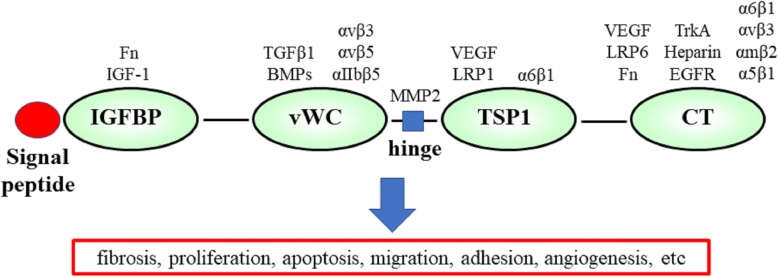
Background: Glomerulonephritis, which causes inflammation in glomeruli, is a common cause of end-stage renal failure. Severe and prolonged inflammation can damage glomeruli and lead to kidney fibrosis. Connective tissue growth factor (CTGF) is a member of the CCN matricellular protein family, consisting of four domains, that regulates the signaling of other growth factors and promotes kidney fibrosis.
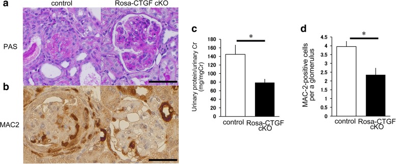
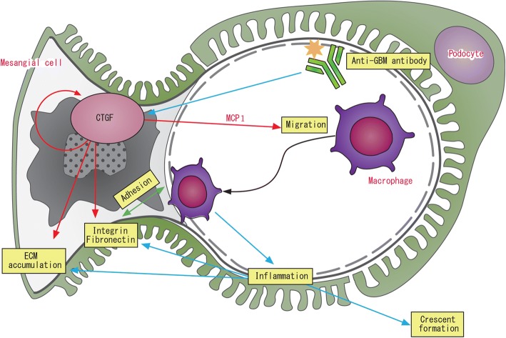
Main body of the abstract: CTGF can simultaneously interact with several factors with its four domains. The microenvironment differs depending on the types of cells and tissues and differentiation stages of these cells. The diverse biological actions of CTGF on various types of cells and tissues depend on this difference in microenvironment. In the kidney, CTGF is expressed at low levels in normal condition and its expression is upregulated by kidney fibrosis. CTGF expression is known to be upregulated in the extra-capillary and mesangial lesions of glomerulonephritis in human kidney biopsy samples. In addition to involvement in fibrosis, CTGF modulates the expression of inflammatory mediators, including cytokines and chemokines, through distinct signaling pathways, in various cell systems. In anti-glomerular basement membrane (GBM) glomerulonephritis, systemic CTGF knockout (Rosa-CTGF cKO) mice exhibit 50% reduction of proteinuria and decreased crescent formation and mesangial expansion compared with control mice. In addition to fibrotic markers, the glomerular mRNA expression of Ccl2 is increased in the control mice with anti-GBM glomerulonephritis, and this increase is reduced in Rosa-CTGF cKO mice with nephritis. Accumulation of MAC2-positive cells in glomeruli is also reduced in Rosa-CTGF cKO mice. These results suggest that CTGF may be required for the upregulation of Ccl2 expression not only in anti-GBM glomerulonephritis but also in other types of glomerulonephritis, such as IgA nephropathy; CTGF expression and accumulation of macrophages in the mesangial area have been documented in these glomerular diseases. CTGF induces the expression of inflammatory mediators and promotes cell adhesion.
Short conclusion: CTGF plays an important role in the development of glomerulonephritis by inducing the inflammatory process. CTGF is a potentiate target for the treatment of glomerulonephritis.
Keywords: CTGF; Chemokine; Glomerulonephritis; Inflammation; Macrophage.
Conflict of interest statement
Not applicable. The authors declare that they have no competing interests. Springer Nature remains neutral with regard to jurisdictional claims in published maps and institutional affiliations.
Figures
References
-
- Bradham DM, Igarashi A, Potter RL, Grotendorst GR. Connective tissue growth factor: a cysteine-rich mitogen secreted by human vascular endothelial cells is related to the SRC-induced immediate early gene product CEF-10. J Cell Biol. 1991;114(6):1285–1294. doi: 10.1083/jcb.114.6.1285. - DOI - PMC - PubMed
Publication types
LinkOut - more resources
Full Text Sources
Other Literature Sources
Molecular Biology Databases
Research Materials
Miscellaneous
