Review
. 2018 Jul;10(Suppl 20):S2333-S2345.
doi: 10.21037/jtd.2018.01.117.

Interfacial tissue engineering of heart regenerative medicine based on soft cell-porous scaffolds

Affiliations
Review

Interfacial tissue engineering of heart regenerative medicine based on soft cell-porous scaffolds

Xiwen Geng et al. J Thorac Dis. 2018 Jul.

Abstract

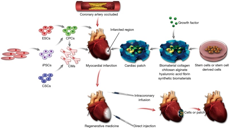
Myocardial infarction (MI), occurs when the coronary artery is occluded resulting in the hypoxia of areas in heart tissue, is increasing in recent years because of the population ageing and lifestyle changes. Currently, there is no ideal therapeutic scheme because of the limitation of MI therapeutic strategies due to the lack of regenerative ability of the heart cells in adult humans. Recent advances in tissue engineering and regenerative medicine brings hope to the MI therapy and current studies are focusing on restoring the function and structure of damaged tissue by delivering exogenous cells or stimulating endogenous heart cells. However, attempts to directly inject stem cells or cardiomyocytes to the infract zone often lead to rapid cell death and abundant cell loss. To address this challenge, various soft repair cells and porous scaffold materials have been integrated to improve cell retention and engraftment and preventing left ventricle (LV) dilatation. In this article, we will review the current method for heart regeneration based on soft cell-porous scaffold interfacial tissue engineering including common stem cell types, biomaterials, and cardiac patch and will discuss potential future directions in this area.

Keywords: Regenerative medicine; biomaterial; cardiac repair; myocardial infarction (MI); tissue engineering.


Conflict of interest statement

Conflicts of Interest: The authors have no conflicts of interest to declare.

Figures

Figure 1
Figure 1
Summary of approaches for cardiac tissue regeneration. The left of this scheme shows the potential cell sources for heart regeneration. The right shows the diagrammatic representation of cardiac patch strategies using biomaterials. In the middle, the diagrammatic representation of the myocardial infarction and the injection methods used in heart regeneration is illustrated. ESCs, embryonic stem cells; CPCs, cardiac progenitor cells; SCs, somatic cells; iPSCs, induced pluripotent stem cells; CMs, cardiomyocytes; CSCs, cardiac stem cells.
Figure 2
Figure 2
Stem cells and heart regeneration. (A) Signal pathways involved in maintaining mouse ESC pluripotency. (B) Model of the transcription factor network in embryonic stem (ES) cells. The arrow indicates the promoting effect and the horizontal line means the blocking effect (21). (C) Embryonic stem cell transplantation improved left ventricular (LV) function (24). (C1) Sham-operated rats; (C2) postinfarcted rats injected with cell-free medium; (C3) postinfarcted rats transplanted with embryonic stem cells. (D) Human embryonic stem cells enhance function of infracted rat hearts (10). The bright field microscopic images were acquired from recipient hearts 4 weeks post-transplantation. (D1) The human pan-centromeric in situ hybridization (brown chromagen) and β-myosin-positive cardiomyocytes (red) indicated the formation of a large cell graft within infarct scar tissue. Scale bar =100 µm. (D2) High magnification of boxed part in D1. (D3) Hematoxylin and eosin stain showed the graft cells have a vacuolated appearance because of the glycogen. Scale bar =50 µm. [(A) reprinted with permission of reference (20), (B) reprinted with permission of reference (21), (C) reprinted with permission of reference (24) and (D) reprinted with permission of reference (10)]. LVP, left ventricular potential; dP/dt, peak LV systolic pressure.
Figure 3
Figure 3
The collagen patch in cardiac regeneration. (A) Coronary artery perfusion of rat hearts (48). (A1) Infarcted heart without collagen patch. (A2) Infarcted heart with collagen patch. The Evans blue was used in the experiment to show the neo-vasculature. (B) The in vitro properties (B1–3) and in vivo effect on cell mobilization (B4–6) of porous collagen cardiac patch (49). (C) Schematic representation of the plastic compression of collagen gels (C1) and inducing MI via left anterior descending (LAD) artery ligation (C2) which was either treated with collagen patch (C3,4) or untreated (C5,6) (50). [Figure (A) reprinted with permission of reference (48), (B) reprinted with permission of reference (49), and (C) reprinted with permission of reference (50)].
Figure 4
Figure 4
The cardiac patch and heart regeneration. (A) Diagrammatic representation of vascularization strategies in heart regeneration (72); (B) schematic of the macroporous iron oxide frameworks in the repair of infracted heart (73). [Figure (A) reprinted with permission of reference (72) and (B) reprinted with permission of reference (73)].

References

    1. Bui AL, Horwich TB, Fonarow GC. Epidemiology and risk profile of heart failure. Nat Rev Cardiol 2011;8:30-41. 10.1038/nrcardio.2010.165 - DOI - PMC - PubMed
    1. Ibáñez B, Heusch G, Ovize M, et al. Evolving therapies for myocardial ischemia/reperfusion injury. J Am Coll Cardiol 2015;65:1454-71. 10.1016/j.jacc.2015.02.032 - DOI - PubMed
    1. Artenie R, Artenie A, Ungureanu D, et al. Myocardial remodeling. Rev Med Chir Soc Med Nat Iasi 2003;107:35-9. - PubMed
    1. Gaudron P, Eilles C, Ertl G, et al. Compensatory and noncompensatory left ventricular dilatation after myocardial infarction: Time course and hemodynamic consequences at rest and during exercise. AM Heart J 1992;123:377-85. 10.1016/0002-8703(92)90649-G - DOI - PubMed
    1. Domenech M, Polo-Corrales L, Ramirez-Vick JE, et al. Tissue engineering strategies for myocardial regeneration: acellular versus cellular scaffolds? Tissue Eng Part B Rev 2016;22:438-58. 10.1089/ten.teb.2015.0523 - DOI - PMC - PubMed

LinkOut - more resources