. 2018 Apr:2:42-50.
doi: 10.1016/j.cophys.2017.12.007. Epub 2017 Dec 23.

Dravet Syndrome: A Sodium Channel Interneuronopathy

Affiliations

Dravet Syndrome: A Sodium Channel Interneuronopathy

William A Catterall. Curr Opin Physiol. 2018 Apr.

Abstract

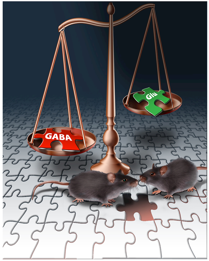
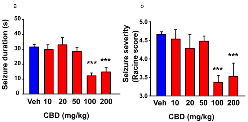
Dravet Syndrome is a devastating childhood epilepsy disorder with high incidence of premature death plus comorbidities of ataxia, circadian rhythm disorder, impaired sleep quality, autistic-like social-interaction deficits and severe cognitive impairment. It is primarily caused by heterozygous loss-of-function mutations in the SCN1A gene that encodes brain voltage-gated sodium channel type-1, termed NaV1.1. Here I review experiments on mouse genetic models that implicate specific loss of sodium currents and action potential firing in GABAergic inhibitory interneurons as the fundamental cause of Dravet Syndrome. The resulting imbalance of excitatory to inhibitory neurotransmission in neural circuits causes both epilepsy and co-morbidities. Promising therapeutic approaches involving atypical sodium channel blockers, novel drug combinations, and cannabidiol give hope for improved outcomes for Dravet Syndrome patients.

Keywords: Epilepsy; autism; cognitive deficit; interneurons; sodium channels.


Conflict of interest statement

The author declares no conflicts of interest.

Figures

Figure 1.
Figure 1.. Spontaneous and thermally induced seizures in DS mice.
The body core temperature of mice was raised to the indicated levels by a thermal feedback circuit and heat lamp in steps of 0.5°C per two minutes, and seizures were recorded by video camera and implanted EEG electrodes. (a) Representative EEG traces during a spontaneous seizure in a DS mouse. (b) Representative EEG traces of F/+:Dlx-Cre- and F/+:Dlx-Cre+ mice during thermal induction of seizures at P24. Top, F/+:Dlx-Cre- mouse at 39.5°C. Bottom, F/+:Dlx-Cre+ mouse during a seizure at 39.5°C. (c) Temperature dependence of thermal induction of seizures in DS mice. Thermally-induced seizures were evoked in DS mice at P30–46 (circles, mean temperature of 39°C) and P20–22 (squares, mean temperature of induction 40.2°C), but no seizures were induced in P17–18 mice (triangles). (d) Temperature dependence of thermal induction of seizures in Dlx-Cre+ mice. Thermally-induced seizures were evoked in all F/+:Dlx-Cre+ animals with a mean temperature of 39°C. No F/+:Dlx-Cre- mice had a seizure.
Figure 2.
Figure 2.. Spontaneous inhibitory and excitatory synaptic activity in WT and DS mice.
Hippocampal slices were prepared from WT and DS mice, and spontaneous, AP-dependent synaptic events were recorded by whole-cell voltage clamp of CA1 neurons. (a) Top. Representative recording of inhibitory postsynaptic currents (IPSCs) from a CA1 neuron from a DS mouse. Bottom. Histogram of inter-event intervals between IPSCs. Inset. Mean frequency of IPSCs. (b) Top. Representative recording of excitatory postsynaptic currents (EPSCs) from a CA1 neuron from a DS mouse. Bottom. Histogram of inter-event intervals between EPSCs. Inset. Mean frequency of EPSCs.
Figure 3.
Figure 3.. Autism and cognitive deficit in DS mice.
(a) A test mouse was placed in the center chamber (C) of a three-chamber apparatus with connecting passageways, and its movement among the three chambers was recorded and digitally analyzed. WT mice spend more time in the side chamber containing a stranger mouse in a small inverted wire cup (M) compared to the side chamber containing an empty wire cup (E). In contrast, DS mice spend equal time in the E and M chambers. (b) In the contextual fear-conditioning test, a mouse was placed in a well-marked cage with an electric grid as its floor, and the movement of the mouse was recorded and digitally analyzed. The % of time that the mouse displays fear-induced freezing behavior was quantified. WT and DS test mice freeze infrequently while exploring the cage (Control), but they freeze in fear during and immediately after a mild foot shock (Train, 2s, 0.5 mA). Test mice were transported to their home cage. Upon return to the context of the fearful shock after 30 min and 24 h, WT mice freeze in anticipation of a shock, even though none is administered. In contrast, DS mice display a profound deficit in short-term (30 min) and long term (24 hr) memory of the spatial context of the foot shock (0.5 mA), (c) In the 3-chamber test, Flox control mice prefer to interact with the stranger mouse (M) compared to the empty wire cup, whereas Dlx1/2-Scn1a+/− mice have no preference for the stranger mouse. (d) In the contextual fear-conditioning test, Dlx1/2-Scn1a+/− mice have a normal fear response during and immediately after the foot shock (Train) but display a profound deficit in short-term (30 min) and long-term (24 hr) memory of the spatial context of the foot shock compared to Flox control mice.
Figure 4.
Figure 4.. Impaired action potential generation in PV and SST interneurons from from Cre+ mice.
DS Flox mice were bred with mice expressing PV-Cre (PV mice) or with mice expressing SST-Cre (SST mice). Cerebral cortical slices were prepared, PV and SST interneurons were identified by specific labeling, and APs were recorded in whole-cell current clamp mode in response to injection of the indicated amounts of depolarizing current. (a) Representative trains of action potentials recorded in whole-cell current clamp during injection of 240 pA of depolarizing current. WT, black; PV, blue. (b) Action potential number plotted vs. the level of depolarizing current. (c) Representative trains of action potentials recorded in whole-cell current clamp during injection of 160 pA of depolarizing current. WT, black; SST, yellow. (d) Action potential number plotted vs. the level of depolarizing current.
Figure 5.
Figure 5.. Imbalance of excitatory vs. inhibitory neurotransmission in DS.
Failure of AP firing in GABAergic interneurons in DS causes a reduction in GABA release and an imbalance of excitatory vs. inhibitory neurotransmission in circuits in the brain. This imbalance leads to epilepsy, cognitive deficit, and autistic-like behaviors plus other co-morbidities in DS.
Figure 6.
Figure 6.. CBD reduced seizure duration and severity in DS mice.
The core body temperature of mice was warmed to 38.5°C as in Fig. 1, and the duration and severity of the resulting seizures were video recorded. (a) Dose dependence of CBD reduction in duration of thermally induced seizures. (b) Dose dependence of CBD reduction in seizure severity based on Racine Score.

References

    1. Hille B: Ionic Channels of Excitable Membranes edn 3rd Ed Sunderland, MA: Sinauer Associates Inc; 2001.
    1. Catterall WA: From ionic currents to molecular mechanisms: The structure and function of voltage-gated sodium channels. Neuron 2000, 26:13–25. - PubMed
    1. Hille B: Ionic Channels of Excitable Membranes, 3rd Ed Sunderland, MA: Sinauer Associates Inc; 2001.
    1. Vilin YY, Ruben PC: Slow inactivation in voltage-gated sodium channels: molecular substrates and contributions to channelopathies. Cell Biochem Biophys 2001, 35:171–190. - PubMed
    1. Dravet C: The core Dravet syndrome phenotype. Epilepsia 2011, 52 Suppl 2:3–9. - PubMed