. 2018 Oct;233(4):478-495.
doi: 10.1111/joa.12860. Epub 2018 Aug 19.

Force-transmitting structures in the digital pads of the tree frog Hyla cinerea: a functional interpretation

Affiliations

Force-transmitting structures in the digital pads of the tree frog Hyla cinerea: a functional interpretation

Julian K A Langowski et al. J Anat. 2018 Oct.

Abstract

The morphology of the digital pads of tree frogs is adapted towards attachment, allowing these animals to attach to various substrates and to explore their arboreal habitat. Previous descriptions and functional interpretations of the pad morphology mostly focussed on the surface of the ventral epidermis, and little is known about the internal pad morphology and its functional relevance in attachment. In this study, we combine histology and synchrotron micro-computer-tomography to obtain a comprehensive 3-D morphological characterisation of the digital pads (in particular of the internal structures involved in the transmission of attachment forces from the ventral pad surface towards the phalanges) of the tree frog Hyla cinerea. A collagenous septum runs from the distal tip of the distal phalanx to the ventral cutis and compartmentalises the subcutaneous pad volume into a distal lymph space and a proximal space, which contains mucus glands opening via long ducts to the ventral pad surface. A collagen layer connects the ventral basement membrane via interphalangeal ligaments with the middle phalanx. The collagen fibres forming this layer curve around the transverse pad-axis and form laterally separated ridges below the gland space. The topological optimisation of a shear-loaded pad model using finite element analysis (FEA) shows that the curved collagen fibres are oriented along the trajectories of the maximum principal stresses, and the optimisation also results in ridge-formation, suggesting that the collagen layer is adapted towards a high stiffness during shear loading. We also show that the collagen layer is strong, with an estimated tensile strength of 2.0-6.5 N. Together with longitudinally skewed tonofibrils in the superficial epidermis, these features support our hypothesis that the digital pads of tree frogs are primarily adapted towards the generation and transmission of friction rather than adhesion forces. Moreover, we generate (based on a simplified FEA model and predictions from analytical models) the hypothesis that dorsodistal pulling on the collagen septum facilitates proximal peeling of the pad and that the septum is an adaptation towards detachment rather than attachment. Lastly, by using immunohistochemistry, we (re-)discovered bundles of smooth muscle fibres in the digital pads of tree frogs. We hypothesise that these fibres allow the control of (i) contact stresses at the pad-substrate interface and peeling, (ii) mucus secretion, (iii) shock-absorbing properties of the pad, and (iv) the macroscopic contact geometry of the ventral pad surface. Further work is needed to conclude on the role of the muscular structures in tree frog attachment. Overall, our study contributes to the functional understanding of tree frog attachment, hence offering novel perspectives on the ecology, phylogeny and evolution of anurans, as well as the design of tree-frog-inspired adhesives for technological applications.

Keywords: attachment organ; bioadhesion; collagen; connective tissue; fibre-matrix-composite; material stiffness; shear load; smooth muscle.


Figures

Figure 1
Figure 1
(A) Basic morphological terminology of the digital tip and the distal interphalangeal joint of the tree frog Hyla cinerea. The blue region depicts the largely uncharacterised dermal and subdermal space including connective, muscular, vascular and other tissues. (B) Nomenclature and usage of the digits I–IV in the right forelimb (F) and I–V in the left hindlimb (H) [light blue arcs (FI–III, HI–II): histology/immunohistochemistry, dark blue arc (HV): μ‐CT]. (C) Definition of terms of anatomical location and of the cutting and viewing planes. DE, dermis; DP, distal phalanx; ED, epidermis; IE, intercalary element; MG, mucus gland; MP, middle phalanx.
Figure 2
Figure 2
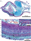
Series of transverse sections of a digital pad of Hyla cinerea (Frog 3, digit FIII) from distal to proximal stained with Crossmon's light green trichrome including Mayer's haematoxylin and Alcian blue [see inset for the approximate locations and the extent of the adhesive ventral epidermis (curved solid line)] through (A) the lymph space, (B) the approximate septum plane, (C) the gland space and (D) the distal end of the base of the distal phalanx. BV, blood vessel; CH, chromatophore; CO, collagen tissue; DE, dermis; DP, distal phalanx; DU, mucus duct; ED, epidermis; LY, lymph space; MG, mucus gland; PB, base of the distal phalanx; SE, septum; SM, smooth muscle.
Figure 3
Figure 3
Series of sagittal sections of a digital pad of Hyla cinerea (Frog 3, digit HI) from lateral to mid‐sagittal stained as in Fig. 2 [see inset for the approximate locations and the extent of the adhesive ventral epidermis (solid line)] (A) close to the lateral epidermis, (B) through the lateral part of the base of the distal phalanx, (C) through the lateral part of the diaphysis of the distal phalanx, and (D) in an approximately mid‐sagittal plane. Abbreviations as in Fig. 2, with the following additions: ET, tendon of the extensor muscle; FT, tendon of the flexor muscle; IE, intercalary element; LI, ligament; MP, middle phalanx; PH, head of the middle phalanx.
Figure 4
Figure 4
Series of horizontal sections of a digital pad of Hyla cinerea (Frog 3, digit FI) from ventral to dorsal stained as in Fig. 2 [see inset for the approximate locations and extent of the adhesive ventral epidermis (curved solid line)] through (A) the apical dermis (stratum spongiosum), (B) the ventral part of the ventral collagen layer, and (C) the dorsal part of the ventral collagen layer. Abbreviations as in Figs 2, 3.
Figure 5
Figure 5
3‐D visualisation of the structures of force transmission in a digital pad of Hyla cinerea (Frog 3, digit HV). (A) 3‐D view of the whole digital tip and internal structures. Only half of the approximately bi‐laterally symmetric smooth muscle fibres are shown (black arrowhead: thin muscle fibre bundle; grey arrowhead: thick muscle fibre bundle; white arrowhead: distal‐cross‐lateral muscle fibre). (BI) Frontal, (BII) lateral and (BIII) ventral view of the joint region: the collateral ligaments (yellow dashed line) give rise to several side arms, which connect to the ventral cutis in the middle digital segment (white arrowheads) and the ventral collagen layer (black arrowheads). The two collateral ligaments are connected via a medial strand that also connects to the intercalary element (grey arrowhead). Abbreviations as in Figs 2, 3, 4. x, longitudinal spatial coordinate; y, lateral spatial coordinate; z, vertical spatial coordinate.
Figure 6
Figure 6
(A) Approximately mid‐sagittal section through a digital pad of Hyla cinerea (Frog 2, digit HI) stained with Crossmon's light green trichrome including Mayer's haematoxylin and Alcian blue. (B) Magnified view of the ventral epidermis [see box in (A)] containing tonofibrils (reddish; black arrowheads), reticular cells (white arrowhead) and reticular connective tissue (grey arrowhead). Layer numbering after Ernst (1973a).
Figure 7
Figure 7
Collagen networks in a digital pad of Hyla cinerea. (A) Loose network of collagen fibres (light turquoise; grey arrowheads) in the ventral stratum spongiosum; horizontal section (Frog 3, digit FI). (B) Loose collagen network in the gland space (white arrowheads) and collagenous septum (black arrowheads); sagittal section (Frog 3, digit FII). (C) Septum (black arrowheads) separating the gland and lymph space; transverse section (Frog 3, digit FIII). Transverse and sagittal sections are oriented upright; in sagittal and horizontal sections, the distal digit side is on the left and at the top, respectively.
Figure 8
Figure 8
Ventral collagen layer and ridges (light turquoise; black arrowheads) in a digital pad of Hyla cinerea in (A) transverse (Frog 3, digit FIII), (B) horizontal (Frog 3, digit FI) and (C) sagittal (Frog 3, digit FII) section. The ridges consist of collagen fibres curved around the lateral pad‐axis. The troughs between the ridges are filled with mucus ducts (grey arrowheads) and blood vessels (white arrowheads). Section orientations as in Fig. 7.
Figure 9
Figure 9
(I) Histochemically (purple‐dark blue) and (II) immunohistochemically (brown) stained smooth muscle fibres in a digital pad of Hyla cinerea. (A) Two thick muscle fibre bundles [black arrowheads in (A) and (B)] run from the distal tip of the distal phalanx towards the ventral epidermis, and distal‐cross‐lateral muscle fibres traverse the lymph space above the ventral dermis [grey arrowhead in (A)]; transverse section (AI: frog 3, digit FIII; AII: frog 1, digit FIII). (B) Thin muscle fibre bundles [grey arrowheads in (B)] traverse the lymph space ventrodorsally; sagittal section (BI: frog 3, digit FII; BII: frog 1, digit HI). Smooth muscle fibres also surround the mucus glands (stars). (C) A fine network of proximal‐cross‐lateral muscle fibres runs laterally through the ventral collagen layer below the widening base of the distal phalanx [black arrowhead in (C) and white arrowhead in (B)]; transverse section (CI: frog 3, digit FIII; CII: frog 1, digit FIII). Section orientations as in Fig. 7. CH, chromatophore.
Figure 10
Figure 10
Topological optimisation of a shear‐loaded pad model. (A) Non‐optimised, ventrally fixed model (Young's modulus E = 20 MPa, Poisson's ratio ν = 0.33) with the approximate shape and size of the ventral collagen layer in a digital pad under a shear load FII,L acting on the proximal surface (red). (B) Topological optimisation leads to the formation of longitudinal ridges, to distal flattening, and to curved stress trajectories, similar to the ridges and the distribution as well as orientation of the collagen fibres, respectively, in the ventral collagen layer in the digital pads of Hyla cinerea (see insets). (I) Geometrical models in dorsoproximal view. The non‐optimised model is dimensioned in mm. (II) Von Mises stresses (dorsal view) indicating regions of low mechanical loading. (III) Vector plot of the maximum principal stresses (lateral view) showing the trajectories of force transmission.
Figure 11
Figure 11
Schematic representation of a digital pad of Hyla cinerea in the midsagittal plane. (AI) Proposed mechanism of shear load transmission during proximal pulling on the middle phalanx. (AII) Equilibrium of the external forces and moments (free body diagram) acting on the shear‐loaded ventral cutis and collagen layer with a hypothetical distribution of shear and normal loads acting on the ventral pad surface during steady attachment. In reality, shear loads will be orders of magnitude higher than normal loads. (B) Hypothesised mechanism of normal load transmission and the induction of peeling during extension of the distal phalanx. (C) Proposed functions of the smooth muscle fibres (m.).

References

    1. Afferante L, Heepe L, Casdorff K, et al. (2016) A theoretical characterization of curvature controlled adhesive properties of bio‐inspired membranes. Biomimetics 1, 3.
    1. Autumn K, Dittmore A, Santos D, et al. (2006) Frictional adhesion: a new angle on gecko attachment. J Exp Biol 209, 3569–3579. - PubMed
    1. Barnes WJP (2012) Adhesion in wet environments: frogs In: Encyclopedia of Nanotechnology. (ed. Bhushan B.), pp. 70–83. New York, NY: Springer.
    1. Barnes WJP, Goodwyn PJP, Nokhbatolfoghahai M, et al. (2011) Elastic modulus of tree frog adhesive toe pads. J Comp Physiol A 197, 969–978. - PMC - PubMed
    1. Barnes WJP, Baum M, Peisker H, et al. (2013) Comparative cryo‐SEM and AFM studies of hylid and rhacophorid tree frog toe pads. J Morphol 274, 1384–1396. - PubMed

Publication types

LinkOut - more resources