. 2018 Dec 1;27(23):4036-4050.
doi: 10.1093/hmg/ddy290.

Hypermorphic and hypomorphic AARS alleles in patients with CMT2N expand clinical and molecular heterogeneities

Affiliations

Hypermorphic and hypomorphic AARS alleles in patients with CMT2N expand clinical and molecular heterogeneities

Marian A J Weterman et al. Hum Mol Genet. .

Abstract

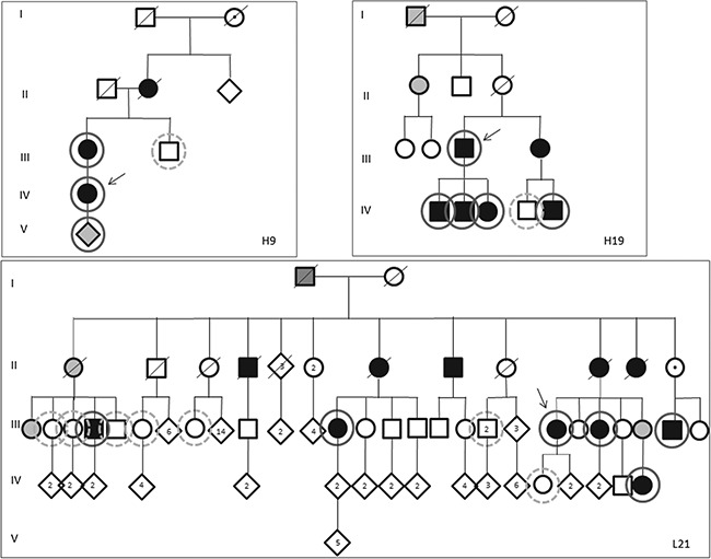
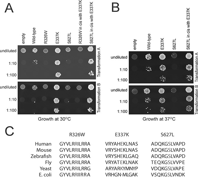
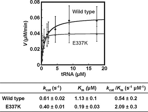
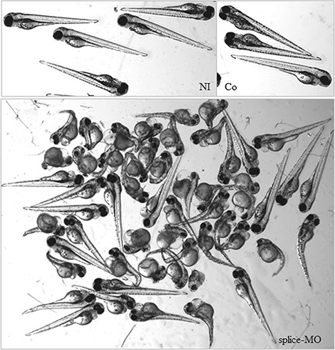
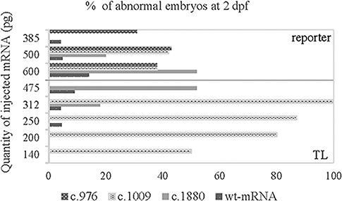
Aminoacyl-tRNA synthetases (ARSs) are ubiquitously expressed enzymes implicated in several dominant and recessive disease phenotypes. The canonical function of ARSs is to couple an amino acid to a cognate transfer RNA (tRNA). We identified three novel disease-associated missense mutations in the alanyl-tRNA synthetase (AARS) gene in three families with dominant axonal Charcot-Marie-Tooth (CMT) disease. Two mutations (p.Arg326Trp and p.Glu337Lys) are located near a recurrent pathologic change in AARS, p.Arg329His. The third (p.Ser627Leu) is in the editing domain of the protein in which hitherto only mutations associated with recessive encephalopathies have been described. Yeast complementation assays demonstrated that two mutations (p.Ser627Leu and p.Arg326Trp) represent loss-of-function alleles, while the third (p.Glu337Lys) represents a hypermorphic allele. Further, aminoacylation assays confirmed that the third mutation (p.Glu337Lys) increases tRNA charging velocity. To test the effect of each mutation in the context of a vertebrate nervous system, we developed a zebrafish assay. Remarkably, all three mutations caused a pathological phenotype of neural abnormalities when expressed in zebrafish, while expression of the human wild-type messenger RNA (mRNA) did not. Our data indicate that not only functional null or hypomorphic alleles, but also hypermorphic AARS alleles can cause dominantly inherited axonal CMT disease.


Figures

Figure 1
Figure 1
Pedigrees of families. Pedigrees of families with hereditary neuropathies as indicated in the figure. Tested individuals are marked by circles; solid line: tested positive for respective AARS mutation, dashed line: tested negative for AARS mutation. III.41,42: only one of the two individuals were tested. The mutations found were the following: c.1880C > T, p.Ser627Leu (H9), c.1009G > A, p.Glu337Lys (H19) and c.976GC > T, p.Arg326Trp (L21). Filled symbols indicate affected patients. Grey symbols: probably affected, said to be affected or no detailed neurologic examination. Open symbols: not affected. The family individuals are numbered consecutively from left to right. The index patient of each family is indicated by an arrow.
Figure 2
Figure 2
CMT-associated AARS mutations affect cellular growth in yeast. (A) Yeast lacking endogenous ALA1 (the yeast ortholog of AARS) were transformed with vectors containing wild-type AARS or mutant AARS or a vector without AARS insert (‘empty’). Transformation was performed twice (transformation A,B). Resulting cultures were plated undiluted or diluted (1:10 or 1:100) on media containing 5-FOA and grown at 30°C. (B) Cultures resulting from the same transformations as in A were plated undiluted or diluted on media containing 5-FOA and grown at 37°C. (C) Conservation of the affected amino acid residues. The position of each variant is shown along with flanking AARS amino acid residues from multiple, evolutionarily diverse species. The position of the affected residue is shown in red for each species.
Figure 3
Figure 3
In vitro aminoacylation assays are suggestive of a gain-of-function mechanism for the p.p.Glu337Lys mutation. Steady state assays of aminoacylation of tRNAAla with 3H-alanine were performed at 37oC with each enzyme at 1.67 nm, and tRNAAla in varying concentrations. Error bars show standard deviations derived from averaging at least three independent sets of experiments.
Figure 4
Figure 4
Morphology of zebrafish embryos at 72 hpf. Normal morphology was observed for control embryos at 72 hpf that were not injected (NI) or injected with control morpholino [GeneTools (Co)]. Splice-MO injected zebrafish showed abnormal morphology mostly consisting of shortened body axis, smaller eyes and curved bodies. Severe cases hardly developed a normal tail.
Figure 5
Figure 5
Injection of zebrafish embryos with splice-MO results in a concentration-dependant aberrant splicing. RT-PCR of splice MO-injected zebrafish with the position of the primers depicted by arrows and the position of the splice MO just preceding exon 14 of the zebrafish aars gene. Exons are indicated by numbered blocks. Sequence analysis of both products demonstrated that the expected wild-type product consisted of part of exon 13, exon 14 and part of exon 15 (black arrow, bottom panel; 268 bp) while the larger product consisted of part of exon 13, intron 13, exon 14 and part of exon 15 (white arrow, bottom panel; 360 bp). The bottom panel shows RT-PCR products of non-injected (A) or splice-MO injected zebrafish with 1 nl of 550 (B) and 650 um (C) solutions. As a molecular size marker (m) we used the 1 kb ladder (Invitrogen).
Figure 6
Figure 6
Whole mount immunohistochemical staining demonstrated aberrant patterns of synaptotagmin expression. Expression of synaptotagmin (blue staining) as detected by antibody znp-1 in wild-type zebrafish at 3 dpf (A) and in zebrafish injected at 1-cell stage with control MO (B,C) or with splice-MO (D–G) at 3 dpf. The immunostaining shows a less defined and increasingly irregular pattern as morphology grows worse.
Figure 7
Figure 7
Western blot and RNA analysis of injected zebrafish embryos of 2 dpf shows no major changes in quantities of AARS mRNA and protein. Comparable quantities of AARS mRNA were obtained after IVT. For quality control, 500 ng of each RNA was run on an agarose gel (A). Analysis of AARS protein in zebrafish lysates of wild-type- (wt) or mutant-mRNA-injected or not injected controls (NI) is shown in (B). For loading control, anti-tubulin was used (C). A quantification of the expression of AARS protein corrected for loading differences expressed in arbitrary units as compared to the expression in non-injected controls (D). Molecular markers were dual precision protein marker (BioRad) and 1 kb ladder (Invitrogen).
Figure 8
Figure 8
Zebrafish embryos injections of mutant mRNA (c.1009G > A, c.1880 C > T) lead to highly abnormal phenotypes. Embryos at 24 hpf (A,C,E,G,I,K) or 2 dpf (B,D,F,H,J,L) injected with 250 pg c.1880C > T, p.Ser627Leu (A–D), 200 pg c.1009G > A, p.Glu337Lys (E,F), 140 pg c.1009G > A and p.Glu337Lys (G,H) of mutant mRNAs or 475 pg wt mRNA (I,J) or non-injected controls (K,L). Wild-type mRNA injections were performed with 140–475 pg mRNA. The highest amount still did not affect the morphology of the embryos (K,L). At 2 dpf, a significant part of the surviving embryos injected with mutant mRNA is still affected.
Figure 9
Figure 9
Injection of all three mutant mRNAs give aberrant morphologies after injection in embryos of zebrafish reporter lines while equal or larger amounts of wild-type mRNAs do not. Pictures were taken at 2 dpf after injection of 500 pg of mutant (A–C) mRNA or wt mRNA (E) in 1-cell stage embryos of reporter strains or of non-injected controls (D). Mutant injected mRNAs: c.1009G > A, p.Glu337Lys (A), c.1880C > T, p.Ser627Leu (B), c.976C > T, p.Arg326Trp (C).
Figure 10
Figure 10
Increase of the number of abnormal embryos after injection of mutant mRNAs. Graphic representation of the percentage of abnormal embryos (number of abnormal embryos as compared to the total number of surviving embryos at 2 dpf) of injections with mutant or wild-type mRNA as indicated in the figure. The percentage of abnormal embryos of the non-injected embryos were not shown since no abnormalities were seen. Injections were performed in the wild-type strain (TL; below line) or reporter strain (above line).
Figure 11
Figure 11
Aberrant neural development in mutant mRNA injected embryos. Red fluorescence is depicted as white (A,B,E,F,G,J,K,L,N,P,Q,R) or red (C,D,H,I,M,O) driven by the olig2 promoter in the reporter strain. The green fluorescence reflects expression driven by the nkx2.2a promoter. Pictures were taken at 2 dpf of embryos injected with mutant or wt-mRNA (D,E,J; 500 pg) or of non-injected controls (N,O). Mutant injected mRNAs: c.976C > T, p.Arg326Trp (A–C), c.1009G > A, p.Glu337Lys (F–I), c.1880C>T, p.Ser627Leu (P–R,K,L).

References

    1. Meyer-Schuman R. and Antonellis A. (2017) Emerging mechanisms of aminoacyl-tRNA synthetase mutations in recessive and dominant human disease. Hum. Mol. Genet., 26, R114–R127. - PMC - PubMed
    1. Oprescu S.N., Griffin L.B., Beg A.A. and Antonellis A. (2017) Predicting the pathogenicity of aminoacyl-tRNA synthetase mutations. Methods, 113, 139–151. - PMC - PubMed
    1. Antonellis A., Ellsworth R.E., Sambuughin N., Puls I., Abel A., Lee-Lin S.Q., Jordanova A., Kremensky I., Christodoulou K., Middleton L.T. et al. (2003) Glycyl tRNA synthetase mutations in Charcot-Marie-Tooth disease type 2D and distal spinal muscular atrophy type V. Am. J. Hum. Genet., 72, 1293–1299. - PMC - PubMed
    1. Jordanova A., Irobi J., Thomas F.P., Van Dijck P., Meerschaert K., Dewil M., Dierick I., Jacobs A., De Vriendt E., Guergueltcheva V. et al. (2006) Disrupted function and axonal distribution of mutant tyrosyl-tRNA synthetase in dominant intermediate Charcot-Marie-Tooth neuropathy. Nat. Genet., 38, 197–202. - PubMed
    1. Latour P., Thauvin-Robinet C., Baudelet-Mery C., Soichot P., Cusin V., Faivre L., Locatelli M.C., Mayencon M., Sarcey A., Broussolle E. et al. (2010) A major determinant for binding and aminoacylation of tRNA(Ala) in cytoplasmic Alanyl-tRNA synthetase is mutated in dominant axonal Charcot-Marie-Tooth disease. Am. J. Hum. Genet., 86, 77–82. - PMC - PubMed

Publication types