. 2018 Sep 28;46(17):8993-9010.
doi: 10.1093/nar/gky733.

The suppressive cap-binding complex factor 4EIP is required for normal differentiation

Affiliations

The suppressive cap-binding complex factor 4EIP is required for normal differentiation

Monica Terrao et al. Nucleic Acids Res. .

Abstract

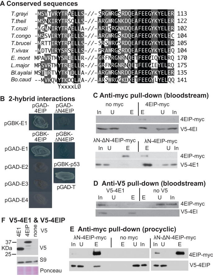
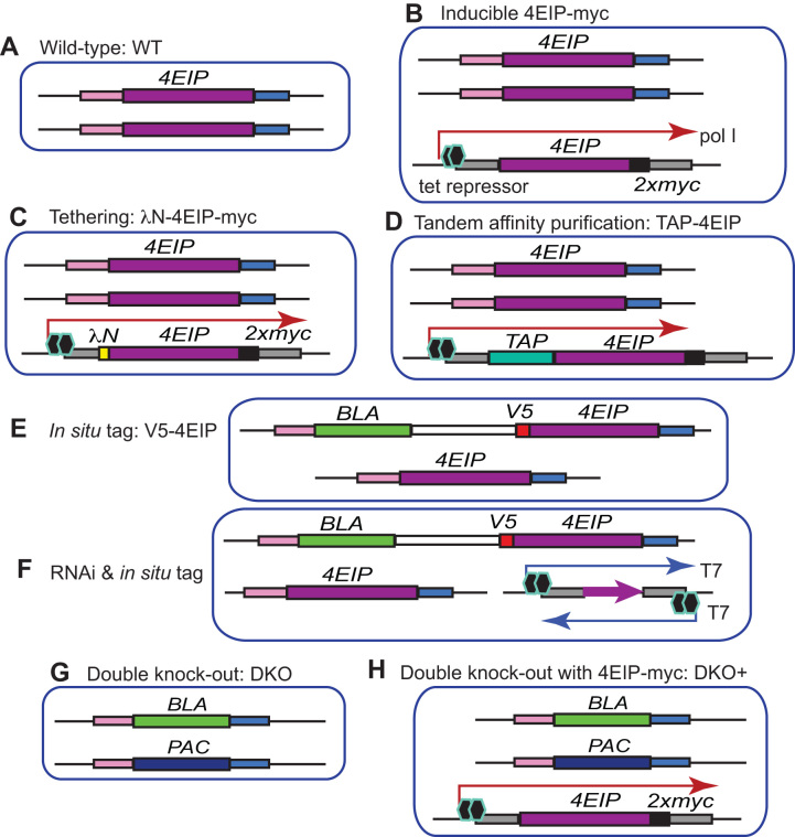
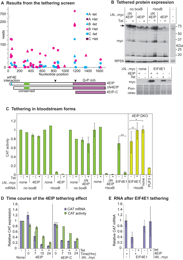
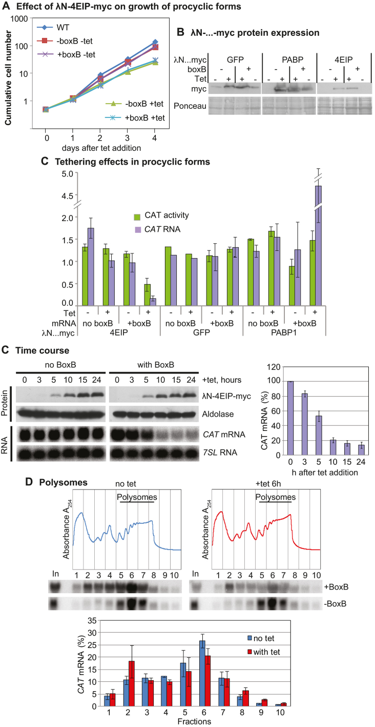
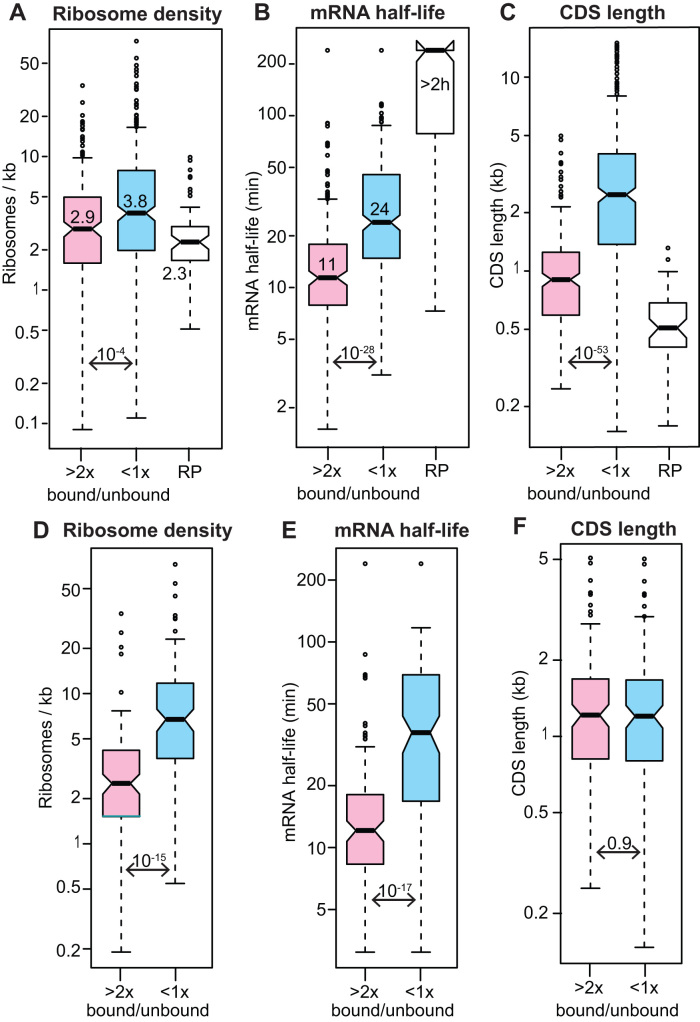
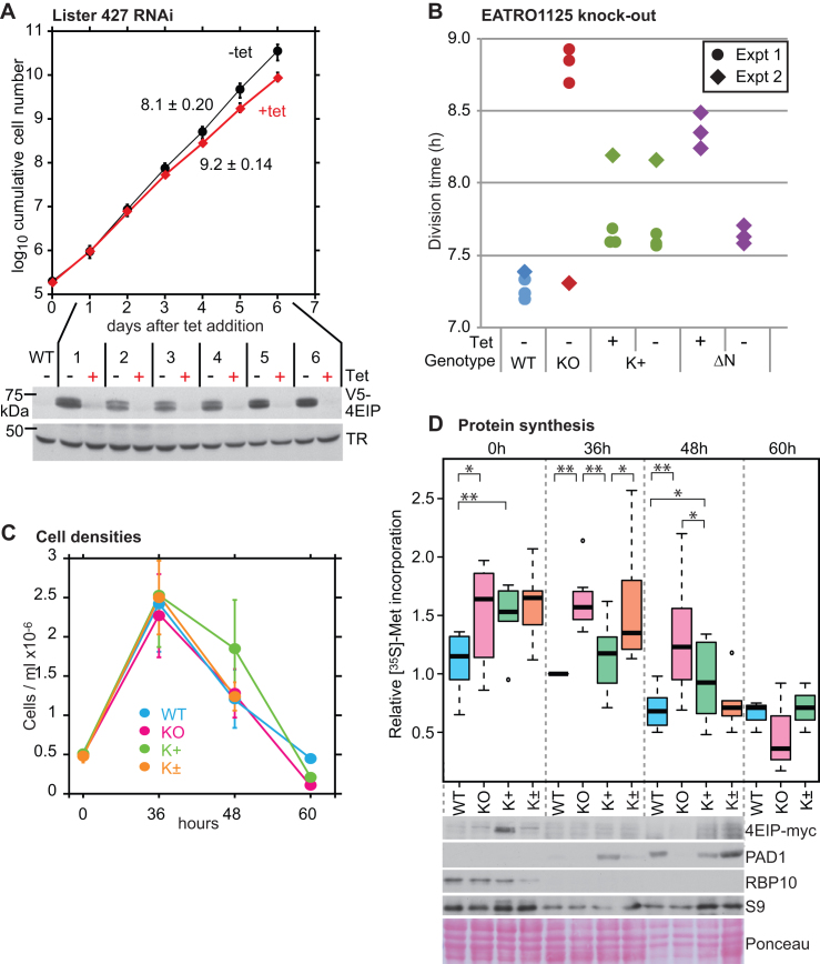
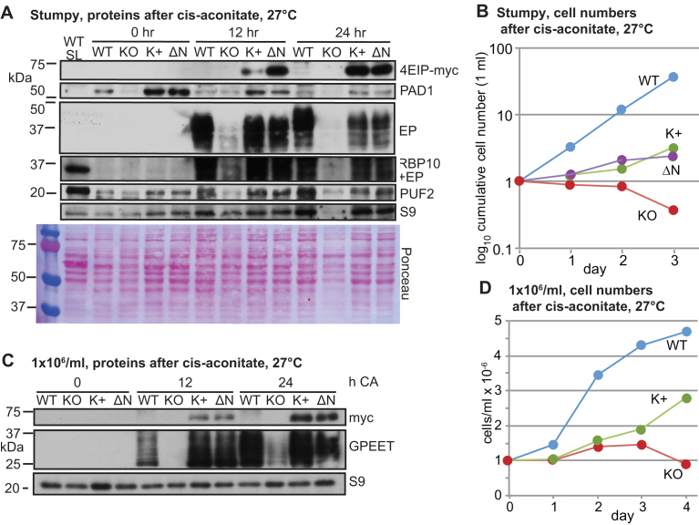
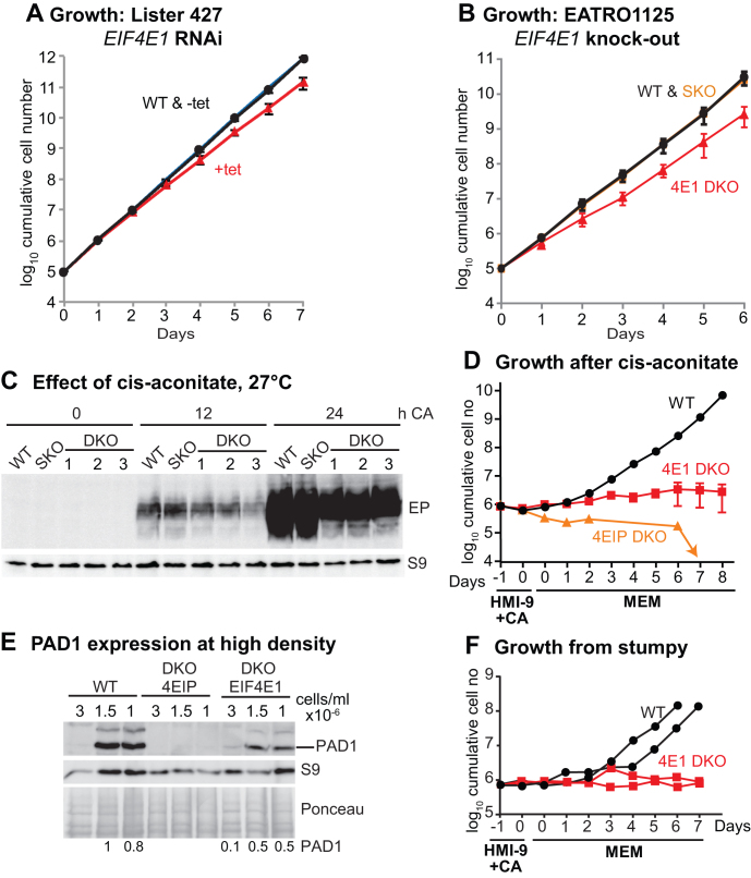
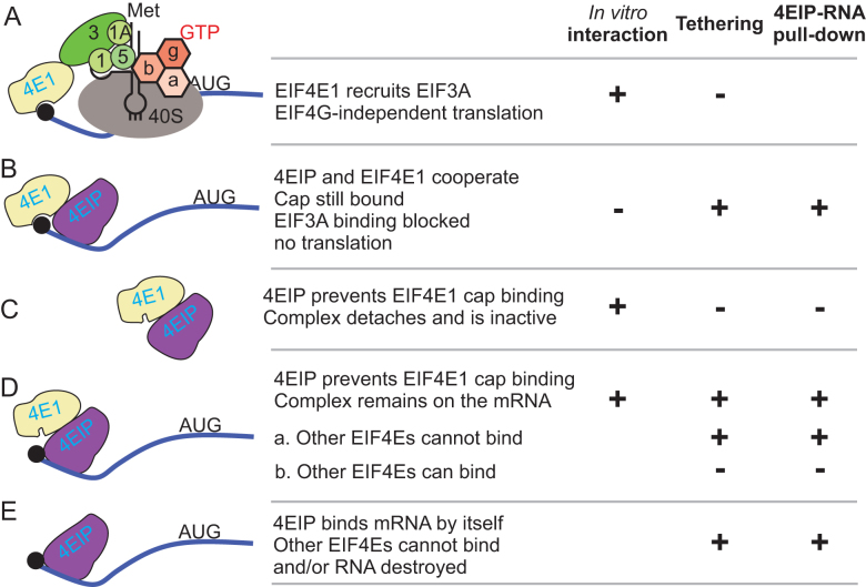
Trypanosoma brucei live in mammals as bloodstream forms and in the Tsetse midgut as procyclic forms. Differentiation from one form to the other proceeds via a growth-arrested stumpy form with low messenger RNA (mRNA) content and translation. The parasites have six eIF4Es and five eIF4Gs. EIF4E1 pairs with the mRNA-binding protein 4EIP but not with any EIF4G. EIF4E1 and 4EIP each inhibit expression when tethered to a reporter mRNA, but while tethered EIF4E1 suppresses only when 4EIP is present, suppression by tethered 4EIP does not require the interaction with EIF4E1. In growing bloodstream forms, 4EIP is preferentially associated with unstable mRNAs. Bloodstream- or procyclic-form trypanosomes lacking 4EIP have only a marginal growth disadvantage. Bloodstream forms without 4EIP are, however, defective in translation suppression during stumpy-form differentiation and cannot subsequently convert to growing procyclic forms. Intriguingly, the differentiation defect can be complemented by a truncated 4EIP that does not interact with EIF4E1. In contrast, bloodstream forms lacking EIF4E1 have a growth defect, stumpy formation seems normal, but they appear unable to grow as procyclic forms. We suggest that 4EIP and EIF4E1 fine-tune mRNA levels in growing cells, and that 4EIP contributes to translation suppression during differentiation to the stumpy form.


Figures

Figure 1.
Figure 1.
Trypanosoma brucei 4EIP interacts with EIF4E1. (A) Conserved regions in kinetoplastid 4EIPs. Amino acids highlighted in black are identical in all aligned sequences. Residues highlighted in gray are partially conserved—either identical or chemically similar in a majority of sequences. Sequences are (in vertical order): Trypanosoma grayi DQ04_01981000; Trypanosoma theileri ORC92063.1; Trypanosoma cruzi TcCLB.508461.290; Trypanosoma congolense TcIL3000_9_4530 Tryanosoma brucei Tb927.9.11050; Trypanosoma vivax TvY486_0905070; Endotrypanum_monterogeii EMOLV88_350043200; Leishmania major LmjF.35.3980; Blechomonas_ayalai Baya_047_0070; Bodo caudatus CUG36708.1. (B) The photos show growth of yeast on plates with stringent selection for the interaction between bait and prey plasmids. The identities of the plasmids used are shown next to the streaked yeast. Full results with controls are shown in Supplementary Figure S1A. (C) Extracts were made from bloodstream-form trypanosomes with or without a V5- in situ tagged eIF4E1 and myc-tagged 4EIP. Anti-myc immunoprecipitates were subjected to sodium dodecylsulphate-polyacrylamide gel electrophoresis (SDS-PAGE) and western blotting. In: input extract from 5 × 10⁶ cells; U: unbound fraction from 5 × 10⁶ cells; E: eluate from immunoprecipitating beads, from 1 × 10⁸ cells. Upper panel: anti-myc pull-down, with cells lacking a myc-tagged protein as control. Lower panel: anti-V5 pull-down, with cells lacking V5-tagged protein as the control. (D) As in (C) but with anti-V5 as the precipitating antibody. (E) As in (C) but with procyclic-form trypanosomes. (F) Western blot showing relative amounts of V5-tagged 4EIP and EIF4E1 in bloodstream forms. The only band from 4EIP migrated at ∼37 kDa. The control is ribosomal protein S9.
Figure 2.
Figure 2.
Cell lines. Diagrams are not to scale. Each picture represents the relevant gene loci. The different DNA segments are labeled on the figure.
Figure 3.
Figure 3.
Tethering of 4EIP to an mRNA suppresses expression. (A) Results for 4EIP from the published tethering screen (67). The trypanosomes used inducibly expressed an mRNA encoding the glycolytic enzyme PGKB, which is toxic in bloodstream forms (86). They were transfected with a library for inducible expression of lambdaN fusion proteins created from randomly sheared trypanosome DNA. After induction of both PGKB-boxB and the lamdaN fusions, cells were grown and the polymerase chain reaction products from the integrated lambdaN fusion proteins were sequenced (67). Results from three independent selections (A, B and C) are shown using different symbols. Counts that are higher in the presence of the inducer, tetracycline (magenta symbols), than without tetracycline (cyan symbols) indicate suppression of PGKB expression. Cartoons of 4EIP showing the two conserved regions (cyan and green) and the PQ-rich C-terminal region are below the x-axis. The two downward arrowheads indicate the cleavage positions that would generate the tagged proteins that were observed by western blotting in panel (B) and Figure 1F. (B) Expression of lambdaN…myc fusion proteins for the experiments in panels (C) and (E). The full-length lambdaN-4EIP (upper panel, arrow) was always found to be degraded; the two C-terminal pieces (30 and 20 kDa) will lack the lambda-N portion, so will be inactive in the assay. (C) Tethering assay using CAT reporter. Bloodstream-form trypanosomes expressing a CAT mRNA with five copies of boxB in the 3′-untranslated region (3′-UTR) were used; cells in which the CAT mRNA lacked boxB served as control. In the left-hand panel, cells were transfected with plasmids designed for tetracycline-inducible expression of either LambdaN-4EIP-myc or LambdaN-4EIP-myc with an N-terminal deletion that eliminated the interaction with eIF4E1 (ΔN4EIP). CAT activity was measured in the presence and absence of tetracycline. Results are expressed as arithmetic mean ± standard deviation of at least three measurements. Right-hand panel (with yellow bars in online version) — cells lacking 4EIP were used to test the activity of tethered EIF4E1, with PUF3 as a control. For the EIF4E1, tests were done six times. Results of Student’s t-tests were *P = 0.04; 〈?P = 0.07; **P = 0.009. Expression of myc-tagged proteins is shown on the right. (D) Time course of the tethering effect in bloodstream forms, using either 4EIP or its C-terminal domain. Results are mean and standard deviation of three measurements. (E) Effect on the CAT reporter mRNA of tethering EIF4E1 in the presence or absence of 4EIP. Results are mean and standard deviation of three measurements. In the paired bars, mRNA measurements are on the left and CAT activity on the right.
Figure 4.
Figure 4.
4EIP tethering suppresses translation and induces mRNA destruction in procyclic forms. (A) Effect of LambdaN-4EIP-myc expression on proliferation. A cumulative growth curve is shown. Results are arithmetic mean with error bars showing standard deviation from three assays. (B) Expression of LambdaN-…-myc fusion proteins. (B) Effects of different tethered reporters on CAT activity (left bars, green in online version) and mRNA (right bars, mauve in online version). Results are arithmetic mean with error bars showing standard deviation. (C) Time course of LambdaN-4EIP-myc expression in cells expressing CAT mRNA with or without boxB. A northern blot showing the effect on CAT mRNA is shown below, and quantitation of replicates is to the right. (D) Polysomes were separated on sucrose gradients, either without tetracycline (blue) or 6 h after tetracycline addition (red). Typical gradient profiles and northern blots are shown at the top, and quantitation of three replicates beneath with “+tet” on the right.
Figure 5.
Figure 5.
RNAs bound to 4EIP are relatively unstable mRNAs and were classified as ‘bound’ if the reads per million in the fraction bound to TAP-4EIP were at least twice those in the flow-though in both biological replicates, and the minimum count number was 10. ‘Unbound’ mRNAs has a ration of <1 in both replicates. The mRNAs encoding ribosomal proteins (RP) were mostly not bound but were considered separately. All results are for bloodstream forms. (A) Ribosome density for all ‘bound’ and ‘unbound’ mRNAs, as judged by ribosome profiling (49). Medians are indicated, as are the Student’s t-test values. (B) mRNA half-lives for all ‘bound’ and ‘unbound’ mRNAs (47). (C) Open reading frame or coding sequence (CDS) lengths of all ‘bound’ and ‘unbound’ mRNAs. (D) As (A), but for a subset of length-matched mRNAs (Supplementary Table S3, sheet 4). (E) As (B), but for the subset of length-matched mRNAs. (F) As (C), but for the subset of length-matched mRNAs.
Figure 6.
Figure 6.
Cells without 4EIP differentiate poorly into stumpy forms. (A) Depletion of 4EIP in monomorphic bloodstream forms (Lister 427) results in a very mild growth defect. Cumulative growth of cultures with tetracycline-inducible RNAi is shown, with mean and standard deviation for three different cell lines. Expression of V5-4EIP in one of the three different RNAi lines, analyzed by western blotting, is shown below. TR is trypanothione reductase. Results for the other two cell lines are in Supplementary Figure S7A. (B) Division times of different EATRO1125 cell lines. The division times of the same cell lines were measured in two independent experiments. Results for the individual replicates are shown. KO: no 4EIP; K+: knockout line with inducible 4EIP-myc, with or without tetracycline; ΔN—knockout line with inducible lambdaN-ΔN-4EIP-myc, with or without tetracycline. Tetracycline was added 48 h prior to the start of measurements. In the second experiment (diamonds), the knockout line appeared to have adapted to the lack of 4EIP during continuous culture. (C) Growth of different EATRO1125 cell lines allowed to reach high density. Cells with a starting density of 5 × 10⁵/ml were allowed to grow in HMI-9 media containing 1.1% methylcellulose for 60 h. Results are mean and standard deviation of three replicates. K+ are the complemented cells and K± are the K+ cells grown without tetracycline. (D) [35S]-Methionine incorporation into proteins during differentiation of EATRO1125 at high density. Trypanosomes were cultured as shown in (B), washed and pre-incubated for 1 h in labeling medium at 37°C. Methionine (35S) was then added for 20 min and proteins were analyzed by SDS-PAGE (Supplementary Figure S8B). Results for seven independent experiments are shown; four of the experiments included time = 0 h and three included time = 60 h. The color code is a paler version of (B). Student’s t-test results are: ** P < 0.01, * P < 0.05, both as paired or unpaired tests. Expression of various proteins, by western blotting, is shown under the box plot. For 4EIP-myc, the full-length protein is shown; degradation products are shown in Supplementary Figure S6F. The PAD1 signals from WT and K± are comparable.
Figure 7.
Figure 7.
Cells without 4EIP cannot differentiate into growing procyclic forms. (A) Expression of various proteins in EATRO1125 cells cultured to high density for 60 h, then treated with cis-aconitate (CA) and transferred to 27°C. WT SL is long slender bloodstream forms; KO: no 4EIP; K+: knockout line with inducible 4EIP-myc and tetracycline; ΔN—knockout line with inducible lambdaN-ΔN-4EIP-myc, with tetracycline. In this experiment, full-length 4EIP-myc was not detected at time = 0. (B) Cell counts for cells treated as in (A) (to the end of day 1), then transferred to procyclic medium. (C) Expression of various proteins in cells cultured to 1 × 10⁶/ml, then treated with cis-aconitate and transferred to 27°C. Replicates are in Supplementary Figure S8C. (D) Cell counts for cells from (C) transferred to procyclic medium on day 1.
Figure 8.
Figure 8.
Bloodstream forms without EIF4E1 cannot differentiate into growing procyclic forms. (A) Depletion of EIF4E1 in Lister 427 bloodstream forms results in a very mild growth defect. Cumulative growth of cultures with tetracycline-inducible RNAi is shown, with mean and standard deviation for three independent experiments. (B) Cumulative growth of different EATRO1125 cell lines. For WT and SKO (single replacement), the division times of the same cell lines were measured in two independent experiments. For DKO (both genes replaced), results for three different lines are shown. (C) Effect of adding cis-aconitate (CA) and shifting the temperature to 27°C (EATRO1125). The starting cell density was 1 × 10⁶/ml. EP procyclin expression was measured by western blotting, with ribosomal protein S9 as the loading control. Although it is not evident from the image, the last three lanes are somewhat under-loaded, and quantitation revealed no significant differences between DKO lines and the SKO or WT. (D) Cell numbers in cultures that were treated as in (C) (HMI-9 +CA, 27°C), then transferred to medium suitable for procyclic forms (MEM, 27°C). The results shown are mean and standard deviation for three EIF4E1 cultures. Controls included two wild-type cultures (mean shown) and a 4EIP culture. (E) Expression of PAD1 after incubation at high density. Cells were incubated until they attained 3 × 10⁶/ml, then the first samples were taken. To ensure complete comparability, subsequent samples were taken when the cell numbers had dropped to precisely 1.5 × 10⁶ and 1.0 × 10⁶/ml. Amounts of PAD1 relative to the Ponceau red stain are indicated. (F) Cells were left at high density for 48 h, then treated as in (D). Cumulative cell counts are shown.
Figure 9.
Figure 9.
Models for 4EIP function. For details see the ‘Discussion’ section.

References

    1. Firczuk H., Kannambath S., Pahle J., Claydon A., Beynon R., Duncan J., Westerhoff H., Mendes P., McCarthy J.E.. An in vivo control map for the eukaryotic mRNA translation machinery. Mol. Syst. Biol. 2013; 9:635. - PMC - PubMed
    1. Shah P., Ding Y., Niemczyk M., Kudla G., Plotkin J.B.. Rate-limiting steps in yeast protein translation. Cell. 2013; 153:1589–1601. - PMC - PubMed
    1. Richter J.D., Coller J.. Pausing on Polyribosomes: Make way for elongation in translational control. Cell. 2015; 163:292–300. - PMC - PubMed
    1. Hanson G., Coller J.. Codon optimality, bias and usage in translation and mRNA decay. Nat. Rev. Mol. Cell Biol. 2018; 19:20–30. - PMC - PubMed
    1. Pestova T.V., Kolupaeva V.G., Lomakin I.B., Pilipenko E.V., Shatsky I.N., Agol V.I., Hellen C.U.. Molecular mechanisms of translation initiation in eukaryotes. Proc. Natl. Acad. Sci. U.S.A. 2001; 98:7029–7036. - PMC - PubMed

Publication types

MeSH terms