. 2018 Sep 11;57(36):5359-5369.
doi: 10.1021/acs.biochem.8b00653. Epub 2018 Aug 28.

Structural Characterization of Competence-Stimulating Peptide Analogues Reveals Key Features for ComD1 and ComD2 Receptor Binding in Streptococcus pneumoniae

Affiliations

Structural Characterization of Competence-Stimulating Peptide Analogues Reveals Key Features for ComD1 and ComD2 Receptor Binding in Streptococcus pneumoniae

Yifang Yang et al. Biochemistry. .

Abstract

Streptococcus pneumoniae is an important pathogen that utilizes quorum sensing (QS) to regulate genetic transformation, virulence, and biofilm formation. The competence-stimulating peptide (CSP) is a 17-amino acid signal peptide that is used by S. pneumoniae to trigger QS. S. pneumoniae strains can be divided into two main specificity groups based on the CSP signal they produce (CSP1 or CSP2) and their compatible receptors (ComD1 or ComD2, respectively). Modulation of QS in S. pneumoniae can be achieved by targeting the CSP:ComD interaction using synthetic CSP analogues. However, to rationally design CSP-based QS modulators with enhanced activities, an in-depth understanding of the structural features that are required for receptor binding is needed. Herein, we report a comprehensive in-solution three-dimensional structural characterization of eight CSP1 and CSP2 analogues with varied biological activities using nuclear magnetic resonance spectroscopy. Analysis of these structures revealed two distinct hydrophobic patches required for effective ComD1 and ComD2 binding.


Figures

Figure 1.
Figure 1.. S. pneumoniae CSP-mediated QS circuit.
The ComC gene encodes a pre-CSP peptide, which is processed and secreted by the ABC transporter (ComAB). As the bacteria grow, the concentration of CSP increases until it reaches a threshold. Upon reaching the threshold concentration, CSP activates a transmembrane histidine kinase receptor (ComD), which, after being activated, transfers a phosphate group to its cognate response regulator (ComE). After phosphorylation ComE triggers the transcription of numerous genes including comX, the effector molecule of the circuitry that regulates QS-mediated phenotypes.
Figure 2.
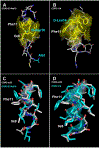
Figure 2.
(A) Heavy atom average structure of CSP1 (BMRB accession ID: 30416, and reference 25). (B) Heavy atom average structure of CSP1-E1A (BMRB accession ID: 30427). (C) Overlay of CSP1 (silver) and CSP1-E1A (cyan) structures. Residues E1-R3 and Q14-K17, as well as the side chains of S5, K6, R9 and D10 residues are hidden in the structures in (C) for clarity. Yellow surfaces in (A) and (B) represent the hydrophobic patch formed by the critical residues. See SI for overlay of the full structures.
Figure 3.
Figure 3.
(A) Heavy atom average structure of CSP1-K6A (BMRB accession ID: 30429). (B) Overlay of CSP1 (silver) and CSP1-K6A (cyan) structures. (C) Overlay of CSP1-E1A (silver) and CSP1-K6A (cyan) structures. Residues E1-R3 and Q14-K17, as well as the side chains of S5, K6 (A6 for CSP1-K6A), R9 and D10 residues are hidden in the structures in (B) and (C) for clarity. Yellow surface in (A) represents the hydrophobic patch formed by the critical residues. See SI for overlay of the full structures.
Figure 4.
Figure 4.
(A) Heavy atom average structure of CSP1-R3A (BMRB accession ID: 30428). (B) Heavy atom average structure of CSP1-f11 (BMRB accession ID: 30431). (C) Heavy atom average structure of CSP1-F11A (BMRB accession ID: 30430). (D) Overlay of CSP1 (silver) and CSP1-R3A (cyan) structures. (E) Overlay of CSP1 (silver) and CSP1-f11 (cyan) structures. (F) Overlay of CSP1 (silver) and CSP1-F11A (cyan) structures. Residues E1-R3 and Q14-K17, as well as the side chains of S5, K6, R9 and D10 residues are hidden in the structures in (D), (E) and (F) for clarity. Yellow surfaces in (A), (B) and (C) represent the hydrophobic patch formed by the critical residues. See SI for overlay of the full structures.
Figure 5.
Figure 5.
(A) Heavy atom average structure of CSP2-d10 (BMRB accession ID: 30432). (B) Overlay of CSP1-K6A (silver) and CSP2-d10 (cyan) structures. (C) Overlay of CSP1 (silver) and CSP2-d10 (cyan) structures. Residues El-R3 and Q14-K17, as well as the side chains of S5, K6 (A6 for CSP1-K6A), R9 and DIO residues for CSP1-K6A and CSP1 are hidden in the structures in (B) and (C) for clarity. Residues E1-R3 and L14-K17, as well as the side chains of S5, R6 and DIO for CSP2-d10 are hidden in the structures in (B) and (C) for clarity. Yellow surface in (A) represents the hydrophobic patch formed by the critical residues. See SI for overlay of the full structures.
Figure 6.
Figure 6.
(A) Heavy atom average structure of CSP2-ElAd10 (BMRB accession ID: 30433). (B) Heavy atom average structure of CSP2-114 (BMRB accession ID: 30434). (C) Overlay of CSP2-d10 (silver) and CSP2-ElAd10 (cyan) structures. (D) Overlay of CSP2-d10 (silver) and CSP2-114 (cyan) structures. Residues E1-R3 and L14-K17, as well as the side chains of S5, R6 and DIO are hidden in all structures in (C) and (D) for clarity. Yellow surfaces in (A) and (B) represent the hydrophobic patch formed by the critical residues. See SI for overlay of the full structures.
Figure 7.
Figure 7.
Predicted minimal structural requirements for CSP1 and CSP2 analogues based on the NMR analysis.
Figure 8.
Figure 8.
CD spectra of the three CSP1 and CSP2 analogues in membrane mimicking conditions. The two CSP1 analogues were found to be unstructured while the CSP2 analogue was found to exhibit an α-helix fiber characteristics. The spectra on the top right corner are the spectra of the two CSP1 analogues with adjusted y axis range to show their shapes more clearly. CD spectra of CSP1, CSP2, and CSP2-d10 were taken from reference . All CD spectra were acquired using the same conditions (20% TFE in PBS), with the exception that the concentration of CSP1, CSP2 and CSP2-d10 was 200 μM, while the concentration of the new CSP1 and CSP2 analogues was 20 μM.

References

    1. Mehr S, and Wood N (2012) Streptococcus pneumoniae--A Review of Carriage, Infection, Serotype Replacement and Vaccination, Paediatr Respir Rev 13, 258–264. - PubMed
    1. Huang SS, Johnson KM, Ray GT, Wroe P, Lieu TA, Moore MR, Zell ER, Linder JA, Grijalva CG, Metlay JP, and Finkelstein JA (2011) Healthcare Utilization and Cost of Pneumococcal Disease in the United States, Vaccine 29, 3398–3412. - PubMed
    1. Linares J, Ardanuy C, Pallares R, and Fenoll A (2010) Changes in Antimicrobial Resistance, Serotypes and Genotypes in Streptococcus pneumoniae Over a 30-Year Period, Clin Microbiol Infect 16, 402–410 - PubMed
    1. Mitchell PK, Lipsitch M, and Hanage WP (2015) Carriage Burden, Multiple Colonization and Antibiotic Pressure Promote Emergence of Resistant Vaccine Escape Pneumococci, Philos Trans R Soc Lond, B, Biol Sci 370, 20140342. - PMC - PubMed
    1. Rutherford ST, and Bassler BL (2012) Bacterial Quorum Sensing: Its Role in Virulence and Possibilities for Its Control, Cold Spring Harb Perspect Med 2. - PMC - PubMed

Publication types

MeSH terms

Substances