. 2019 Jan;95(1):364-377.
doi: 10.1111/php.13002. Epub 2018 Oct 13.

Nanolipid Formulations of Benzoporphyrin Derivative: Exploring the Dependence of Nanoconstruct Photophysics and Photochemistry on Their Therapeutic Index in Ovarian Cancer Cells

Affiliations

Nanolipid Formulations of Benzoporphyrin Derivative: Exploring the Dependence of Nanoconstruct Photophysics and Photochemistry on Their Therapeutic Index in Ovarian Cancer Cells

Girgis Obaid et al. Photochem Photobiol. 2019 Jan.

Abstract

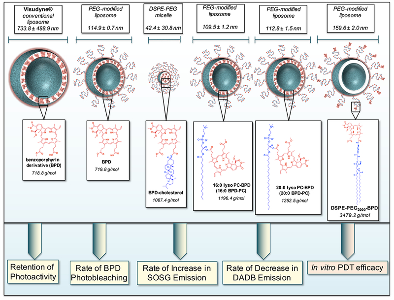
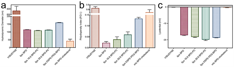
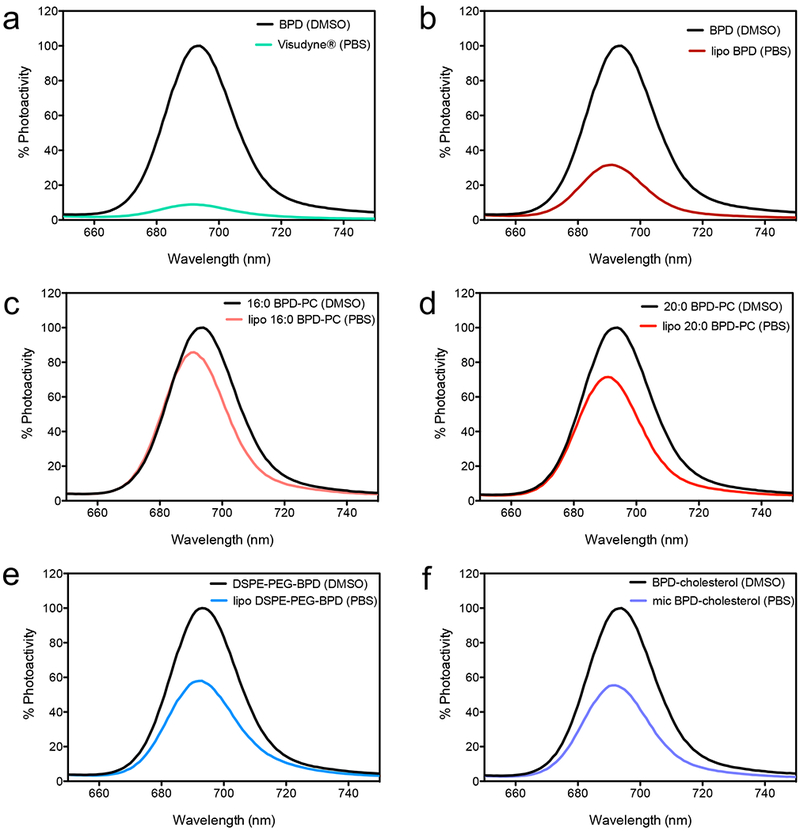
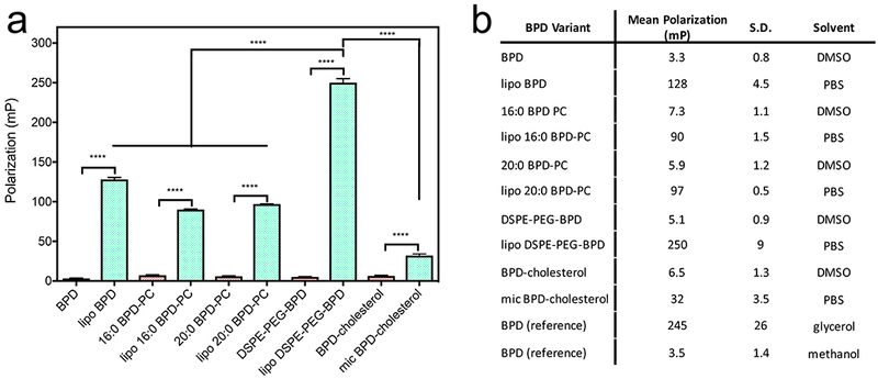
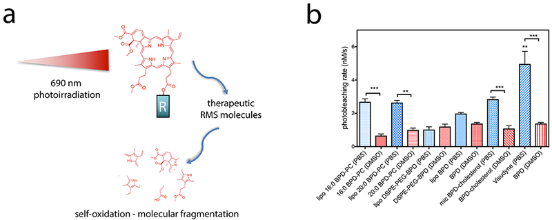
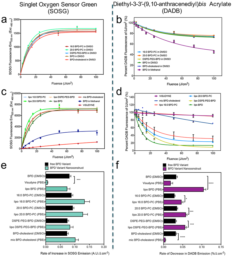
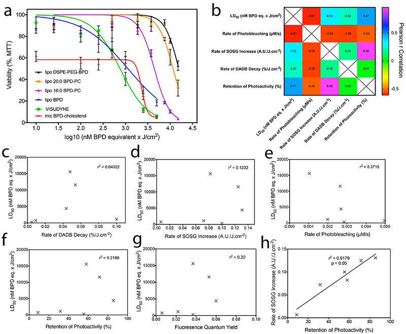
With the rapidly emerging designs and applications of light-activated, photodynamic therapy (PDT)-based nanoconstructs, photonanomedicines (PNMs), an unmet need exists to establish whether conventional methods of photochemical and photophysical characterization of photosensitizers are relevant for evaluating new PNMs in order to intelligently guide their design. As a model system, we build on the clinical formulation of benzoporphyrin derivative (BPD), Visudyne® , by developing a panel of nanolipid formulations entrapping new lipidated chemical variants of BPD with differing chemical, photochemical and photophysical properties. These are 16:0 and 20:0 lysophosphocholine-BPD (16:0/20:0 BPD-PC), DSPE-PEG-BPD and BPD-cholesterol. We show that Visudyne® was the most phototoxic formulation to OVCAR-5 cells, and the least effective was liposomal DSPE-PEG-BPD. However, these differences did not match their optical, photophysical and photochemical properties, as the static BPD quenching was highest in Visudyne, which also exhibited the lowest generation of singlet oxygen. Furthermore, we establish that OVCAR-5 cell phototoxicity also does not correlate with rates of photosensitizer photobleaching and fluorescence quantum yields in any nanolipid formulations. These findings warrant critical future studies into subcellular targets and molecular mechanisms of phototoxicity of photodynamic nanoconstructs, as more reliable prognostic surrogates for predicting efficacy to appropriately and intelligently guide their design.


Figures

Figure 1.
Figure 1.
Graphical representation of the panel of BPD-based nanolipid formulations investigated in this study with the respective hydrodynamic diameters (nm) determined using dynamic light scattering. Chemical structures of BPD, BPD-cholesterol, 16:0 BPD-PC, 20:0 BPD-PC and DSPE-PEG-BPD entrapped in the nanolipid formulations are presented for the respective constructs. The photoactivity, photobleaching, singlet oxygen production (as a function of increased Singlet Oxygen Sensor Green (SOSG) emission and decreased diethyl-3-3′-(9,10-anthracenediyl)bis Acrylate (DADB) emission) of the BPD-based nanolipid formulations will be evaluated with respect to their in vitro PDT efficacy in OVCAR-5 cells.
Figure 2.
Figure 2.
Dynamic Light Scattering analysis of the nanolipid formulations of BPD and its lipidated variants portraying hydrodynamic diameter (a), polydispersity indices (P.D.I.’s; b) and ζ-potential (c) of all constructs.
Figure 3.
Figure 3.
Fluorescence emission spectra of BPD and its lipid conjugates in DMSO or as nanolipid formulation. An excitation wavelength at 435 nm was used for all preparations and the spectra are mean emission intensities (n=4).
Figure 4.
Figure 4.
a) Fluorescence polarization of BPD and its lipidated variants in DMSO and of the respective nanoformulations in PBS. (Values are mean ± S.D., n=3) b) Summary of all fluorescence polarization measurements of BPD and its lipidated variants including reference measurements of BPD in glycerol and methanol.
Figure 5.
Figure 5.
a) Graphical representation of self-oxidation and photobleaching of BPD and its lipidated derivatives by the production of reactive molecular species (RMS) following photoirradiation using 690 nm laser light. ‘R’ indicates either a hydroxyl group of the carboxylate in native BPD, or the lipids conjugated in the lipidated variants. Self-oxidation leads to molecular fragmentation of the PS molecule, resulting in permanent photobleaching. b) Rates of photobleaching of BPD or its lipidated derivatives free in DMSO or entrapped in nanolipid formulations in PBS. (Statistical analysis performed by One-way ANOVA with a Tukey Post-Test. Values are mean ± S.E.M., n=4)
Figure 6.
Figure 6.
SOSG Emission with increasing fluences of 690 nm light irradiation of BPD and its lipid variants (a), in addition to their respective nanolipid formulations (b). Diethyl-3-3′-(9,10-anthracenediyl)bis Acrylate (DADB) (mean = ± SEM)
Figure 7.
Figure 7.
a) Survival curves of OVCAR-5 cells obtained using the MTT viability assay 72h following PDT with varying fluences of 690 nm light at an irradiance of 150 mW/cm2 to deliver PDT dose products of 10 – 15,000 nM BPD equivalent × J/cm2. b) Pearson’s r correlation matrix of nanolipid formulation LD50 values in OVCAR-5 cells with their respective photochemical and photophysical properties. Individual plots are represented for LD50 values with rates of DADB decay (c), rate of SOSG increase (d), rate of photobleaching (e), degree of retention of photoactivity (f), and fluorescence quantum yield (g). The only statistically significant correlation exists between the degree of retention of photoactivity and rate of SOSG increase (h, r2 = 0.9179, p<0.05).

References

    1. Label VV f. I. F. (NDA 21-119/S-022) Reference ID: 3151429.
    1. Huggett MT, Jermyn M, Gillams A, Illing R, Mosse S, Novelli M, Kent E, Bown SG, Hasan T, Pogue BW and Pereira SP (2014) Phase I/II study of verteporfin photodynamic therapy in locally advanced pancreatic cancer. Br. J. Cancer 110, 1698–1704. - PMC - PubMed
    1. Schmidt-Erfurth U, Hasan T, Gragoudas E, Michaud N, Flotte TJ and Birngruber R (1994) Vascular targeting in photodynamic occlusion of subretinal vessels. Ophthalmology 101, 1953–1961. - PubMed
    1. Richter AM, Waterfield E, Jain AK, Canaan AJ, Allison BA and Levy JG (1993) Liposomal delivery of a photosensitizer, benzoporphyrin derivative monoacid ring A (BPD), to tumor tissue in a mouse tumor model. Photochem. Photobiol 57, 1000–1006. - PubMed
    1. Rizvi I, Obaid G, Bano S, Hasan T and Kessel D (2018) Photodynamic therapy: Promoting in vitro efficacy of photodynamic therapy by liposomal formulations of a photosensitizing agent. Lasers Surg. Med - PMC - PubMed

Publication types