Review
. 2018 Sep 21;13(9):2375-2386.
doi: 10.1021/acschembio.8b00520. Epub 2018 Sep 5.

Genetic Code Expansion in Animals

Affiliations
Review

Genetic Code Expansion in Animals

Wes Brown et al. ACS Chem Biol. .

Abstract

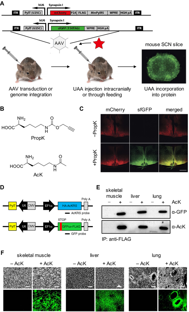
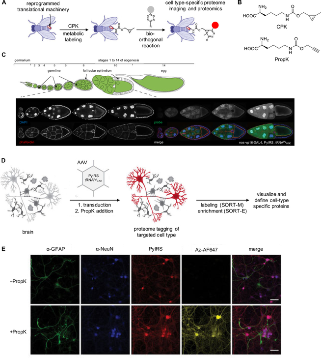
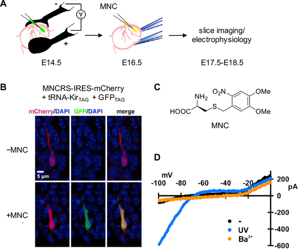
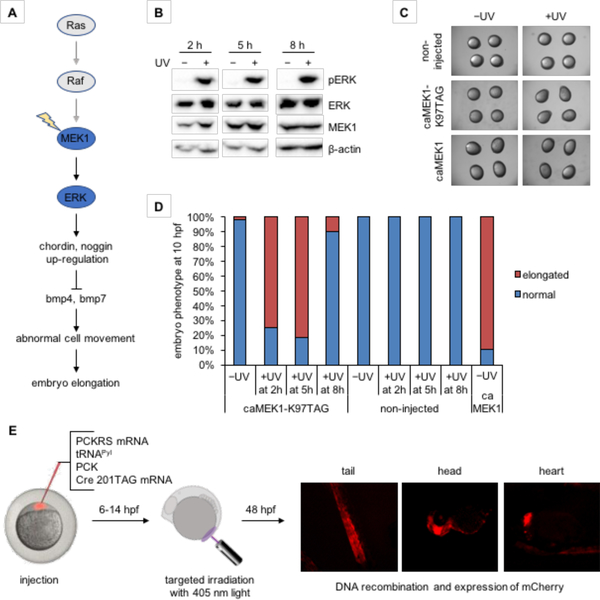
Expanding the genetic code to enable the incorporation of unnatural amino acids into proteins in biological systems provides a powerful tool for studying protein structure and function. While this technology has been mostly developed and applied in bacterial and mammalian cells, it recently expanded into animals, including worms, fruit flies, zebrafish, and mice. In this review, we highlight recent advances toward the methodology development of genetic code expansion in animal model organisms. We further illustrate the applications, including proteomic labeling in fruit flies and mice and optical control of protein function in mice and zebrafish. We summarize the challenges of unnatural amino acid mutagenesis in animals and the promising directions toward broad application of this emerging technology.


Conflict of interest statement

Competing Interests

The authors declare no competing financial interests.

Figures

Figure 1.
Figure 1.
Overview of genetic code expansion. (A) Major milestones in the development of genetic code expansion and its translation into increasingly complex systems. (B) The tRNA synthetase (aaRS) aminoacylates its cognate tRNA with the unnatural amino acid (UAA). The aminoacylated tRNA is transported to the ribosome, where the UAA is site-specifically incorporated into the growing polypeptide chain in response to the UAG codon. (C) Structure of the M. mazei PylRS reveals key residues for substrate recognition in wild-type (PDB 2ZCE) and mutant (PDB 4BWA and 3QTC) enzymes. Substrate binding pocket residues in the wild-type PylRS are labeled green and mutant residues are shown in red.
Figure 2.
Figure 2.
Expanding the genetic code of Caenorhabditis elegans. (A) Diagram of UAA incorporation into mCherry154TAG in C. elegans body-wall muscle using a genome-integrated EcLeuRS/tRNALeu. (B) Expression of the mCherry154TAG reporter in the absence or presence of O-methyl tyrosine (OMeY) or an alanine-dansylalanine dipeptide (A-DanA). Dotted lines indicate the outline of the worms. (C) Incorporation of OMeY is dependent on both UAA concentration and tRNA copy number in the genome, as demonstrated by measurement of luciferase activity using the Luc185TAG reporter (left). Incorporation efficiency is also influenced by the number of generations exposed to OMeY (right). Adapted with permission from ACS Chem. Biol. 2012, 7, 1292. Copyright 2012 American Chemical Society.
Figure 3.
Figure 3.
Expanding the genetic code in Drosophila melanogaster. (A) Diagram of UAA incorporation into proteins in flies via an expression cassette integrated into the genome using P-element insertion. (B) Structures of BocK and PropK. (C) Western blots of lysates from ovary and other body tissue demonstrating tissue-specific incorporation of PropK. The full-length protein, including the HA tag, is only expressed when the amber stop codon is suppressed. Adapted with permission from Nat. Chem. Biol. 2012, 8, 748. Copyright 2012 Nature Publishing Group.
Figure 4.
Figure 4.
Expanding the genetic code in Danio rerio embryos. (A) Diagram of UAA incorporation into proteins in zebrafish embryos through injection of the UAA, tRNAPyl, and mRNAs encoding the PylRS and Renilla luciferase (Rluc) L95TAG. (B) Structures of the UAAs incorporated into proteins in zebrafish embryos. (C) Genetic encoding of the photocaged lysine PCK allows for optical control of firefly luciferase activity in zebrafish embryos through blocking of the active site of the enzyme until UV exposure (left), as demonstrated by luminescence recovery after irradiation (right). Adapted with permission from J. Am. Chem. Soc. 2017, 139, 9100. Copyright 2017 American Chemical Society.
Figure 5.
Figure 5.
Expanding the genetic code in Mus musculus. (A) Diagram of UAA incorporation into proteins in mice via AAV transduction of the expression cassette or generation of stable lines through genome integration, with the UAA introduced either intracranially or through the drinking water. (B) Chemical structure of PropK and AcK. (C) Fluorescence imaging showing incorporation of PropK into sfGFP151TAG in coronal sections of the hypothalamus and suprachiasmatic nucleus. Scale bar: 500 μm. (D) DNA constructs used for generation of a transgenic mouse line for UAA incorporation. (E) Western blotting analysis of anti-FLAG-immunoprecipitated proteins from AcK-encoding sfGFP151TAG mice, demonstrating successful genetic code expansion. (F) Fluorescence imaging showing incorporation of AcK into GFP39TAG in skeletal muscle, liver, and lung tissues of live mice. Scale bar: 200 μm. Adapted with permission from Nat. Chem. Biol. 2016, 12, 776 and Nat. Commun. 2017, 8, 14568. Copyright 2016 and 2017 Nature Publishing Group.
Figure 6.
Figure 6.
Proteome labeling using Drosophila and mice with an expanded genetic code. (A) Diagram of proteome labeling in fruit flies using cyclopropene-lysine (CPK) incorporation into fly proteins. (B) Structure of CPK and PropK. (C) Stochastic incorporation of CPK into proteins was controlled by expression of the PylRS/tRNAPyl under control of nos-vp16, limiting expression of the PylRS/tRNAPyl to germline cells in the ovary. When labeled, fluorescence was restricted to germline cells with no incorporation of CPK in the somatic follicular epithelium. (D) Diagram of proteome labeling in the mice brain using PropK. (E) Cell-type specific incorporation of PropK and labeling with an azido-fluorophore. α-GFAP is a glial cell specific protein and α-NeuN is a neuron specific nuclear protein. Adapted with permission from Nat. Biotechnol. 2014, 32, 445, Nat. Biotechnol. 2014, 32, 465, and Nat. Biotechnol. 2017, 36, 156. Copyright 2014 and 2017 Nature Publishing Group.
Figure 7.
Figure 7.
Application of genetic code expansion to the optical control of a potassium channel in the mouse neocortex. (A) Electroporation of genetic constructs for UAA mutagenesis into the mouse neocortex and injection of photocaged cysteine (MNC) into the embryonic lateral ventricle after two days. (B) Fluorescence imaging showing incorporation of MNC into GFP182TAG and Kir2.1TAG in mice embryonic cortical neurons. (C) Structure of MNC. (D) I-V plot of currents recorded from mouse neocortical neurons showing light-dependent activation of Kir2.1. Adapted with permission from Neuron 2013, 80, 358. Copyright 2013 Elsevier.
Figure 8.
Figure 8.
Optical control of a signaling pathway and of Cre recombinase in zebrafish embryos. (A) In zebrafish, activation of caged MEK1, as part of the Ras/MAPK signaling pathway, induces an elongated phenotype through the secreted bmp inhibitors chordin and noggin. (B) Time-course analysis of ERK phosphorylation by optically activated MEK1. (C) Embryos expressing caged MEK1 displayed an elongation phenotype only when activated through light exposure. (D) Temporal control of MEK1 activity reveals a time window when embryo morphogenesis is most susceptible to MEK1 induced elongation. (E) Optical Cre recombinase activation in targeted cell populations in the zebrafish embryo at early developmental periods using a genetically encoded photocaged lysine (PCK, see Figure 4B) allowed for lineage tracing of embryonic cells that formed the tail, head, and heart of the 48 hpf embryo. Adapted with permission from J. Am. Chem. Soc. 2017, 139, 9100 and ChemBioChem 2018, 19, 1244. Copyright 2017 American Chemical Society and copyright 2018 John Wiley and Sons.

References

    1. Romagne F; Santesmasses D; White L; Sarangi GK; Mariotti M; Hubler R; Weihmann A; Parra G; Gladyshev VN; Guigo R; Castellano S, SelenoDB 2.0: annotation of selenoprotein genes in animals and their genetic diversity in humans. Nucleic Acids Res. 2014, 42, 437–443. - PMC - PubMed
    1. Chuh Kelly N.; Batt Anna R.; Pratt Matthew R., Chemical methods for encoding and decoding of posttranslational modifications. Cell Chemical Biology 2016, 23, 86–107. - PMC - PubMed
    1. Heckler TG; Zama Y; Naka T; Hecht SM, Dipeptide formation with misacylated tRNAPhes. J. Biol. Chem 1983, 258, 4492–4495. - PubMed
    1. Noren CJ; Anthonycahill SJ; Griffith MC; Schultz PG, A General-method for site-specific incorporation of unnatural amino-acids into proteins. Science 1989, 244, 182–188. - PubMed
    1. Ibba M; Hennecke H, Relaxing the substrate specificity of an aminoacyl-tRNA synthetase allows in vitro and in vivo synthesis of proteins containing unnatural amino acids. FEBS Lett. 1995, 364, 272–275. - PubMed

Publication types

LinkOut - more resources