Review
. 2018 Aug 19;19(8):2445.
doi: 10.3390/ijms19082445.

The Pathophysiology of Post-Traumatic Glioma

Affiliations
Review

The Pathophysiology of Post-Traumatic Glioma

Donata Simińska et al. Int J Mol Sci. .

Abstract

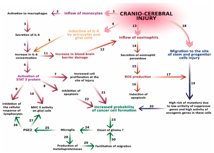
Malignant glioma is a brain tumor with a very high mortality rate resulting from the specific morphology of its infiltrative growth and poor early detection rates. The causes of one of its very specific types, i.e., post-traumatic glioma, have been discussed for many years, with some studies providing evidence for mechanisms where the reaction to an injury may in some cases lead to the onset of carcinogenesis in the brain. In this review of the available literature, we discuss the consequences of breaking the blood⁻brain barrier and consequences of the influx of immune-system cells to the site of injury. We also analyze the influence of inflammatory mediators on the expression of genes controlling the process of apoptosis and the effect of chemical mutagenic factors on glial cells in the brain. We present the results of experimental studies indicating a relationship between injury and glioma development. However, epidemiological studies on post-traumatic glioma, of which only a few confirm the conclusions of experimental research, indicate that any potential relationship between injury and glioma, if any, is indirect.

Keywords: IL-6; STAT3; blood–brain barrier; brain; injury; pathophysiology; post-traumatic glioma.


Conflict of interest statement

The authors declare no conflict of interest.

Figures

Figure 1
Figure 1
A 78-year-old woman with hypertension and hypercholesterolemia was admitted to the hospital due to her head injury (after falling in moving bus), small changes were revealed in the computed tomography (CT) scan as well as in the radiographic examination that did not require surgical treatment. (a) (arrow) A little amount of blood along the falx of the brain, on the tent of the cerebellum on the right side and in a fissura of the right parietal lobe with scant concussion in it. In the white matter of corona radiata, a number of dimly defined, diminished-density areas were observed, most likely corresponding to degenerative vascular changes. The patient, without neurological deficits except diminishing post-traumatic headaches, was discharged after the observation period in a good condition. Two years later, the patient was readmitted to the Department of Neurosurgery with a diagnosed brain tumor, manifested by a 1.5-month history of memory and orientation disorders. (b) (arrow) In the MRI scans, the right brain hemisphere tumor was found, the position of which corresponded to previous post-traumatic lesions. Right temporoparietal craniotomy was performed and the cytoreduction of the tumor was extended. Histopathology revealed Glioblastoma multiforme (facultatively). The histopathological pictures show (c) characteristic necrosis, surrounded by a pseudopalisade (arrow 1), visible cell mitosis (arrow 2), (d) large cell density, mitosis (arrow 1), lumen of the vessel with erythrocytes (arrow 2). The patient was accepted for radiotherapy with chemotherapy and discharged home in good condition, without additional neurological deficits, walking with little assistance. The study was approved by the Local Ethical Committee at the Pomeranian Medical University in Szczecin, Poland (approval No. KB-0012/96/14; Szczecin, Poland, 24.11.2014).
Figure 1
Figure 1
A 78-year-old woman with hypertension and hypercholesterolemia was admitted to the hospital due to her head injury (after falling in moving bus), small changes were revealed in the computed tomography (CT) scan as well as in the radiographic examination that did not require surgical treatment. (a) (arrow) A little amount of blood along the falx of the brain, on the tent of the cerebellum on the right side and in a fissura of the right parietal lobe with scant concussion in it. In the white matter of corona radiata, a number of dimly defined, diminished-density areas were observed, most likely corresponding to degenerative vascular changes. The patient, without neurological deficits except diminishing post-traumatic headaches, was discharged after the observation period in a good condition. Two years later, the patient was readmitted to the Department of Neurosurgery with a diagnosed brain tumor, manifested by a 1.5-month history of memory and orientation disorders. (b) (arrow) In the MRI scans, the right brain hemisphere tumor was found, the position of which corresponded to previous post-traumatic lesions. Right temporoparietal craniotomy was performed and the cytoreduction of the tumor was extended. Histopathology revealed Glioblastoma multiforme (facultatively). The histopathological pictures show (c) characteristic necrosis, surrounded by a pseudopalisade (arrow 1), visible cell mitosis (arrow 2), (d) large cell density, mitosis (arrow 1), lumen of the vessel with erythrocytes (arrow 2). The patient was accepted for radiotherapy with chemotherapy and discharged home in good condition, without additional neurological deficits, walking with little assistance. The study was approved by the Local Ethical Committee at the Pomeranian Medical University in Szczecin, Poland (approval No. KB-0012/96/14; Szczecin, Poland, 24.11.2014).
Figure 2
Figure 2
Schematic representation of the involvement of carcinogenic factors after cerebral injury. An injury causes the influx of monocytes to the site of damage (1), where they are activated to macrophages (2) that produce interleukin 6 (IL-6) (3). The injury also induces enhanced IL-6 secretion by astrocytes and microglial cells (4). Together, these processes cause a significant increase in IL-6 concentration (5). The increased concentration of IL-6 activates STAT3 (6), which increases cell proliferation at the site of injury (7) and inhibition of apoptosis (8). STAT3 inhibits the activity of T cells (LcT) and (9) decreases the activity of major histocompatibility complex (MHC) particles on cells of the immune system and glial cells (10). The increase in IL-6 concentration also affects the blood–brain barrier (BBB) (11), facilitating the penetration by eosinophils (12) flowing to the site of injury (13). The eosinophils, which flow through the damaged barrier, secrete eosinophil peroxidase, which generates reactive oxygen species (ROS). ROS contribute to the induction of apoptosis (16), but may also contribute to the formation of mutations (17) in stem and progenitor cells that migrate to the site of injury (18). Low activity of suppressor genes in these cells and high activity of oncogenic genes may additionally increase the risk of mutations (19). The risk of these mutations (20), an increase in cell proliferation at the site of injury (21) and the inhibition of apoptosis (22) may jointly contribute to the formation of a cancer cell, and start the process of carcinogenesis (23). The glioma resulting from these processes affects microglia (24), which secrete PGE2 (25), increasing the inhibition of LcT activity (26) and causing a decrease in MHC molecules activity on cells of the immune system and glial cells (27). In addition, microglia secrete metalloproteinases (28) in the tissues adjacent to the tumor, facilitating its migration (29) and thus facilitating its development (30).

References

    1. Li W., Graeber M.B. The molecular profile of microglia under the influence of glioma. Neuro Oncol. 2012;14:958–978. doi: 10.1093/neuonc/nos116. - DOI - PMC - PubMed
    1. Tyagi V., Theobald J., Barger J., Bustoros M., Bayin N.S., Modrek A.S., Kader M., Anderer E.G., Donahue B., Fatterpekar G., et al. Traumatic brain injury and subsequent glioblastoma development: Review of the literature and case reports. Surg. Neurol. Int. 2016;26:77–78. doi: 10.4103/2152-7806.189296. - DOI - PMC - PubMed
    1. Ostrom Q.T., Gittleman H., Liao P., Rouse C., Chen Y., Dowling J., Wolinsky Y., Kruchko C., Barnholtz-Sloan J. CBTRUS statistical report: Primary brain and central nervous system tumors diagnosed in the United States in 2007–2011. Neuro Oncol. 2014;16:61–63. doi: 10.1093/neuonc/nou223. - DOI - PMC - PubMed
    1. Schwartzbaum J.A., Fisher J.L., Aldape K.D., Wrensch M. Epidemiology and molecular pathology of glioma. Nat. Clin. Pract. Neurol. 2006;2:494–503. doi: 10.1038/ncpneuro0289. - DOI - PubMed
    1. Munch T.N., Gørtz S., Wohlfahrt J., Melbye M. The long-term risk of malignant astrocytic tumors after structural brain injury—A nationwide cohort study. Neuro Oncol. 2015;17:718–724. doi: 10.1093/neuonc/nou312. - DOI - PMC - PubMed

LinkOut - more resources