

Suprachoroidal hemorrhage followed by swept-source optical coherence tomography: a case report
- PMID: 30126397
- PMCID: PMC6102832
- DOI: 10.1186/s12886-018-0881-4
Suprachoroidal hemorrhage followed by swept-source optical coherence tomography: a case report
Abstract
Background: To report a case of Suprachoroidal Hemorrhage followed by Swept-Source Optical Coherence Tomography.
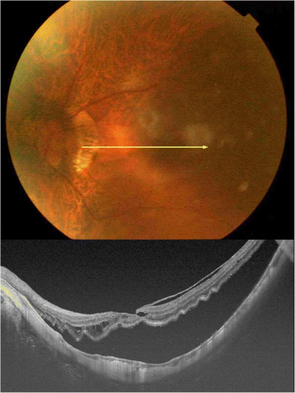
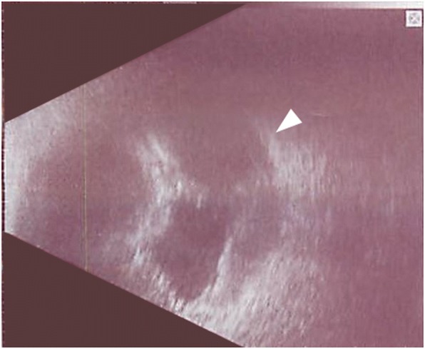
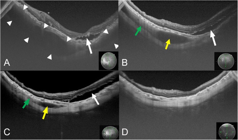
Case presentation: A 66-year-old woman with a rhegmatogenous retinal detachment in her left eye underwent pars plana vitrectomy. During the intraocular photocoagulation for a retinal tear after fluid-air exchange, a vitreous hemorrhage and suprachoroidal hemorrhage (SCH) developed. The surgical incisions were closed after filling the vitreous cavity with silicone oil. Two weeks later, the hemolyzed hemorrhage was removed, and new silicone oil was injected. After the surgery, a low reflective region was detected near the macula in the swept-source optical coherence tomographic (SS-OCT) images. The low reflective region was caused by the residual hemorrhage. The size of the reflective region gradually decreased and was not present at 3 months. We conclude that SS-OCT can be used to follow the resolution of a suprachoroidal hemorrhage.
Conclusion: SS-OCT can be used to detect and follow the natural course of a suprachoroidal hemorrhage including the absorptive processes.
Keywords: High myopia; SS-OCT; Suprachoroidal hemorrhage.
Conflict of interest statement
Ethics approval and consent to participate
Not applicable.
Consent for publication
Written informed consent was obtained from the patient for publication of this case report and any accompanying images.
Competing interests
The authors declare that they have no competing interests.
Publisher’s Note
Springer Nature remains neutral with regard to jurisdictional claims in published maps and institutional affiliations.
Figures
References
Publication types
MeSH terms
LinkOut - more resources
Full Text Sources
Other Literature Sources
Miscellaneous
