Review
. 2018 Aug 20;16(1):80.
doi: 10.1186/s12958-018-0391-5.

A novel and compact review on the role of oxidative stress in female reproduction

Affiliations
Review

A novel and compact review on the role of oxidative stress in female reproduction

Jiayin Lu et al. Reprod Biol Endocrinol. .

Abstract

In recent years, the study of oxidative stress (OS) has become increasingly popular. In particular, the role of OS on female fertility is very important and has been focused on closely. The occurrence of OS is due to the excessive production of reactive oxygen species (ROS). ROS are a double-edged sword; they not only play an important role as secondary messengers in many intracellular signaling cascades, but they also exert indispensable effects on pathological processes involving the female genital tract. ROS and antioxidants join in the regulation of reproductive processes in both animals and humans. Imbalances between pro-oxidants and antioxidants could lead to a number of female reproductive diseases. This review focuses on the mechanism of OS and a series of female reproductive processes, explaining the role of OS in female reproduction and female reproductive diseases caused by OS, including polycystic ovary syndrome (PCOS), endometriosis, preeclampsia and so on. Many signaling pathways involved in female reproduction, including the Keap1-Nrf2, NF-κB, FOXO and MAPK pathways, which are affected by OS, are described, providing new ideas for the mechanism of reproductive diseases.

Keywords: Antioxidants; Female fertility; Imbalance; Oxidative stress; ROS; Reproductive diseases; Signaling pathways.


Conflict of interest statement

Ethics approval and consent to participate

Not applicable.

Consent for publication

Not applicable.

Competing interests

The authors declare that they have no competing interests.

Publisher’s Note

Springer Nature remains neutral with regard to jurisdictional claims in published maps and institutional affiliations.

Figures

Fig. 1
Fig. 1
The development of ovarian follicles. Primary follicle: The center has an oocyte, and there is a flat layer of follicular cells on its periphery. Growing follicle: Including the primary growth follicle and secondary growth follicles. Primary growth follicle: One or more layers of cuboidal follicular cells between the egg cells, and follicular cells demonstrate red-stained zona pellucida, while the follicular periphery appears like connective tissue follicular membrane. Secondary growth follicle: Follicular cells appear in the follicular cavity, and some follicular cavities are large, forming a cumulus of oophores. Follicular cells are located on the inner wall of follicles and are arranged in layers, called granular layers. The follicular membrane includes the inner and outer membrane layers. Mature follicle: The follicle cavity is very large, and cumulus oophores are obvious. Follicular endometrial cells appear close to the follicular granule layer. There is a layer of basement membrane between the granule layer cells and follicular endometrial cells; endometrial cells are polygonal, with clear cytoplasm and round nuclei; cells can be seen between many capillaries, and outer membrane cells are located in the outermost layer, mostly spindle shaped with the surrounding connective tissue boundaries not obvious. Ovulation: Mature follicles develop to a certain stage, obviously protruding from the ovarian surface; with the follicular fluid increasing sharply, the pressure increases so that the prominent part of the ovarian tissue becomes thinner and finally ruptures; secondary oocytes and their peripheral zona pellucida and radiation crowns are discharged together with the follicular fluid. Empty follicle: At this time, the follicle is empty, indicating that the corpus luteum starts to form. Corpus luteum: The residual follicle wall collapses after ovulation; the connective tissue of the follicular membrane and capillaries stretches into the particle layer, and as the role of LH evolves, it evolves into a larger volume cell cluster, rich in capillaries and endocrine function and fresh yellow in color
Fig. 2
Fig. 2
The changes of biology during different estrus cycle. Estrogen and progesterone are secreted via the ovary or uterus and undergo changes during the estrus cycle. In addition, the basal body temperature also changes, while the thickness of the endometrium has corresponding transformations. After menstruation, the new estrus cycle starts to develop. During the follicular period, the level of the basal body temperature and estrogen gradually rise. The thickness of the endometrium also increases. The levels of basal body temperature and estrogen maintain certain concentrations until the ovulation period. Thus, progesterone starts to increase. With the appearance of the luteal phase, all changes are restored until the end of menstruation
Fig. 3
Fig. 3
Fertilization processes of most viviparous and ovoviviparous animals. In most viviparous and ovoviviparous animals, the sperm and oocyte combine at the fallopian tube ampulla. In the picture, the first zygote shows a radiation crown dissolving; the second zygote shows the zona pellucida dissolving; the last zygote shows fertilized eggs and cortical response
Fig. 4
Fig. 4
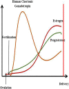
The trends of HCG, estrogen and progesterone during pregnancy. The yellow line represents the change in HCG. The green line represents the change in progesterone. The red line represents the level of estrogen. The final results of ovulation include two impacts, one of which is output in the body, called menstruation, and the other of which is combines with sperm, called fertilization. The level of hormones changes after fertilization; in particular, hCG immediately increases to the highest level. However, the levels of estrogen and progesterone slowly increase to stable concentrations, while hCG begins to drop to a certain extent
Fig. 5
Fig. 5
The process of implantation (including invasive implantation with decidualization and non-invasive implantation of non-decidualizing species) a: Zygote; b: 2 cells; c: 4 cells; d: 8 cells; e: Morula; f: Blastocysts; g: Endometrium; h: Uterine cavity; i: Trophoblast cells; j: Microvilli. Estradiol (E2) produced by the developing ovarian follicles interacts with progesterone produced by the CL to prepare the endometrium receptivity necessary for embryo implantation. The meeting of the oocyte and sperm and subsequent fertilization occur in the ampulla of the oviduct, followed by early embryo development within the oviduct, and the morula migrates to the uterus, where implantation occurs. The appearance of a fluid-filled inner cavity (blastocoel) is accompanied by cellular differentiation: the surface cells become the trophoblast and give rise to the extra-embryonic tissues, including the placenta, while the inner cell mass gives rise to the embryo and finally shedding of the zona pellucida, followed by orientation, apposition, attachment and adhesion of the blastocyst to the endometrium. If the blastocyst was not present, the CL would regress, and the uterus would start the cycle again. The time and chronological events of implantation differ among mammalian species irrespective of the length of gestation. In contrast to humans, horses, primates and rodents, in which implantation occurs shortly after the hatching of the blastocyst, the blastocyst in domestic ruminants and pigs elongates before implantation (the time to implantation: in pigs, the 14th day; in sheep, the 16th day; and in cattle, the 18th day), and this unique developmental event does not occur in the laboratory or in rodents or humans
Fig. 6
Fig. 6
The defense mechanism against oxygen free radicals. SOD: Superoxide dismutase; GPx: Glutathione peroxidase; GSSG: Glutathione oxidase; GSH: Glutathione reductase; ROS: Reactive oxygen species; O2•: Superoxide; H2O2: Hydrogen peroxide; •OH: Hydroxyl
Fig. 7
Fig. 7
The signaling pathway of OS and pregnancy (a brief view). When the body, especially the maternal body, suffers from an imbalance between oxidation and antioxidant levels during pregnancy, in addition to changes in TNF-α, changes in progesterone cannot be ignored. First, TNF-α activates a series of signaling pathways in cells through cAMP, such as stimulation of the Keap1-Nrf2 signaling pathway, NF-κB signaling pathway, MAPK signaling pathway, etc., then promoting an increase in cytokines and changes in antioxidant-related genes. However, FOXO3 is involved in these signaling pathways. When FOXO3 is increased, it promotes the binding of Keap1-Nrf2, lowering the level of antioxidants and promoting the release of NF-κB by IKKβ by stimulating BCL10, thereby promoting the increase in cytokines and apoptosis. Finally, the mechanism underlying the changes in the FOXO family under the combined effects of both reproductive and oxidative stress remains unclear. It can only be demonstrated that JNK undergoes dephosphorylation of FOXO1 under the action of cAMP and ROS when oxidative stress occurs to induce it to enter the nucleus and promote apoptosis. When progesterone is reduced, nuclear translocation occurs in FOXO1, and it is phosphorylated

Similar articles

Cited by

References

    1. Burton GJ, Jauniaux E. Oxidative stress. Best Practice & Research Clinical Obstetrics & Gynaecology. 2011;25:287–299. doi: 10.1016/j.bpobgyn.2010.10.016. - DOI - PMC - PubMed
    1. Cindrova-Davies T, Yung HW, Johns J, Spasic-Boskovic O, Korolchuk S, Jauniaux E, Burton GJ, Charnock-Jones DS. Oxidative stress, gene expression, and protein changes induced in the human placenta during labor. Am J Pathol. 2007;171:1168–1179. doi: 10.2353/ajpath.2007.070528. - DOI - PMC - PubMed
    1. Ruder EH, Hartman TJ, Goldman MB. Impact of oxidative stress on female fertility. Current Opinion in Obstetrics & Gynecology. 2009;21:219–222. doi: 10.1097/GCO.0b013e32832924ba. - DOI - PMC - PubMed
    1. Attaran M, Pasqualotto E, Falcone T, Goldberg JM, Miller KF, Agarwal A, Sharma RK. The effect of follicular fluid reactive oxygen species on the outcome of in vitro fertilization. International Journal of Fertility and Womens Medicine. 2000;45:314–320. - PubMed
    1. Szczepanska M, Kozlik J, Skrzypczak J, Mikolajczyk M. Oxidative stress may be a piece in the endometriosis puzzle. Fertil Steril. 2003;79:1288–1293. doi: 10.1016/S0015-0282(03)00266-8. - DOI - PubMed

Substances