. 2018 Oct 25;86(11):e00511-18.
doi: 10.1128/IAI.00511-18. Print 2018 Nov.

Histopathology of Bordetella pertussis in the Baboon Model

Affiliations

Histopathology of Bordetella pertussis in the Baboon Model

Lindsey I Zimmerman et al. Infect Immun. .

Abstract

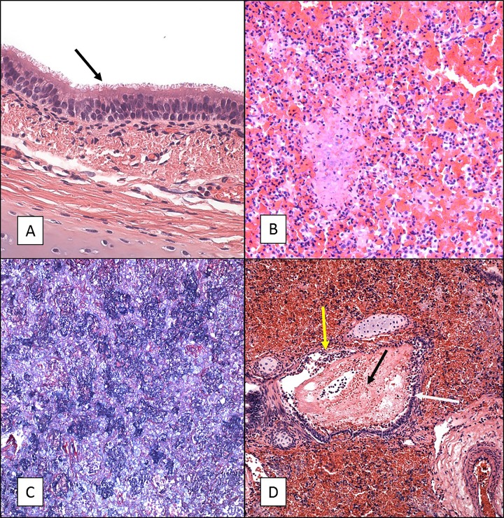
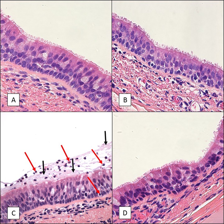
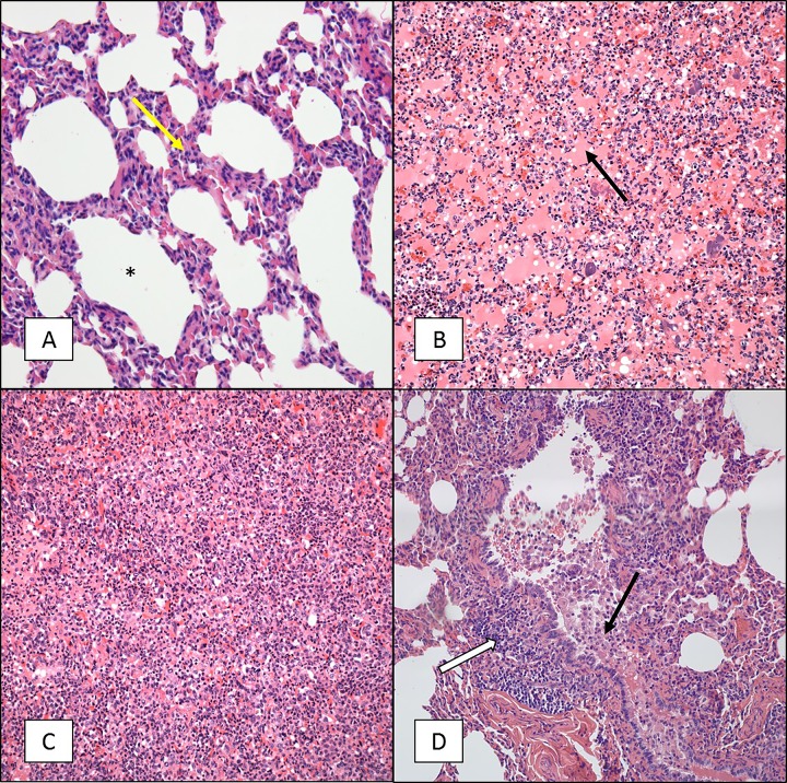
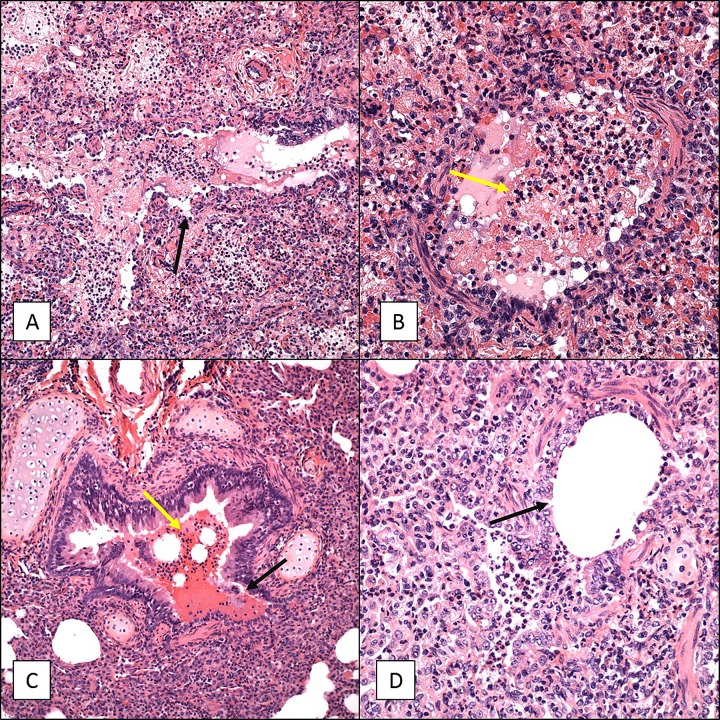
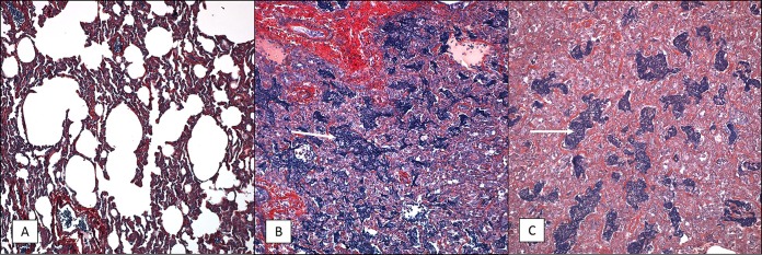
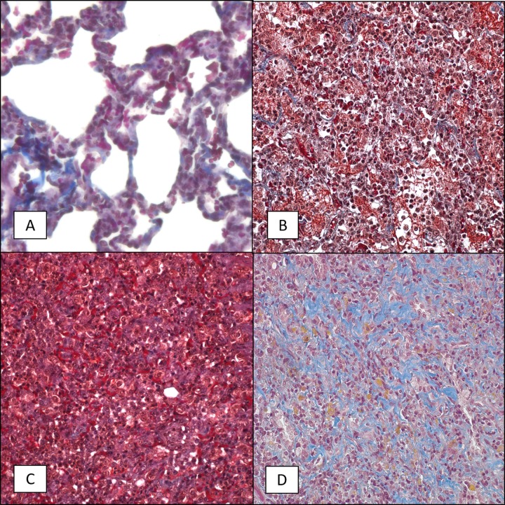
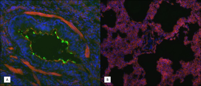
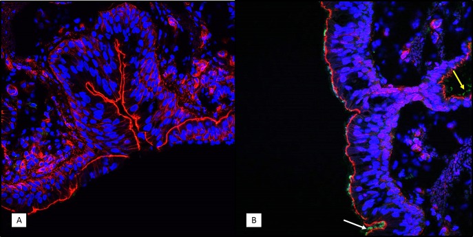
Pertussis is a severe respiratory disease caused by Bordetella pertussis The classic symptoms of pertussis include paroxysmal coughing with an inspiratory whoop, posttussive vomiting, cyanosis, and persistent coryzal symptoms. Infants under 2 months of age experience more severe disease, with most deaths occurring in this age group. Most of what is known about the pathology of pertussis in humans is from the evaluation of fatal human infant cases. The baboon model of pertussis provides the opportunity to evaluate the histopathology of severe but nonfatal pertussis. The baboon model recapitulates the characteristic clinical signs of pertussis observed in humans, including leukocytosis, paroxysmal coughing, mucus production, heavy colonization of the airway, and transmission of the bacteria between hosts. As in humans, baboons demonstrate age-related differences in clinical presentation, with younger animals experiencing more severe disease. We examined the histopathology of 5- to 6-week-old baboons, with the findings being similar to those reported for fatal human infant cases. In juvenile baboons, we found that the disease is highly inflammatory and concentrated to the lungs with signs of disease that would typically be diagnosed as acute respiratory distress syndrome (ARDS) and bronchopneumonia. In contrast, no significant pathology was observed in the trachea. Histopathological changes in the trachea were limited to cellular infiltrates and mucus production. Immunohistostaining revealed that the bacteria were localized to the surface of the ciliated epithelium in the conducting airways. Our observations provide important insights into the pathology of pertussis in typical, severe but nonfatal pertussis cases in a very relevant animal model.

Keywords: Bordetella pertussis; Papio; baboon; bronchiolitis; histopathology; infant; infectious disease; juvenile; nonhuman primates; pneumonia.


Figures

FIG 1
FIG 1
H&E and PTAH staining of tissue sections from 5- to 6-week-old baboons infected with B. pertussis. Five- to 6-week-old baboons were challenged with B. pertussis. H&E- and PTAH-stained slides were prepared from tissues as described in Materials and Methods. Representative images are presented. (A) Tracheal tissue stained with H&E showing intact cilia (arrow) and a normal appearance. (B) Lung tissue stained with H&E showing severe acute vascular leakage and mostly acute inflammation. (C) PTAH staining of lung tissue indicating intra-alveolar fibrin deposition in blue. (D) H&E staining of lung tissue showing necrotizing bronchitis/bronchiolitis. White arrow, bronchus, intact mucosa; yellow arrow, damaged mucosa; black arrow, intralumenal blood and edematous fluid. Magnifications, ×600 (A) and ×200 (B to D).
FIG 2
FIG 2
Numbers of bacterial CFU in airway tissues. Six- to 9-month-old baboons were challenged with B. pertussis, and samples were collected on days 2, 7, and 28 postchallenge. Tissues were weighed prior to homogenization in PBS, and the CFU were enumerated after dilution and plating on Regan-Lowe plates. The numbers of CFU per gram of tissue are reported. Direct comparison of bacterial numbers between tissue types is not possible due to the differences in the densities of the different tissues.
FIG 3
FIG 3
Pathology scores for 6- to 9-month-old baboons challenged with B. pertussis. Six- to 9-month-old baboons were challenged with B. pertussis as described in Materials and Methods. Animals were euthanized at days 2 (n = 3), 7 (n = 3), and 28 (n = 2). H&E-stained sections of the tracheal and lung tissues of all animals were examined by a board-certified veterinary pathologist and scored for vascular leakage, airway obstruction, BALT hyperplasia, and lung consolidation. Scores on a scale ranging from 0 to 4 were assigned as described in Materials and Methods. P values are reported relative to the naive group.
FIG 4
FIG 4
H&E staining of tracheal tissue sections from 6- to 9-month-old baboons infected with B. pertussis. Six- to 9-month-old baboons were challenged with B. pertussis. Tracheal tissue samples were collected on days 2, 7, and 28 postchallenge, and H&E-stained slides were prepared as described in Materials and Methods. Representative images are presented. (A) Sample collected from an uninfected baboon. (B) Sample collected from a baboon at day 2 postchallenge. (C) Sample collected from a baboon at day 7 postchallenge demonstrating influx of neutrophils (red arrows) and excess mucus (black arrows) lining the mucosa. (D) Sample collected from a baboon at day 28 postchallenge. Magnifications, ×600.
FIG 5
FIG 5
H&E staining of lung tissue sections from 6- to 9-month-old baboons infected with B. pertussis. Six- to 9-month-old baboons were challenged with B. pertussis. Lung tissue samples were collected on days 2, 7, and 28 postchallenge, and H&E-stained slides were prepared as described in Materials and Methods. Representative images are presented. (A) Sample collected from an uninfected baboon. Note that the alveoli (*) are empty and there are few inflammatory cells in the interalveolar septum (yellow arrow). (B) Sample collected from a baboon at day 2 postchallenge revealing marked vascular leakage consisting primarily of intra-alveolar edema (which stains pink; black arrow). (C) Sample collected from a baboon at day 7 postchallenge revealing moderate vascular leakage and marked lung consolidation. Note the almost complete loss of alveolar space and the influx of inflammatory cells and edema. (D) Sample collected from a baboon at day 28 postchallenge revealing marked lung consolidation and BALT hyperplasia (white arrow) with the conducting airways obstructed by macrophages and sloughed mucosal epithelial cells (black arrow). Magnifications, ×200.
FIG 6
FIG 6
Necrotizing bronchitis/bronchiolitis of 6- to 9-month-old baboon lungs infected with B. pertussis. Six- to 9-month-old baboons were challenged with B. pertussis. Lung tissue samples were collected on days 2 and 7, and H&E-stained slides were prepared as described in Materials and Methods. Representative images showing necrotizing bronchitis/bronchiolitis are presented. (A and B) Samples collected from a baboon at day 2 postchallenge. (C and D) Samples collected from a baboon at day 7 postchallenge. For all panels, the lumen of the bronchus is almost completely filled with edema, fibrin, and inflammatory cells (yellow arrows), and the mucosa (black arrows) has been severely damaged. Magnifications, ×200 (A and C) and ×400 (B and D).
FIG 7
FIG 7
PTAH staining of lung tissue sections from 6- to 9-month-old baboons infected with B. pertussis. Six- to 9-month-old baboons were challenged with B. pertussis, and PTAH-stained slides were prepared as described in Materials and Methods. Representative images are presented. Lung tissue samples were collected from uninfected animals (A) and from infected animals at day 2 postchallenge (B) and day 7 postchallenge (C), and intra-alveolar fibrin deposition is shown in dark blue/purple (white arrows). Magnifications, ×200.
FIG 8
FIG 8
Trichrome staining of lung tissue sections from 6- to 9-month-old baboons infected with B. pertussis. Six- to 9-month-old baboons were challenged with B. pertussis, and trichrome-stained slides were prepared as described in Materials and Methods. Representative images of lung tissue samples collected from uninfected animals (A) and from infected animals at day 2 postchallenge (B), day 7 postchallenge (C), and day 28 postchallenge (D) showing collagen deposition in blue are presented. Magnifications, ×400.
FIG 9
FIG 9
Immunohistochemistry (IHC) staining of B. pertussis in lung tissue sections. Frozen lung tissue sections were prepared from infected animals on day 7 postchallenge as described in Materials and Methods. B. pertussis bacteria were visualized using rabbit polyclonal antibodies specific for a surface-exposed antigen. Representative images are shown. Blue indicates the DAPI staining of the nucleus, red indicates the staining of F actin, and the green in panel A indicates B. pertussis bacterial cells. (A) Bronchiole showing bacteria lining the mucosal surface. (B) Alveolar space of an infected animal showing an absence of bacteria. Magnifications, ×200 (A) and ×100 (B).
FIG 10
FIG 10
Immunohistochemistry (IHC) staining of B. pertussis in tracheal tissue sections. Frozen tracheal tissue sections were prepared from uninfected animals (A) and from infected animals on day 7 postchallenge (B), as described in Materials and Methods. B. pertussis bacteria were visualized using rabbit polyclonal antibodies specific for a surface-exposed antigen. Representative images are shown. Blue indicates the DAPI staining of the nucleus, red indicates the staining of F actin, and the green in panel B indicates B. pertussis bacterial cells. Bacteria are observed on the mucosal surface in the tracheal lumen (white arrow) and submucosal ducts (yellow arrow). Magnifications, ×400.

Similar articles

Cited by

References

    1. Yeung KHT, Duclos P, Nelson EAS, Hutubessy RCW. 2017. An update of the global burden of pertussis in children younger than 5 years: a modelling study. Lancet Infect Dis 17:974–980. doi:10.1016/S1473-3099(17)30390-0. - DOI - PubMed
    1. Melvin JA, Scheller EV, Miller JF, Cotter PA. 2014. Bordetella pertussis pathogenesis: current and future challenges. Nat Rev Microbiol 12:274–288. doi:10.1038/nrmicro3235. - DOI - PMC - PubMed
    1. Kilgore PE, Salim AM, Zervos MJ, Schmitt HJ. 2016. Pertussis: microbiology, disease, treatment, and prevention. Clin Microbiol Rev 29:449–486. doi:10.1128/CMR.00083-15. - DOI - PMC - PubMed
    1. Pinto MV, Merkel TJ. 2017. Pertussis disease and transmission and host responses: insights from the baboon model of pertussis. J Infect 74(Suppl 1):S114–S119. doi:10.1016/S0163-4453(17)30201-3. - DOI - PubMed
    1. Wood N, McIntyre P. 2008. Pertussis: review of epidemiology, diagnosis, management and prevention. Paediatr Respir Rev 9:201–211. doi:10.1016/j.prrv.2008.05.010. - DOI - PubMed

Publication types