. 2018 Oct 1;201(7):1843-1854.
doi: 10.4049/jimmunol.1800293. Epub 2018 Aug 20.

Testosterone Decreases House Dust Mite-Induced Type 2 and IL-17A-Mediated Airway Inflammation

Affiliations

Testosterone Decreases House Dust Mite-Induced Type 2 and IL-17A-Mediated Airway Inflammation

Hubaida Fuseini et al. J Immunol. .

Abstract

As adults, women are twice as likely as men to have asthma; however, the mechanisms explaining this sexual dimorphism remain unclear. Increased type 2 cytokines and/or IL-17A, leading to increased airway eosinophils and neutrophils, respectively, are associated with asthma. Previous studies showed that testosterone, signaling through the androgen receptor (AR), decreased Th2-mediated allergic inflammation and type 2 innate immune responses during allergic inflammation. Therefore, we hypothesized that testosterone and AR signaling attenuate type 2 and IL-17A-mediated airway inflammation. To test our hypothesis, sham-operated and gonadectomized female and male mice were intranasally challenged with house dust mite (HDM) or vehicle (PBS) for 3 wk. Testosterone decreased and ovarian hormones increased HDM-induced eosinophilic and neutrophilic inflammation, IgE production, and airway hyperresponsiveness, as well as decreased the numbers of IL-13+ CD4 Th2 cells and IL-17A+ CD4 Th17 cells in the lung. Next, using wild-type male and female mice and ARtfm male mice that are unable to signal through the AR, we determined AR signaling intrinsically attenuated IL-17A+ Th17 cells but indirectly decreased IL-13+ CD4 Th2 cells in the lung by suppressing HDM-induced IL-4 production. In vitro Th2 and Th17 differentiation experiments showed AR signaling had no direct effect on Th2 cell differentiation but decreased IL-17A protein expression and IL-23R mRNA relative expression from Th17 cells. Combined, these findings show AR signaling attenuated type 2 and IL-17A inflammation through different mechanisms and provide a potential explanation for the increased prevalence of asthma in women compared with men.


Figures

Figure 1:
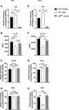
Figure 1:. HDM-induced BAL eosinophils and neutrophils were decreased in male compared to female mice.
(A) Experimental design for HDM-induced airway inflammation. (B-C) IL-13 and IL-17A protein expression in the lung homogenates as measured by ELISA. (D-H) Total BAL cells and total BAL eosinophils, neutrophils, macrophages, and lymphocytes. * p< 0.05, 1-way ANOVA with Tukey post-hoc analysis, n= 6–9 mice/group. Data pooled from 2 independent experiments.
Figure 2:
Figure 2:. Testosterone decreased and ovarian hormones increased HDM-induced airway eosinophilic and neutrophilic inflammation, IgE production, and AHR.
WT gonadectomized and sham-operated male and female BALB/c mice were challenged with HDM or PBS. (A-B) Total BAL eosinophils and neutrophils. (C) Total serum IgE levels. (A-C) *p<0.05, 1-way ANOVA with Tukey post hoc analysis, n=3–7 mice/group. Representative sample of 3 independent experiments. (D) AHR in response to increasing concentrations of methacholine 24 hours after the last HDM challenge. * p<0.05 ANOVA of repeated measures with Bonferroni post hoc analysis. n=12–14/group. Data pooled from 2 independent experiments.
Figure 3:
Figure 3:. Testosterone decreased and ovarian hormones increased HDM-induced total numbers IL-13 and IL-17A producing CD45+ cells.
WT gonadectomized and sham-operated male and female BALB/c mice were challenged with HDM or PBS. Lungs were harvested 24 hours after the last challenge, and lung cells were restimulated with PMA, ionomycin and golgi stop. (A) Representative dot plots of viable, CD45+ IL-13+ or IL-17A+ cells. (B-C) Percent and total numbers of IL-13+ CD45+ cells. (D-E) Percent and total numbers of IL-17A+ CD45+ cells. *p<0.05 1-way ANOVA with Tukey post hoc analysis n=6–10 mice/group. Data pooled from two independent experiments. n.s. means not statistically significant.
Figure 4:
Figure 4:. Testosterone decreased and ovarian hormones increased HDM-induced total numbers of IL-13+ Th2 and IL-17A+ Th17 cells.
WT gonadectomized and sham-operated male and female BALB/c mice were challenged with HDM or PBS. Lungs were harvested 24 hours after the last challenge, and lung cells were restimulated with PMA, ionomycin and golgi stop. (A) Representative dot plots of viable, CD45+ CD3+ CD4+IL-13+ or IL-17A+ cells. (B-D) Percent and total numbers of IL-13+ Th2 cells (defined as CD45+, CD3+ CD4+ IL-13+ cells) and IL-13+ ILC2 cells (defined as Lin- CD45+ CD25+ CD90+ IL-13+ cells). (E-G) Percent and total numbers of IL-17A+ Th17 cells and IL-17A+ γδ T cells (defined as CD45+ CD3+ CD4-γδTCR+) cells. *p<0.05 1-way ANOVA with Tukey post hoc analysis n=6–10 mice/group. Data pooled from two independent experiments. n.s. means not statistically significant.
Figure 5:
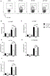
Figure 5:. Androgen receptor signaling attenuated HDM-induced eosinophilic and neutrophilic inflammation.
WT female, WT male, and androgen receptor testicular feminized mice (ARtfm ) male C57BL/6 mice were challenged with HDM or PBS. (A-B) BAL fluid eosinophils and neutrophils. (C) Representative flow staining of IL-13 and IL-17A expressing T cells. (D-E) Total numbers of IL-13+ Th2 cells and IL-17A+ Th17 cells. * p<0.05, ANOVA with Tukey post hoc test. n=3–5 mice/group. Data shown is from one representative experiment of three independent experiments. n.s. means not statistically significantly.
Figure 6:
Figure 6:. Androgen receptor signaling intrinsically decreased IL-17+ Th17 cells and indirectly decreased IL-13+ Th2 cells.
(A) Experimental design of mixed bone marrow chimera experiments with 1:1 bone marrow mixture from WT (CD90.1) or ARtfm (CD90.2) male C57BL/6J mice transferred into lethally irradiated heterozygous CD90.1+CD90.2+ recipient C57BL/6J mice. After 6 weeks of reconstitution, recipient mice underwent HDM or PBS challenge and IL-17A+ and IL-13+ CD4+ T cells were determined for WT and ARtfm mice. (B) Representative dot plots showing gating strategy. (C-D) Total numbers of IL-13+ Th2 and IL-17A+ Th17 cells derived from WT CD90.1 or ARtfm CD90.2 lineages.* p<0.05 ANOVA, with Tukey post hoc analysis, n=5–10 mice/group. Data pooled from 2 combined experiments.
Figure 7:
Figure 7:. Androgen receptor signaling attenuated Th17 but not Th2 differentiation in vitro.
Naïve CD4+ T cells from WT female, WT male, and ARtfm male mice were activated and differentiated into Th2 or Th17 cells. Cell culture supernatants and RNA were collected 4 days after differentiation. (A) and (E) AR mRNA expression in Th2 cells (left column) and Th17 cells (right column). (B-D) IL-13 protein expression from Th2 cell culture supernatants and Gata3 and Il4rα mRNA relative expression from in differentiated Th2 cells. (F-H) IL-17A protein expression in Th17 cell culture supernatants and Rorc and Il23r mRNA relative expression in differentiated Th17 cells. IL-13 and IL-17A protein expression were measured by ELISA and mRNA relative expression measured by qRT-PCR with relative expression in Th2 or Th17 cells were normalized to Gapdh. * p<0.05, ANOVA with Tukey post hoc analysis. n=9–12 mice/group. Data pooled from 2 independent experiments. n. s. means not statistically significant.
Figure 8:
Figure 8:. Androgen receptor signaling decreased total numbers of IL-4 producing cells.
WT female, WT male and ARtfm male mice were challenged with HDM or PBS. (A) Representative dot plots of IL-4 producing CD45+ cells. (B-C) Percentage and total numbers of viable IL-4+ CD45+ cells. (D-F) Number of IL-4 producing Th2 cells, mast cells, and basophils. Th2 cells were defined as CD45+ CD3+ CD4+ cells, mast cells were defined as CD45+ FcεRI+ CD117+ DX5- cells, and basophils were defined as CD45+ FcεRI+ CD117- DX5+ cells. * p<0.05, ANOVA with Tukey post hoc test. n=3–9 mice/group with data pooled from 2 independent experiments. n.s. means not statistically significant.

References

    1. Hastie AT, Moore WC, Meyers DA, Vestal PL, Li H, Peters SP, and Bleecker ER 2010. Analyses of asthma severity phenotypes and inflammatory proteins in subjects stratified by sputum granulocytes. J. Allergy Clin Immunol 125: 1028–1036. - PMC - PubMed
    1. Wenzel SE, and Busse WW 2007. Severe asthma: lessons from the Severe Asthma Research Program. J. Allergy Clin Immunol 119: 14–21. - PubMed
    1. Wu W, Bleecker E, Moore W, Busse WW, Castro M, Chung KF, Calhoun WJ, Erzurum S, Gaston B, Israel E, Curran-Everett D, and Wenzel SE 2014. Unsupervised phenotyping of Severe Asthma Research Program participants using expanded lung data. J. Allergy Clin Immunol 133: 1280–1288. - PMC - PubMed
    1. Agarwal AK, and Shah A 1997. Menstrual-linked asthma. J. Asthma 34: 539–545. - PubMed
    1. Murphy VE, Clifton VL, and Gibson PG 2006. Asthma exacerbations during pregnancy: incidence and association with adverse pregnancy outcomes. Thorax 61: 169–176. - PMC - PubMed

Publication types

MeSH terms