. 2018 Aug 20;10(8):446.
doi: 10.3390/v10080446.

The Morphology and Assembly of Respiratory Syncytial Virus Revealed by Cryo-Electron Tomography

Affiliations

The Morphology and Assembly of Respiratory Syncytial Virus Revealed by Cryo-Electron Tomography

Zunlong Ke et al. Viruses. .

Abstract

Human respiratory syncytial virus (RSV) is the leading cause of lower respiratory tract disease in young children. With repeat infections throughout life, it can also cause substantial disease in the elderly and in adults with compromised cardiac, pulmonary and immune systems. RSV is a pleomorphic enveloped RNA virus in the Pneumoviridae family. Recently, the three-dimensional (3D) structure of purified RSV particles has been elucidated, revealing three distinct morphological categories: spherical, asymmetric, and filamentous. However, the native 3D structure of RSV particles associated with or released from infected cells has yet to be investigated. In this study, we have established an optimized system for studying RSV structure by imaging RSV-infected cells on transmission electron microscopy (TEM) grids by cryo-electron tomography (cryo-ET). Our results demonstrate that RSV is filamentous across several virus strains and cell lines by cryo-ET, cryo-immuno EM, and thin section TEM techniques. The viral filament length varies from 0.5 to 12 μm and the average filament diameter is approximately 130 nm. Taking advantage of the whole cell tomography technique, we have resolved various stages of RSV assembly. Collectively, our results can facilitate the understanding of viral morphogenesis in RSV and other pleomorphic enveloped viruses.

Keywords: cryo-electron microscopy (cryo-EM); cryo-electron tomography (cryo-ET); enveloped virus; human respiratory syncytial virus (RSV); viral morphogenesis; virus assembly.


Conflict of interest statement

Martin L. Moore is an employee of Meissa Vaccines, Inc. Other authors declare no competing financial interests.

Figures

Figure 1
Figure 1
Assessment of sample preparation conditions for cryo-EM data collection. (A) One-step growth curve of RSV A2 in BEAS-2B cells over a period of 36 h at a M.O.I. of 10. Infected cells were harvested at indicated time points and titrated in Vero cells. (BD) Flow cytometry, viral titer, and RNA quantification analysis of RSV-infected BEAS-2B cells, at indicated M.O.I.s after 24 h.p.i. These data indicate that at a M.O.I. of 10, infectious particles are produced at 24 h.p.i., suggesting that the particles being imaged by cryo-EM are infectious. (B) Relative ratio of cells infected at indicated M.O.I. by flow cytometry using mK+ signal, represented by the ratio of infected cells over the total cells. (C) Viral titer at 24 h.p.i. determined by FFU using mK+ signal. (D) Relative amount of viral RNA from infected cells determined by real time RT-PCR represented by inverse CT values (1/CT). CT value is the number of cycles needed to pass the background level. For all 4 experiments, error bars represent the standard deviation of 3 independent experiments.
Figure 2
Figure 2
Representative montages showing 2D projections of RSV-infected cells. (A) Representative montages showing released viruses from RSV-infected cells. Montage view of released viral particles captured from RSV A2 infected A549 cells. Sample was prepared as described in the method section. RSV morphology varies, with the overall structure being filamentous. Filaments may be straight, bent, or branched. (B) Montage view of the assembly site from RSV-infected cells with the F glycoprotein immunogold labeled. The region imaged is an active assembly site, with RSV filaments extending from the cell plasma membrane (dashed yellow line). (C) Zoomed-in view of the boxed region in B. Note the presence of 6-nm immunogold on the RSV filaments (white arrows), and the absence of gold particles on cell extension (black arrows). Scale bars are 2 µm (A), 1 µm (B), and 500 nm (C).
Figure 3
Figure 3
Cryo-ET of released RSV filaments from RSV-infected cells. (AF) Cell lines including HeLa (A,F) and lung derived cell lines A549 (B), MRC-5 (C), and BEAS-2B (D,E), were infected with RSV A2 strain (AD) or RSV TN strain (E,F). Among these samples, filamentous particles were consistently observed under frozen-hydrated conditions. Scale bars are 200 nm.
Figure 4
Figure 4
RSV morphology quantification in multiple virus strains and infected cell lines. (A) Overall quantification of RSV filament length and diameter. (B) RSV filament length distribution in indicated virus-infected cells. (C) RSV filament diameter distribution. Note, the mean diameter of RSV filaments is around ~130 nm. (D,E) Filament length (D) and diameter (E) distribution from A2-infected BEAS-2B cells.
Figure 5
Figure 5
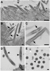
RSV morphology is filamentous from infected-NHBE cells. (AC) Filamentous RSV particles were observed at virus assembly sites on infected cells. Black arrows indicate filamentous RSV. (D,E) Cellular filaments observed from the polarized NHBE cells, such as cilia (black arrowheads) and microvilli (white arrowhead). Scale bars are 200 nm.
Figure 6
Figure 6
Structural analysis of RSV filamentous particles. (AC) Representative tomographic slices and linear density profiles of RSV ultrastructure. White boxes indicate representative regions used to generate linear density plots. Linear density profiles are averaged results of multiple line profiles from several regions. (DF) Segmentation illustration of the filamentous particles from indicated virus-infected cells. Note that the segmentation areas do not correspond to the regions in (AC), but representative regions of samples from (AC). Density peaks 1 through 6 represent: 1, Viral glycoproteins (Red); 2, Viral membrane (Green); 3, Matrix (Cyan); 4, M2-1 (Blue); 5 and 6, RNP (Magenta). Scale bars are 20 nm.
Figure 7
Figure 7
Structural comparison between filamentous and spherical RSV particles. (A,B) Representative tomographic slices of a filamentous (A, black outline) and spherical (B, red outline) particle. (C,D) Model fitting of representative regions of the isosurface rendering of the tomographic data for filamentous (C, black outline) and spherical (D, red outline) particles. Prefusion F (PDB ID: 4JHW) and postfusion F (PDB ID: 3RRT) were used for (C) and (D), respectively. (E) Linear density profiles of a filamentous particle (black line) and spherical particle (red line). Density peaks 1 through 6 represent: 1, Viral glycoproteins; 2, Viral membrane; 3, Matrix; 4, M2-1; 5 and 6, RNP. (F) Glycoprotein length quantification from filamentous and spherical particles. **** indicates the student t-test p-value is below 0.0001. Scale bars are 100 nm (A,B) and 10 nm (C,D).
Figure 8
Figure 8
RSV assembly steps revealed by cryo-ET. (A) Representative cryo-EM images of RSV-infected cells. Note the assembly site indicated by the dashed white box and the released viral particles. The dashed yellow line indicates the cell plasma membrane. (B) Segmentation of the assembly site indicated in the white dashed box in (A), illustrating initiation (C), elongation (D), and scission (E) events. See also Supplementary Movie S1. Green is membrane, red is glycoprotein, and magenta is the RNP complex. Scale bars are 1 µm (A) and 200 nm (BE).
Figure 9
Figure 9
RSV assembly in the presence of fusion inhibitor. RSV-infected HeLa cells were infected at a M.O.I. of 10, washed twice with PBS prior to adding 600 nM RSV fusion inhibitor (BMS-433771), and incubated for the indicated length of time. (A) Titration results of RSV-infected cells with or without RSV fusion inhibitor at 1 and 4 d.p.i. ** indicates the student t-test p-value is below 0.01. **** indicates the student t-test p-value is below 0.0001. (B) Representative images showing the absence of syncytial formation with inhibitor and syncytial formation without inhibitor. (C) Polygon montage of an assembly site. Micrographs were acquired using cryo-EM/ET, demonstrating the assembly events at 24 h.p.i. (D) Tomographic slice (6.14 nm) of the assembly site collected from the region indicated by the dashed black box in (C). The black arrows indicate the early assembly events prior to RSV filament elongation. Note the presence of matrix layer and the glycoproteins. The white arrows indicate the plasma membrane; note the absence of the matrix layer and the glycoproteins. The dashed white box indicates an elongating RSV filament. Scale bars are 100 μm (B), 1 μm (C), and 200 nm (D).
Figure 10
Figure 10
RSV morphology model. A schematic representation of a filamentous (A) and a spherical (B) RSV viral particle. Enlarged views of the boxed regions in (A) and (B) are shown in (C) and (D), respectively. The structure of G and the ratio of G to F are unknown. In the filamentous particle (A,C), F is in the prefusion form with matrix (M) lining the viral membrane (VM). M2-1 acts as a linker protein between M and the RNP. In the spherical particle (B,D), F is in the postfusion form while M is detached from the viral membrane. RNP is disordered and not linked by M2-1 to the matrix protein.

References

    1. Lamb R.A., Parks G.D. Paramyxoviridae: The viruses and their replication. In: Knipe D.M., Howley P.M., editors. Fields Virology. 5th ed. Volume 1. Wolters Kluwer/Lippincott Williams & Wilkins; Philadelphia, PA, USA: 2007. pp. 1449–1496.
    1. Nair H., Nokes D.J., Gessner B.D., Dherani M., Madhi S.A., Singleton R.J., O’Brien K.L., Roca A., Wright P.F., Bruce N., et al. Global burden of acute lower respiratory infections due to respiratory syncytial virus in young children: A systematic review and meta-analysis. Lancet. 2010;375:1545–1555. doi: 10.1016/S0140-6736(10)60206-1. - DOI - PMC - PubMed
    1. Shi T., McAllister D.A., O’Brien K.L., Simoes E.A.F., Madhi S.A., Gessner B.D., Polack F.P., Balsells E., Acacio S., Aguayo C., et al. Global, regional, and national disease burden estimates of acute lower respiratory infections due to respiratory syncytial virus in young children in 2015: A systematic review and modelling study. Lancet. 2017;390:946–958. doi: 10.1016/S0140-6736(17)30938-8. - DOI - PMC - PubMed
    1. Anderson L.J., Dormitzer P.R., Nokes D.J., Rappuoli R., Roca A., Graham B.S. Strategic priorities for respiratory syncytial virus (RSV) vaccine development. Vaccine. 2013;31:B209–B215. doi: 10.1016/j.vaccine.2012.11.106. - DOI - PMC - PubMed
    1. Griffiths C., Drews S.J., Marchant D.J. Respiratory syncytial virus: Infection, detection, and new options for prevention and treatment. Clin. Microbiol. Rev. 2017;30:277–319. doi: 10.1128/CMR.00010-16. - DOI - PMC - PubMed

Publication types