Review
. 2018 Aug 20;7(8):225.
doi: 10.3390/jcm7080225.

The Emerging Role of Pathogenesis of IgA Nephropathy

Affiliations
Review

The Emerging Role of Pathogenesis of IgA Nephropathy

Meng-Yu Wu et al. J Clin Med. .

Abstract

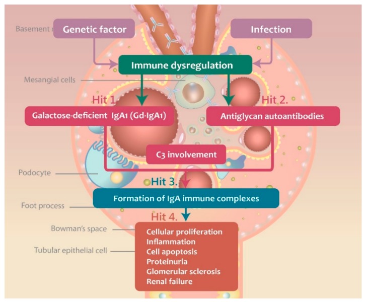
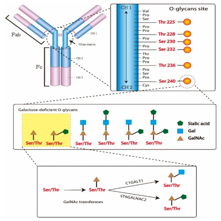
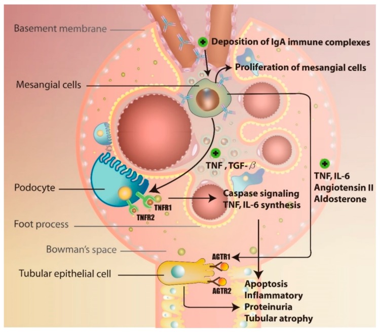
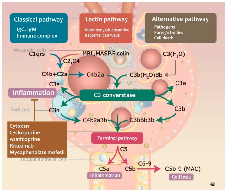
IgA nephropathy is an autoimmune disease induced by fthe ormation of galactose-deficient IgA1 and anti-glycans autoantibody. A multi-hit hypothesis was promoted to explain full expression of IgA nephropathy. The deposition of immune complex resulted in activation of the complement, increasing oxidative stress, promoting inflammatory cascade, and inducing cell apoptosis via mesangio-podocytic-tubular crosstalk. The interlinked signaling pathways of immune-complex-mediated inflammation can offer a novel target for therapeutic approaches. Treatments of IgA nephropathy are also summarized in our review article. In this article, we provide an overview of the recent basic and clinical studies in cell molecular regulation of IgAN for further treatment interventions.

Keywords: IgA nephropathy; anti-glycans autoantibody; galactose-deficient IgA1; inflammation; mesangio-podocytic-tubular crosstalk.


Conflict of interest statement

The authors declare no conflict of interest.

Figures

Figure 1
Figure 1
The multi-“hit” hypothesis explaining the immunopathogenesis of IgAN.
Figure 2
Figure 2
Structure of immunoglobulin A1 (IgA1) hinge region with O‑glycans. In the hinge region, IgA1 potentially has three to six O-glycans. The O-glycan attached to IgA1 via the link with serine/threonine residues and oxygen atoms. The structure of O-glycans consisted of N-acetylgalactosamine (GalNAc), galactose and/or sialic acid. The attachment of GalNAc to serine/threonine residues by GalNAc transferases is first step of glycosylation. Second step, the GalNAc can be added galactose by glycoprotein N-acetylgalactosamine 3-β-galactosyltransferase (C1GALT1) or sialic acid by α-N-acetylgalactosaminide α2,6-sialyltransferase 2 (ST6GALNAC2).
Figure 3
Figure 3
The mechanism of activating B cells to produce the galactose-deficient IgA1.
Figure 4
Figure 4
The mechanism of glomerulo-podocytic-tubular crosstalk in IgAN.
Figure 5
Figure 5
The mechanism of complement activation pathways in IgAN.

References

    1. Geddes C.C. A Tricontinental View of IgA Nephropathy. Nephrol. Dial. Transplant. 2003;18:1541–1548. doi: 10.1093/ndt/gfg207. - DOI - PubMed
    1. Silva F.G. Disappearance of glomerular mesangial IgA deposits after renal allograft transplantation. Transplantation. 1982;33:241–246. - PubMed
    1. Suzuki H. The Pathophysiology of IgA Nephropathy. J. Am. Soc. Nephrol. 2011;22:1795–1803. doi: 10.1681/ASN.2011050464. - DOI - PMC - PubMed
    1. McGrogan A., Franssen C.F., De Vries C.S. The incidence of primary glomerulonephritis worldwide: A systematic review of the literature. Nephrol. Dial. Transplant. 2011;26:414–430. doi: 10.1093/ndt/gfq665. - DOI - PubMed
    1. Barbour S.J. Individuals of Pacific Asian origin with IgA nephropathy have an increased risk of progression to end-stage renal disease. Kidney Int. 2013;84:1017–1024. doi: 10.1038/ki.2013.210. - DOI - PubMed