. 2018 Sep;25(9):805-813.
doi: 10.1038/s41594-018-0108-7. Epub 2018 Aug 20.

Structure and gating mechanism of the transient receptor potential channel TRPV3

Affiliations

Structure and gating mechanism of the transient receptor potential channel TRPV3

Appu K Singh et al. Nat Struct Mol Biol. 2018 Sep.

Abstract

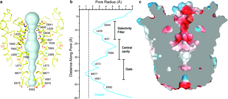
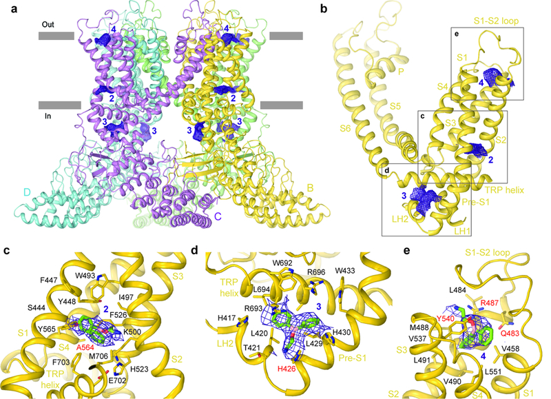
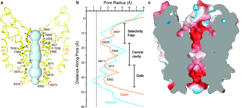
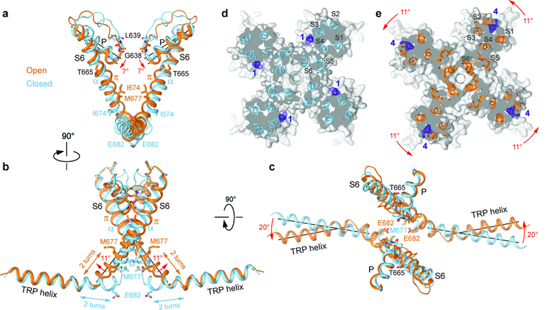
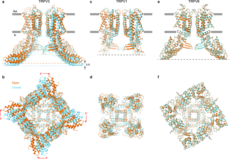
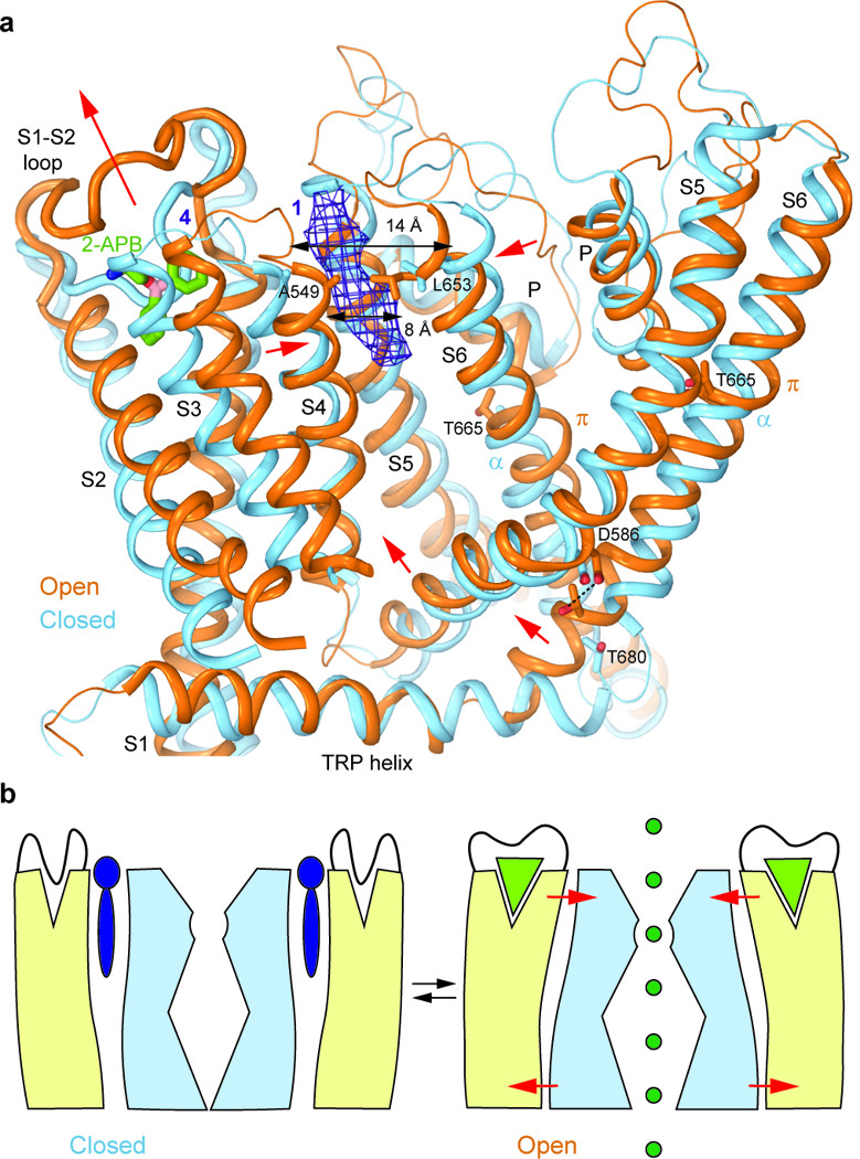
Transient receptor potential vanilloid subfamily member 3 (TRPV3) channel plays a crucial role in skin physiology and pathophysiology. Mutations in TRPV3 are associated with various skin diseases, including Olmsted syndrome, atopic dermatitis, and rosacea. Here we present the cryo-electron microscopy structures of full-length mouse TRPV3 in the closed apo and agonist-bound open states. The agonist binds three allosteric sites distal to the pore. Channel opening is accompanied by conformational changes in both the outer pore and the intracellular gate. The gate is formed by the pore-lining S6 helices that undergo local α-to-π helical transitions, elongate, rotate, and splay apart in the open state. In the closed state, the shorter S6 segments are entirely α-helical, expose their nonpolar surfaces to the pore, and hydrophobically seal the ion permeation pathway. These findings further illuminate TRP channel activation and can aid in the design of drugs for the treatment of inflammatory skin conditions, itch, and pain.


Figures

Figure 1.
Figure 1.. 3D cryo-EM reconstruction and structure of TRPV3 in the apo state.
a,b, Top (a) and side (b) views of 3D cryo-EM reconstruction of TRPV3 in the apo state with subunits coloured green, yellow, violet and cyan and lipid in purple. The densities are shown at a threshold of 0.057 (UCSF Chimera). c, Expanded view of the two putative lipid densities shown as purple mesh at 4σ. d,e, Top (d) and side (e) views of the TRPV3 structure with the same coloring as in panels a-c. f, Expanded view of the boxed region in (e) displaying the interaction of the TRPV3 C-terminal loop domain with the ARD from the neighbouring subunit.
Figure 2:
Figure 2:. Closed pore of TRPV3.
a, Pore-forming domain of TRPV3 in the apo state with residues lining the pore shown as sticks. Only two of four subunits are shown; the front and back subunits are omitted for clarity. The pore profile is shown as a space-filling model (cyan). b, Pore radius calculated using HOLE. The vertical dashed line denotes the radius of a water molecule, 1.4 Å. c, Coronal section of the TRPV3 pore domain, with surface coloured by electrostatic potential.
Figure 3:
Figure 3:. Structure of 2-APB-bound TRPV3(Y564A).
a, Side view of the TRPV3(Y564A)2-APB structure with each subunit coloured differently and non-protein densities shown as purple mesh at 4σ. b-e, Non-protein densities for one subunit (b) with expanded views of densities at site 2 (c), site 3 (d) and site 4 (e). 2-APB molecules in (c-e) and residues surrounding them are shown as sticks.
Figure 4:
Figure 4:. Open pore of TRPV3.
a, Pore-forming domain of TRPV3(Y564A)2-APB with residues lining the pore shown as sticks and the pore profile shown as a space-filling model (cyan). b, Pore radius calculated using HOLE for TRPV3 (blue) and TRPV3(Y564A)2-APB (orange). The vertical dashed line denotes the radius of a water molecule, 1.4 Å. c, Coronal section of the TRPV3(Y564A)2-APB pore-forming domain, with surface coloured by electrostatic potential.
Figure 5:
Figure 5:. Structural changes associated with TRPV3 channel opening.
a-c, Superposition of the P-loop, S6 and TRP helices of TRPV3 (blue) and TRPV3(Y564A)2-APB (orange) viewed parallel to the membrane (a,b) or extracellulary (c). Only two of four subunits are shown for clarity. Residues lining the pore are shown as sticks. Rotations of the pore, S6 and TRP helices in TRPV3(Y564A)2-APB relative to TRPV3 are indicated by red arrows. d-e, Sections through the space filling models of TRPV3 (d, blue) and TRPV3(Y564A)2-APB (e, orange) made perpendicular to the pore axis and viewed extracellularly. Non-protein densities at site 1 (d) and site 4 (e) are shown as purple mesh at 4σ. The structures are aligned based on their pore domains. The 11° rotation of the S1-S4 domains in TRPV3(Y564A)2-APB relative to their position in TRPV3 is indicated by the red arrows.
Figure 6:
Figure 6:. Comparison of gating rearrangements in different TRPV channels.
a-f, Superposition of structures in the open (orange) and closed (blue) states for TRPV3 (a-b, TRPV3(Y564A)2-APB and TRPV3, respectively), TRPV1 (c-d, PDB: 5IRX and PDB: 5IRZ, respectively) and TRPV6 (e-f, PDB: 6BO8 and PDB: 6BOA, respectively) viewed parallel to the membrane (a,c,e) or intracellularly (b,d,f). Only two of four subunits are shown in (a,c,e), with the front and back subunits omitted for clarity. The structures are aligned based on their pore domains. Note, the TRPV3 structure becomes shorter (black arrows) and its intracellular skirt undergoes substantial rotation (red arrows) during channel opening, while the overall architecture of TRPV1 and TRPV6 remains the same.
Figure 7:
Figure 7:. TRPV3 Gating Mechanism.
a, Superposition of the transmembrane domains and TRP helices of TRPV3 (blue) and TRPV3(Y564A)2-APB (orange) viewed parallel to the membrane. Only a single subunit and a fragment of a neighbouring subunit (S5 through S6) are shown for clarity. The putative molecule of 2-APB bound at site 4 in TRPV3(Y564A)2-APB is shown as sticks and the non-protein density at site 1 of TRPV3 is shown as purple mesh at 4σ. Red arrows indicate movements of domains in TRPV3(Y564A)2-APB relative to their positions in TRPV3. b, Cartoon illustrating gating in TRPV3. In the closed state (left), a lipid (purple) is bound between the S1-S4 (beige) and pore (light blue) domains and the pocket at the top of the S1-S4 domain is occupied by the S1-S2 loop. In the open state (right), 2-APB (light green triangle) binds to and expands the top of the S1-S4 domain, squeezes out the lipid and brings the S1-S4 and pore domains closer together. As a result, the S1-S4-pore domain interface rearranges, and S5 and S6 move away from the pore, opening it for permeation of ions (dark green).

References

    1. Clapham DE TRP channels as cellular sensors. Nature 426, 517–524 (2003). - PubMed
    1. Xu HX et al. TRPV3 is a calcium-permeable temperature-sensitive cation channel. Nature 418, 181–186 (2002). - PubMed
    1. Peier AM et al. A heat-sensitive TRP channel expressed in keratinocytes. Science 296, 2046–2049 (2002). - PubMed
    1. Smith GD et al. TRPV3 is a temperature-sensitive vanilloid receptor-like protein. Nature 418, 186–190 (2002). - PubMed
    1. Cheng X. et al. TRP channel regulates EGFR signaling in hair morphogenesis and skin barrier formation. Cell 141, 331–343 (2010). - PMC - PubMed

Publication types