Review
. 2021 Sep;90(3):524-531.
doi: 10.1038/s41390-018-0114-9. Epub 2018 Jul 20.

Point-of-care lung ultrasound in neonatology: classification into descriptive and functional applications

Affiliations
Review

Point-of-care lung ultrasound in neonatology: classification into descriptive and functional applications

Francesco Raimondi et al. Pediatr Res. 2021 Sep.

Abstract

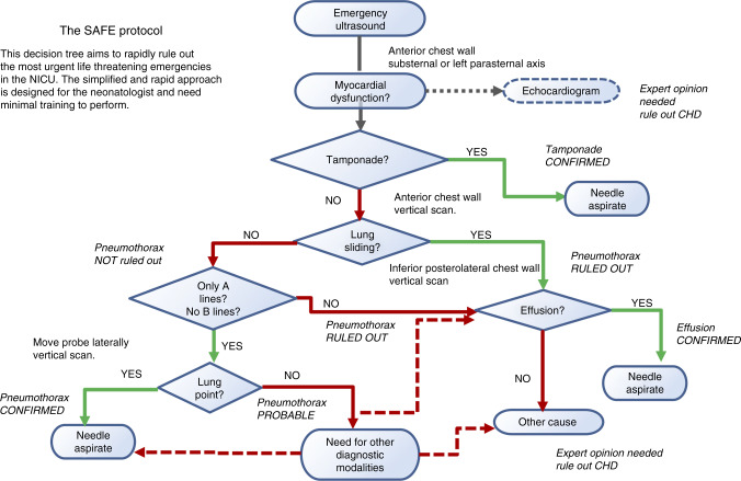
Lung ultrasound (LUS) is the latest amongst imaging techniques: it is a radiation-free, inexpensive, point-of-care tool that the clinician can use at the bedside. This review summarises the rapidly growing scientific evidence on LUS in neonatology, dividing it into descriptive and functional applications. We report the description of the main ultrasound features of neonatal respiratory disorders and functional applications of LUS aiming to help a clinical decision (such as surfactant administration, chest drainage etc). Amongst the functional applications, we propose SAFE (Sonographic Algorithm for liFe threatening Emergencies) as a standardised protocol for emergency functional LUS in critical neonates. SAFE has been funded by a specific grant issued by the European Society for Paediatric Research. Future potential development of LUS in neonatology might be linked to its quantitative evaluation: we also discuss available data and research directions using computer-aided diagnostic techniques. Finally, tools and opportunities to teach LUS and expand the research network are briefly presented.


Conflict of interest statement

The authors declare no competing interests.

Figures

Fig. 1
Fig. 1
Papers published on lung ultrasound in neonatology in 2006 and 2016. Retrieved by searching in PubMed (on 30 Dec 2017), limited to the newborn age, with the following words and MeSH terms: ((“lung”[MeSH Terms]OR“lung”[All Fields])AND (“diagnostic imaging”[Subheading]OR(“diagnostic”[All Fields]AND“imaging”[All Fields])OR“diagnostic imaging”[All Fields]OR“ultrasound”[All Fields]OR“ultrasonography”[MeSH Terms]OR “ultrasonography”[All Fields]OR“ultrasound”[All Fields]OR“ultrasonics”[MeSH Terms]OR“ultrasonics”[All Fields]))AND((“2006/01/01”[PDAT]: “2006/12/31”[PDAT])AND “infant, newborn”[MeSH Terms])OR((“2016/01/01”[PDAT]:“2016/12/31”[PDAT])
Fig. 2
Fig. 2
Lung ultrasound semiology. The basic semiology patterns are illustrated: these patterns may be variably found in different respiratory disorders described in Table 1. Arrows indicate the sub-pleural consolidation, the border of a consolidation, the double lung point or the lung point. The size threshold to distinguish micro-consolidations (sub-pleural) from consolidations (0.5 cm) is arbitrary. Some semiology patterns are also dynamically shown in the videos in Supplementary Material 1
Fig. 3
Fig. 3
SAFE (Sonographic Algorithm for liFe threatening Emergencies) algorithm for critically ill neonates. The algorithm is designed for unexpected severe decompensations (bradycardia or severe desaturation requiring resuscitative manoeuvres or significantly increasing oxygen/ventilator parameters to maintain stable oxygen saturation levels) in formerly stable neonates. SAFE protocol starts with a quick ‘eyeball’ assessment of myocardial contractility (which is accurate enough if there are no arrhythmias, extreme heart rate or ventricular sizes). Then, SAFE screens the more urgent and common causes of life-threatening event: (1) cardiac tamponade, (2) pneumothorax and (3) pleural effusion. The algorithm only takes a few minutes and aims to help diagnosing the most urgent treatable complications whilst awaiting expert help. A paediatric cardiologist evaluation of congenital heart defects is included in the algorithm but only when the most urgent causes have been already ruled out. SAFE is designed for the average neonatologist and may be applied using any probe without losing time to change it

References

    1. Lichtenstein DA, Menu Y. A bedside ultrasound sign ruling out pneumothorax in the critically ill. Lung sliding. Chest. 1995;108:1345–1348. doi: 10.1378/chest.108.5.1345. - DOI - PubMed
    1. Kurepa D, et al. Neonatal lung ultrasound exam guidelines. J. Perinatol. 2018;38:11–22. doi: 10.1038/jp.2017.140. - DOI - PubMed
    1. Escourrou G, De Luca D. Lung ultrasound decreased radiation exposure in preterm infants in a neonatal intensive care unit. Acta Paediatr. 2016;105:e237–e239. doi: 10.1111/apa.13369. - DOI - PubMed
    1. International Liaison Committee on Lung Ultrasound (ILC-LUS) for the International Consensus Conference on Lung Ultrasound (ICC-LUS International evidence-based recommendations for point-of-care lung ultrasound. Intensive Care Med. 2012;38:577–591. doi: 10.1007/s00134-012-2513-4. - DOI - PubMed
    1. Lichtenstein D, et al. The comet-tail artifact. An ultrasound sign of alveolar-interstitial syndrome. Am. J. Respir. Crit. Care Med. 1997;156:1640–1646. doi: 10.1164/ajrccm.156.5.96-07096. - DOI - PubMed

Publication types

Substances

LinkOut - more resources