Review
. 2018 Aug 6:12:242.
doi: 10.3389/fncel.2018.00242. eCollection 2018.

An Overview of in vitro Methods to Study Microglia

Affiliations
Review

An Overview of in vitro Methods to Study Microglia

Raissa Timmerman et al. Front Cell Neurosci. .

Abstract

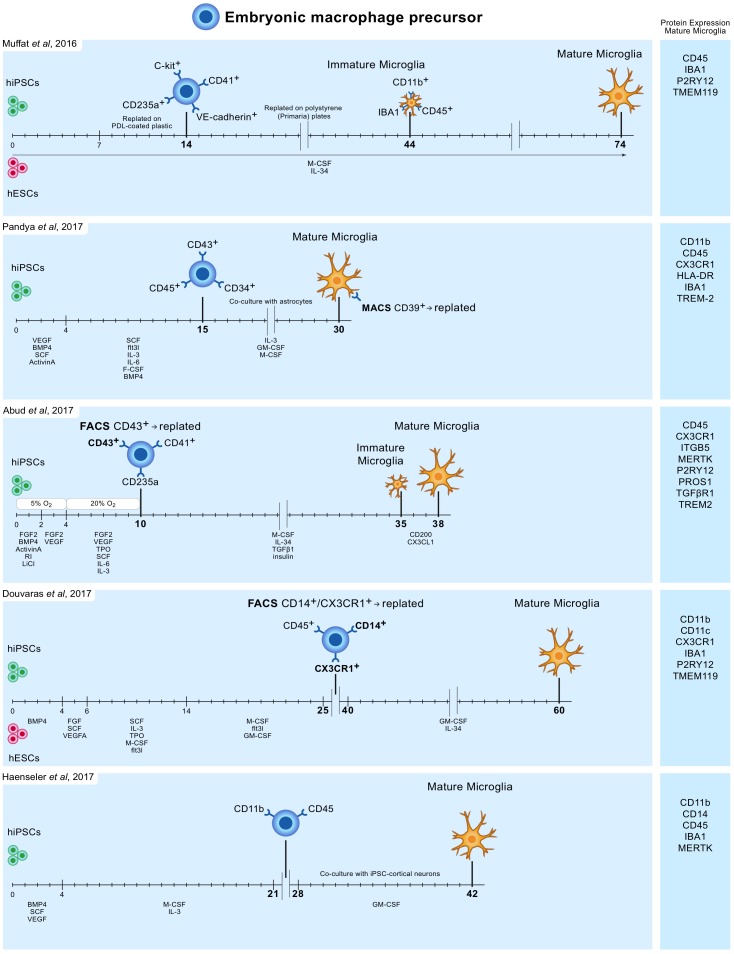
Neuroinflammation is a common feature in neurodegenerative diseases and strategies to modulate neuroinflammatory processes are increasingly considered as therapeutic options. In such strategies, glia cells rather than neurons represent the cellular targets. Microglia, the resident macrophages of the central nervous system, are principal players in neuroinflammation and detailed cellular biological knowledge of this particular cell type is therefore of pivotal importance. The last decade has shed new light on the origin, characteristics and functions of microglia, underlining the need for specific in vitro methodology to study these cells in detail. In this review we provide a comprehensive overview of existing methodology such as cell lines, stem cell-derived microglia and primary dissociated cell cultures, as well as discuss recent developments. As there is no in vitro method available yet that recapitulates all hallmarks of adult homeostatic microglia, we also discuss the advantages and limitations of existing models across different species.

Keywords: cell culture techniques; in vitro models; microglia; neurodegenerative diseases; stem cells.


Figures

FIGURE 1
FIGURE 1
Overview of protocols to differentiate human iPSC and ESC to microglia-like cells. Timelines are indicated in days. hiPSC and hESC are first differentiated to embryonic macrophage precursor cells, which is consistent with the in vivo developmental lineage of microglia. Subsequently, these precursor cells are differentiated into mature microglia-like cells.

Similar articles

Cited by

References

    1. Abud E. M., Ramirez R. N., Martinez E. S., Healy L. M., Nguyen C. H. H., Newman S. A., et al. (2017). iPSC-derived human microglia-like cells to study neurological diseases. Neuron 94 e278.e9–293.e9. 10.1016/j.neuron.2017.03.042 - DOI - PMC - PubMed
    1. Ajami B., Bennett J. L., Krieger C., Tetzlaff W., Rossi F. M. (2007). Local self-renewal can sustain CNS microglia maintenance and function throughout adult life. Nat. Neurosci. 10 1538–1543. 10.1038/nn2014 - DOI - PubMed
    1. Alliot F., Godin I., Pessac B. (1999). Microglia derive from progenitors, originating from the yolk sac, and which proliferate in the brain. Brain Res. Dev. Brain Res. 117 145–152. 10.1016/S0165-3806(99)00113-3 - DOI - PubMed
    1. Alliot F., Marty M. C., Cambier D., Pessac B. (1996). A spontaneously immortalized mouse microglial cell line expressing CD4. Brain Res. Dev. Brain Res. 95 140–143. 10.1016/0165-3806(96)00101-0 - DOI - PubMed
    1. Amit I., Winter D. R., Jung S. (2015). The role of the local environment and epigenetics in shaping macrophage identity and their effect on tissue homeostasis. Nat. Immunol. 17 18–25. 10.1038/ni.3325 - DOI - PubMed