. 2018 Sep;16(3):3895-3900.
doi: 10.3892/ol.2018.9116. Epub 2018 Jul 10.

Neoangiogenesis of gastric submucosa-invasive adenocarcinoma

Affiliations

Neoangiogenesis of gastric submucosa-invasive adenocarcinoma

Hanae Sasaki et al. Oncol Lett. 2018 Sep.

Abstract

Early gastric cancer may be defined as mucosal or submucosal invasive carcinoma, and exhibits a good prognosis: 90% of patients survive >10 years. Early gastric cancer infrequently exhibits lymph node metastasis, although submucosal invasion, the presence of vascular invasion and/or lymphatic permeation are independent risk factors for lymph node metastasis in early gastric cancer. The analysis of tumor lymphangiogenesis and angiogenesis are important to determine the extent of invasive progression and metastasis in patients. Previously, the presence of vessels expressing the D2-40 antibody and the factor-VIII protein has been identified immunohistochemically. The vessels that are immunoreactive for D2-40 and factor-VIII are morphologically similar to lymphatic vessels or small-size veins, also termed venules. In the present study, the association between tumor invasion and neoangiogenesis in early gastric cancer was examined. The D2-40/factor-VIII double-stained vessel (DSV) density was analyzed, in addition to lymphatic and blood vessel (vein and artery) density, using 46 submucosa-invasive and 50 mucosal carcinomas, and 20 non-neoplastic gastric tissues. The lymphatic density and DSV density of submucosa beneath the carcinoma and submucosa of the surrounding region in submucosa-invasive carcinoma were significantly increased (P<0.001) in comparison with those in mucosal carcinoma or non-neoplastic gastric tissue. No significant difference was observed in blood vessel density between non-neoplastic gastric, mucosal carcinoma and submucosa-invasive carcinoma tissues other than that of mucosa. The present study suggests the potential for the presence of D2-40/factor-VIII DSV and the importance of this vessel for neoangiogenesis in early gastric cancer.

Keywords: D2-40; and factor-VIII; early gastric cancer; progression.


Figures

Figure 1.
Figure 1.
(A) Images of a 0.25-µm DSV captured in continuity. DSV was stained with D2-40 (3,3′-Diaminobenzidine, brown) and factor-VIII (FAST RED, red). (B) 3D merges of images were captured in continuity. The color of the vessel walls changed from red to brown. DSV, double-stained vessel.
Figure 2.
Figure 2.
Lymphatic vessels were stained with DAB (arrow head), blood vessel was stained with FAST RED (dashed arrow), and DSV were stained with DAB and FAST RED (arrow) (magnification, ×400). DSV were morphologically similar to lymphatic vessels. DAB, 3,3′-Diaminobenzidine; FAST RED, factor VIII; double-stained vessels, DSV.
Figure 3.
Figure 3.
The countable areas of vessels in an early gastric cancer tissue. The red squares indicated mucosal layer (M). Green squares indicated submucosa beneath the carcinoma (SMb). Blue squares indicated submucosa of the surrounding region (SMs). Three points were selected in each area for counting vessels (×20).
Figure 4.
Figure 4.
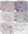
Representative images of double immunohistochemical staining by D2-40 and factor-VIII in mucosal carcinoma and submucosa-invasive carcinoma (×400). (A) and (B) were identified as mucosal layer. (C) and (D) were identifed as submucosa beneath the carcinoma. (E) and (F) were identified as submucosa of the surrounding region. Blood vessels were stained with factor-VIII colored with red. Lymphatics were stained by D2-40 colored with brown. Double stained vessels were stained by both factor-VIII and D2-40, arrow.
Figure 5.
Figure 5.
(A) Lymphatic density of the SMb in submucosa-invasive carcinoma was higher compared with that of mucosal carcinoma tissues (P<0.001) and non-neoplastic gastric tissues (P<0.001). Lymphatic density of SMs in submucosa-invasive carcinoma was also higher compared with that of mucosal carcinoma tissues (P<0.001) and non-neoplastic gastric tissues (P<0.001). (B) Vessel density of the M in submucosa-invasive carcinoma was higher compared with that of mucosal carcinoma tissues (P<0.001). (C) Double stained vessel density of the SMb (P<0.001) and the SMs (P<0.001) in submucosa-invasive carcinoma were higher compared with those of the non-neoplastic gastric and the mucosal carcinoma tissues. *P<0.05. M, mucosa; SMb, mucosa beneath the carcinoma; SMs, submucosa of the surrounding region.

References

    1. Tsukuma H, Mishima T, Oshima A. Prospective study of ‘early’ gastric cancer. Int J Cancer. 1983;31:421–426. doi: 10.1002/ijc.2910310405. - DOI - PubMed
    1. Bogomoletz WV. Early gastric cancer. Am J Surg Pathol. 1984;8:381–391. doi: 10.1097/00000478-198405000-00007. - DOI - PubMed
    1. Kidokoro T. Frequency of resection, metastasis, and five year survival rate of early gastric carcinoma in a surgical clinic: Early Gastric Cancer. Japanese Scientific Press; 1971. pp. 43–49.
    1. Fielding JW, Ellis DJ, Jones BG, Paterson J, Powell DJ, Waterhouse JA, Brookes VS. Natural history of ‘early’ gastric cancer: results of a 10-year regional survey. Br Med J. 1981;281:965–957. doi: 10.1136/bmj.281.6246.965. - DOI - PMC - PubMed
    1. Lauwers GY, Carneiro F, Graham DY, Curado MP, Franceschi S, Montgomery E, Tatematsu M, Hattori T. Gastric carcinoma. In: Bosman FT, Carneiro F, Hruban RH, Theise ND, editors. WHO Classification of Tumours of the Digestive System. 4th edition. World Health Organization; Switzerland: 2010. pp. 48–58.