Review
. 2018 Jul 27:16:279-287.
doi: 10.1016/j.csbj.2018.07.003. eCollection 2018.

Biophysics of Tumor Microenvironment and Cancer Metastasis - A Mini Review

Affiliations
Review

Biophysics of Tumor Microenvironment and Cancer Metastasis - A Mini Review

Bashar Emon et al. Comput Struct Biotechnol J. .

Abstract

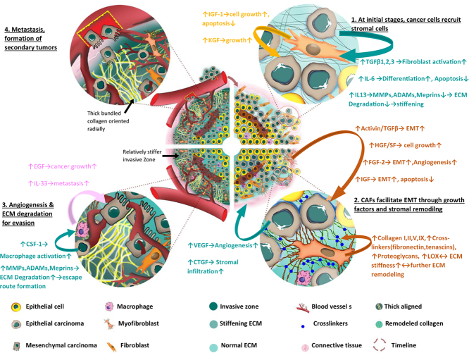
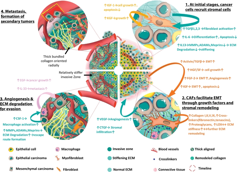
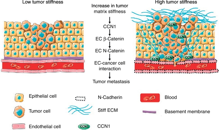
The role of tumor microenvironment in cancer progression is gaining significant attention. It is realized that cancer cells and the corresponding stroma co-evolve with time. Cancer cells recruit and transform the stromal cells, which in turn remodel the extra cellular matrix of the stroma. This complex interaction between the stroma and the cancer cells results in a dynamic feed-forward/feed-back loop with biochemical and biophysical cues that assist metastatic transition of the cancer cells. Although biochemistry has long been studied for the understanding of cancer progression, biophysical signaling is emerging as a critical paradigm determining cancer metastasis. In this mini review, we discuss the role of one of the biophysical cues, mostly the mechanical stiffness of tumor microenvironment, in cancer progression and its clinical implications.

Keywords: ADAMs, Adamalysins; ANGPT2, Angiopoietin 2; Activin/TGFβ; CAF, Cancer associated fibroblast; CSF-1, Colony stimulating factor 1; CTGF, Connective tissue growth factor; CYR61/CCN1, Cysteine-rich angiogenic inducer 61/CCN family member 1; Cancer; ECM stiffness; ECM, Extracellular matrix; EGF, Epidermal growth factor; EMT, Epithelial to mesenchymal transition; FGF, Fibroblast growth factor; Growth factors; HGF/SF, Hepatocyte growth factor/Scatter factor; IGFs, Insulin-like growth factors; IL-13, Interleukin-13; IL-33, Interleukin-33; IL-6, Interleukin-6; KGF, Keratinocyte growth factor, also FGF7; LOX, Lysyl Oxidase; MMPs, Matrix metalloproteinases; Metastasis; NO, Nitric oxide; SDF-1/CXCL12, Stromal cell-derived factor 1/C-X-C motif chemokine 12; TACs, Tumor-associated collagen signatures; TGFβ, Transforming growth factor β; TNF-α, Tumor necrosis factor-α; Tumor biophysics; VEGF, Vascular endothelial growth factor; α-SMA, α-Smooth muscle actin.


Figures

Unlabelled Image
Graphical abstract
Fig. 1
Fig. 1
Chronological development of pro-metastatic stromal architecture and the major factors involved. Segment 1 (top right): At the early stages of cancer, epithelial cancer cells secrete various growth factors that facilitates fibroblast activation, differentiation, downregulates ECM degradation by reducing MMPs and hence increase stiffness. In response, stromal fibroblasts regulate factors such as IGF, KGF etc. that promote cell growth and inhibit apoptosis. Segment 2 (bottom right): Upon activation, fibroblasts manifests myofibroblast (or, CAF) signatures and produce activin/TGFβ, IGF that stimulate EMT; HGF that increases cell growth; FGF-2 that increases angiogenesis and so on. In addition, CAFs continue to remodel and reinforce ECM by depositing collagen I, II, V, IX, increasing crosslinking, upregulating LOX and thus stromal stiffness gradually goes up. Due to excessive cellular proliferation and tumor growth, a region at the core becomes hypoxic and cancer cells increase secretion of VEGF and CTGF that are known to support angiogenesis and infiltration respectively. Segment 3 (bottom left): As carcinoma cells go through EMT, they produce CSF-1 which activates macrophages that in turn produce EGF, IL-33 etc. that promotes metastasis. At some regions of the invasive front, the stromal cells align thick collagen bundles radially that can be used as an escape route by the metastatic cells. Eventually, aggressive cancer cells degrade stiff ECM by upregulating MMPs, ADAMs etc., evade stroma, infiltrate lymph nodes and blood vessels and go through metastasis. Segment 4 (top left): Migrating cancer cells anchor at distant sites and starts the process all over to develop secondary tumors. [[51], [58], [66], [67], [68], [69], [70], [71]].
Fig. 2
Fig. 2
Epithelial cancer cells, perhaps, wait for stromal stiffness to reach an optimum level before they decide for EMT. HCT-8 cells (human colon cancer) adhere to 21 kPa polyacrylamide (PA) gel substrate, functionalized with fibronectin, and form cell islands. On substrate of appropriate rigidity (here 21 kPa), they dissociate from each other and become rounded after 7 days of culture (a) [93]. Within few more days, most of the cell islands become R cells. The R cells are more tumorigenic in mouse models, and express several oncogenes [92].
Fig. 3
Fig. 3
TGFβ from the stromal cells increases the activin ligand in epithelial cells and in serum which is required for an increase in epithelial cell migration leading to metastasis. This process can by blocked by the activin specific ligand trap, follistatin.
Fig. 4
Fig. 4
Influence of tumor stiffness in endothelial CCN1(CYR61) facilitated metastasis [127].

References

    1. American Cancer Society Cancer facts & figures 2016. Cancer Facts Fig. 2016;2016:1–9.
    1. Bissell M.J., Radisky D. Putting tumours in context. Nat Rev Cancer. 2001;1:46–54. - PMC - PubMed
    1. Bissell M.J., Radisky D.C., Rizki A., Weaver V.M., Petersen O.W. The organizing principle: microenvironmental influences in the normal and malignant breast. Differentiation. 2002;70:537–546. - PMC - PubMed
    1. Katira P., Bonnecaze R.T., Zaman M.H. Modeling the mechanics of Cancer: effect of changes in cellular and extra-cellular mechanical properties. Front Oncol. 2013;3 - PMC - PubMed
    1. Hanahan D., Coussens L.M. Accessories to the crime: functions of cells recruited to the tumor microenvironment. Cancer Cell. 2012;21:309–322. - PubMed