. 2018 Jun 25;5(8):1800006.
doi: 10.1002/advs.201800006. eCollection 2018 Aug.

Histone Deacetylase 7-Derived Peptides Play a Vital Role in Vascular Repair and Regeneration

Affiliations

Histone Deacetylase 7-Derived Peptides Play a Vital Role in Vascular Repair and Regeneration

Yiwa Pan et al. Adv Sci (Weinh). .

Abstract

Cardiovascular disease is a leading cause of morbidity and mortality globally. Accumulating evidence indicates that local resident stem/progenitor cells play an important role in vascular regeneration. Recently, it is demonstrated that a histone deacetylase 7-derived 7-amino acid peptide (7A, MHSPGAD) is critical in modulating the mobilization and orientated differentiation of these stem/progenitor cells. Here, its therapeutic efficacy in vascular repair and regeneration is evaluated. In vitro functional analyses reveal that the 7A peptide, in particular phosphorylated 7A (7Ap, MH[pSer]PGAD), could increase stem cell antigen-1 positive (Sca1+) vascular progenitor cell (VPC) migration and differentiation toward an endothelial cell lineage. Furthermore, local delivery of 7A as well as 7Ap could enhance angiogenesis and ameliorate vascular injury in ischaemic tissues; these findings are confirmed in a femoral artery injury model and a hindlimb ischaemia model, respectively. Importantly, sustained delivery of 7A, especially 7Ap, from tissue-engineered vascular grafts could attract Sca1+-VPC cells into the grafts, contributing to endothelialization and intima/media formation in the vascular graft. These results suggest that this novel type of peptides has great translational potential in vascular regenerative medicine.

Keywords: HDAC7‐derived peptide; ischaemia disease; tissue‐engineered vascular grafts (TEVGs); vascular progenitor cells (VPCs).


Figures
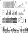
Figure 1
Figure 1
7A and 7Ap peptides increased migration and differentiation toward the EC lineage. A) 7Ap significantly increased VPC migration in a wound healing model. Wound was introduced into confluent VPCs by tip scratching, and incubated with DMEM medium containing 2% FBS and 1 ng mL−1 of 7S, 7Aa, 7A, or 7Ap peptide. Images were taken at 0, 12, 24, and 36 h postscratching (left, Scale bar: 100 µm). The migrated cells in scratched area were counted from three views per scratching, three scratchings per well, and three wells per peptide (right). Data presented are representative images or mean of three independent experiments. *: p < 0.05 (7Ap vs 7S) (n = 6, one‐way‐ANOVA followed by Tukey's post hoc analysis). B,C) 7A/7Ap increased VPC differentiation toward the EC lineage. The 3 d spontaneously differentiated VPCs were incubated with differentiation medium containing 1 ng mL−1 peptides and 10 ng mL−1 VEGF for 4 d, followed by quantitative RT‐PCR analysis with GAPDH as house‐keeping gene (B) or tube formation assay (C, Scale bar: 200 µm). 1% BSA was included as vehicle control. The data presented are representative images or mean of three independent experiments (n = 6, two‐way ANOVA followed by Dunnett's multiple comparison tests). D) MEKK1‐7A‐14‐3‐3γ mediated 7aa‐peptide‐induced VPC differentiation toward EC lineage. The VPCs were transfected with siRNA and cultured in differentiation medium for 3 d, followed by further differentiation with same protocol described above for 4 d, followed by quantitative RT‐PCR analysis of Pecam1, Cdh5, and Tagln mRNA levels with Gapdh as house‐keeping gene. 1% BSA was included as vehicle control. The data presented are representative images or mean of three independent experiments. *: p < 0.05. **: p < 0.01 (n = 6, two‐way ANOVA followed by Dunnett's multiple comparison tests).
Figure 2
Figure 2
The peptide 7Ap increased vascular injury repair and angiogenesis in ischemic tissues in vivo. A) The peptides 7A and 7Ap attenuated neointima formation in a mouse femoral artery wire‐guided injury model. The left panel shows the H&E staining images of the injured vessel sections four weeks postsurgery. Scale bar: 100 µm. The right panel shows the average intima plus media area, with the value for the PBS group set as 1.0 (n = 6, one‐way‐ANOVA followed by Tukey's post hoc analysis). B) The peptide 7Ap increased foot blood perfusion in mice with a hindlimb ischemia model, which was introduced into six‐month‐old C57bl/6 mice, with 200 µL Pluronic‐127 gel containing 1 ng mL−1 peptides applied around the injured vessels. The left panel shows the Doppler Scanner images. The right panel shows the average ratio of blood flow in the right side (injured) to the left side (uninjured) (n = 6, two‐way ANOVA followed by Dunnett's multiple comparison tests). C) The peptide 7Ap increased Sca1+ (green) cell migration into the ischemic tissue and differentiation into CD31+ cells (red). DAPI was included to counterstain the nuclei. The left panel depicts representative immunofluorescence staining images on a skeletal muscle section from the injured leg. Arrow indicates the Sca1+ cell niche. The right panel shows the mean ± SEM CD31+ or Sca1 + cells from six 20× views. Scale bar: 20 µm. *: p < 0.05. **: p < 0.01 (n = 6, two‐way ANOVA followed by Dunnett's multiple comparison tests).
Figure 3
Figure 3
The fabrication of the peptide‐loaded tissue engineered vascular grafts. A) The images show the morphology of the tubular vascular grafts, the microstructure as revealed by scanning electronic microscope, and B) (upper panel) the distribution of the two types of fibers of PCL revealed as red by DiI and collagen revealed as green by DiO. B) (lower panel) The graft parameters are summarized. C) In vitro release of 7A peptide from the TEVG (n = 3). D) Implantation of the graft in a rat to replace the abdominal artery (left), and the ultrasound scanning image of the implanted TEVGs (right). E) The patency rate of implanted TEVGs at different timepoints.
Figure 4
Figure 4
7Ap effectively increased endothelialization of the TEVGs. The TEVGs were implanted into rats to replace the abdominal artery and collected at two and four weeks postimplantation, followed by observation of the endothelialization on the lumen side of the TEVGs using A) scanning electronic microscope or B) CD144 staining of the sections. C) The endothelialized coverage was calculated by the CD144 positive cells covered area as a percentage of the entire lumen area. DAPI was used to counterstain the nuclei. Data are represented as the mean ± SEM for each group. *: p < 0.05. **: p < 0.01 (n = 6, one‐way‐ANOVA followed by Tukey's post hoc analysis).
Figure 5
Figure 5
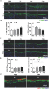
7Ap increased EC‐SMC interactions in the implanted TEVGs. Immunofluorescence staining was performed on TEVGs sections to observe the recruitment of SMCs by using A) anti‐α‐SMA and B) anti‐MHC, and quantification of the average layer thickness of B) α‐SMA and D) SM‐MHC positive cells. E) The interaction between ECs and SMCs was observed by double immunofluorescence staining with anti‐α‐SMA (green) and anti‐vWF (red). DAPI was included to counterstain the nuclei. Data are represented as the mean ± SEM for each group (n = 6 for 4 weeks, and n = 4 for 12 weeks). **: p < 0.01 (one‐way‐ANOVA followed by Tukey's post hoc analysis).
Figure 6
Figure 6
7Ap increased Sca1+‐VPC differentiation toward ECs contributing to endothelialization of TEVGs. A) Immunofluorescence staining was performed to detect the recruitment of Sca1+‐VPCs using anti‐Sca1 antibody on the implanted TEVGs sections at time indicated, and B) quantification of the infiltrated Sca1+ cells within 0–100 µm depth from the lumen by randomly selecting six views (40×) from each cross section. C) The contribution of the Sca1+ progenitor cells‐derived ECs to the endothelialization was detected by double immunofluorescence staining with anti‐Sca1 (green) and anti‐CD144 (red) antibodies on the four weeks implanted TEVGs sections. DAPI was used to counterstain the nuclei. Data are represented as the mean ± SEM for each group (n = 6 at 2 and 4 weeks, and n = 4 at 12 weeks). *: p < 0.05; **: p < 0.01 (one‐way‐ANOVA followed by Tukey's post hoc analysis).
Figure 7
Figure 7
Bi‐layered PCL TEVGs revealed that the Sca1+‐VPCs were mainly derived from the surrounding tissues. A) An illustration of the bi‐layered PCL fabricated TEVGs. B,C) The outer layer with small pores reduced Sca1+‐VPCs recruitment into the TEVGs wall and the lumen endothelialization of the TEVGs. B) The single PCL and bi‐layered PCL fabricated TEVGs were implanted into rats to replace the abdominal arteries, and harvested at two and four weeks, respectively, postimplantation, followed by immunofluorescence staining with anti‐Sca1 antibody to show the recruitment of Sca1+‐VPCs in the TEVGs wall or C) with anti‐CD31 (green, upper) and anti‐CD144 (red, lower) antibodies to show the lumen endothelialization. DAPI was used to counterstain the nuclei. Data are represented as the mean ± SEM. *: p < 0.05; **: p < 0.01; ***: p < 0.001 (n = 6, two‐way ANOVA followed by Dunnett's multiple comparison tests).
Figure 8
Figure 8
7Ap increased in vivo migration and differentiation of Sca1+‐VPCs preseeded in the TEVGs. A) Matrigel containing GFP‐Sca1+‐VPCs was applied surrounding the adventitia of the vascular grafts. B) Immunofluorescence staining was performed to detect the migration of seeded VPCs within 0–200 µm depth from the lumen (dotted area) after 3 d postimplantation, and C) the corresponding quantification on GFP‐Sca1+‐VPC. D) GFP‐Sca1+‐VPCs were seeded in the graft wall directly. E) Double immunofluorescence staining was performed to detect the differentiation of seeded VPCs after 7 d postimplantation using anti‐GFP (green) and anti‐CD31 (red) antibodies, and the corresponding quantification on GFP+CD31+VPC on the lumen side from five randomly selected views of each cross section. F) DAPI was included to counterstain the nuclei. Data are represented as mean ± SEM, *: p < 0.05 (n = 5, two‐tailed unpaired Student's t‐test).

Similar articles

Cited by

References

    1. Park K. H., Park W. J., J. Korean Med. Sci. 2015, 30, 1213. - PMC - PubMed
    1. Davignon J., Ganz P., Circulation 2004, 109, III27. - PubMed
    1. Xu Q., Zhang Z., Davison F., Hu Y., Circ. Res. 2003, 93, e76. - PubMed
    1. Libby P., Arterioscler., Thromb., Vasc. Biol. 2012, 32, 2045. - PMC - PubMed
    1. Yip H. K., Tsai T. H., Lin H. S., Chen S. F., Sun C. K., Leu S., Yuen C. M., Tan T. Y., Lan M. Y., Liou C. W., Lu C. H., Chang W. N., Crit. Care 2011, 15, R40. - PMC - PubMed

LinkOut - more resources