

Case Reports
doi: 10.1016/j.case.2018.04.002.
eCollection 2018 Aug.
Unicuspid Aortic Valve Presenting with Decompensated Critical Aortic Stenosis
Affiliations
- PMID: 30128410
- PMCID: PMC6098169
- DOI: 10.1016/j.case.2018.04.002
Item in Clipboard
Case Reports
Unicuspid Aortic Valve Presenting with Decompensated Critical Aortic Stenosis
CASE (Phila).
.
Abstract
•UAV is a rare congenital anomaly that leads to severe symptomatic stenosis.•Echocardiography plays a critical role in the evaluation of aortic stenosis.•Correctly distinguishing between UAV and BAV is relevant in determining intervention.
Keywords: Aortic stenosis; Aortic valve; Unicommisural; Unicuspid.
Figures

Electrocardiogram on admission to outside hospital showing normal sinus rhythm with J-point elevation and ST-segment changes concerning for left ventricular strain and left ventricular hypertrophy.
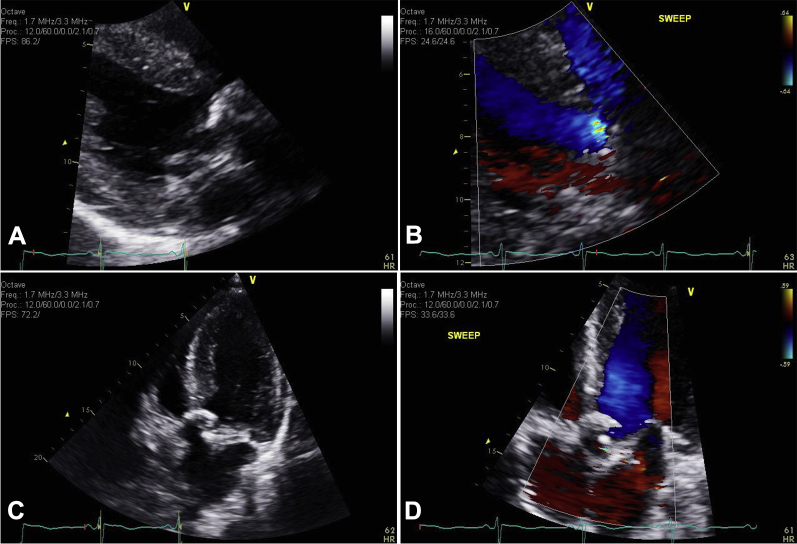
(A) Close-up image from transthoracic echocardiographic parasternal long-axis view showing heavily calcified, minimally mobile aortic valve. (B) Color flow Doppler parasternal long-axis view showing eccentric jet of aortic insufficiency (AI). (C) Transthoracic five-chamber apical view, demonstrating extension of aortic valve calcification from fused cusps into the left ventricular outflow tract. (D) Five-chamber apical view with color Doppler showing AI with main eccentric jet hugging the septum. See Videos 5-8.
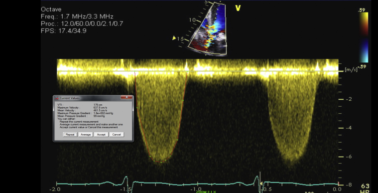
Gradient across aortic valve showing severely elevated maximal velocity of 6.4 m/sec.
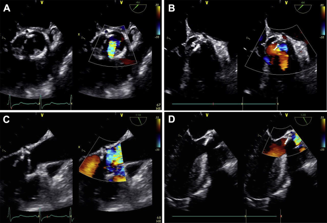
(A) Transesophageal echocardiographic (TEE) midesophageal aortic valve (AV) short-axis view showing en face view of heavily calcified, severely stenotic unicommisural UAV with and without color Doppler. (B) TEE midesophageal AV short-axis view showing a different angulation of the open commissure with a single attachment point. (C) TEE midesophageal long-axis view showing heavily calcified aortic root with a classic calcification pattern into the left ventricular outflow tract (LVOT), as well as an eccentric aortic regurgitation jet. (D) Long-axis midesophageal view taken at 120° also displaying unusual pattern of extended calcification into the LVOT. See Videos 5-8.
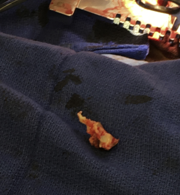
Direct visualization of explanted UAV.

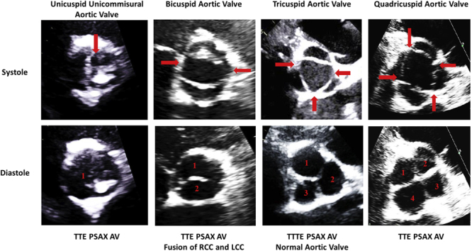
The number of points of commissural contact with the aortic root is a helpful way to identify the number of aortic valve cusps present. Red arrows indicate commissural attachment sites, with cusps numbered in the diastolic views. LCC, Left coronary cusp; RCC, right coronary cusp; TTE PSAX AV, transthoracic parasternal short-axis view focusing on the aortic valve.
References
-
- Edwards J.E. Pathologic aspects of cardiac valvular insufficiencies. AMA Arch Surg. 1958;77:634–649. - PubMed
-
- Novaro G.M., Mishra M., Griffin B.P. Incidence and echocardiographic features of congenital unicuspid aortic valve in an adult population. J Heart Valve Dis. 2003;12:674–678. - PubMed
-
- Subramanian R., Olson L.J., Edwards W.D. Surgical pathology of combined aortic stenosis and insufficiency: a study of 213 cases. Mayo Clin Proc. 1985;60:247–254. - PubMed
-
- Tretter J.T., Spicer D.E., Mori S., Chikkabyrappa S., Redington A.N., Anderson R.H. The significance of the interleaflet triangles in determining the morphology of congenitally abnormal aortic valves: implications for noninvasive imaging and surgical management. J Am Soc Echocardiogr. 2016;29:1131–1143. - PubMed
-
- Anderson R.H. Understanding the structure of the unicuspid and unicommissural aortic valve. J Heart Valve Dis. 2003;12:670–673. - PubMed
Publication types
LinkOut - more resources
Full Text Sources
Other Literature Sources
