Review
. 2018 Sep;17(9):802-815.
doi: 10.1016/S1474-4422(18)30238-2.

Selective autophagy as a potential therapeutic target for neurodegenerative disorders

Affiliations
Review

Selective autophagy as a potential therapeutic target for neurodegenerative disorders

Aurora Scrivo et al. Lancet Neurol. 2018 Sep.

Abstract

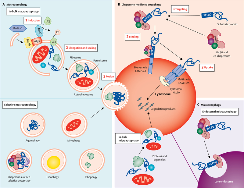
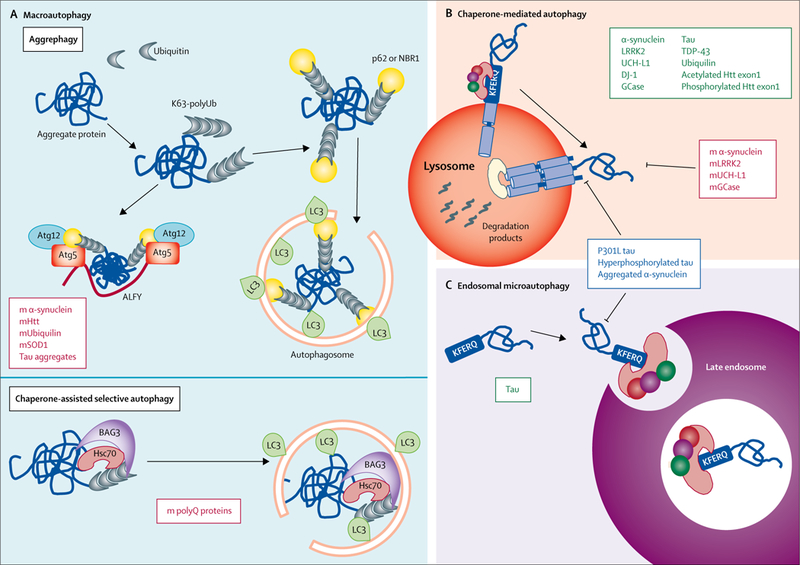
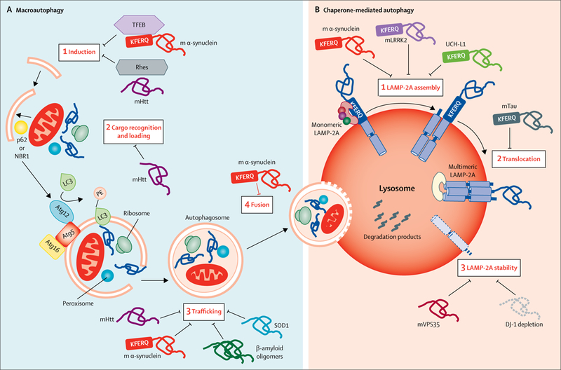
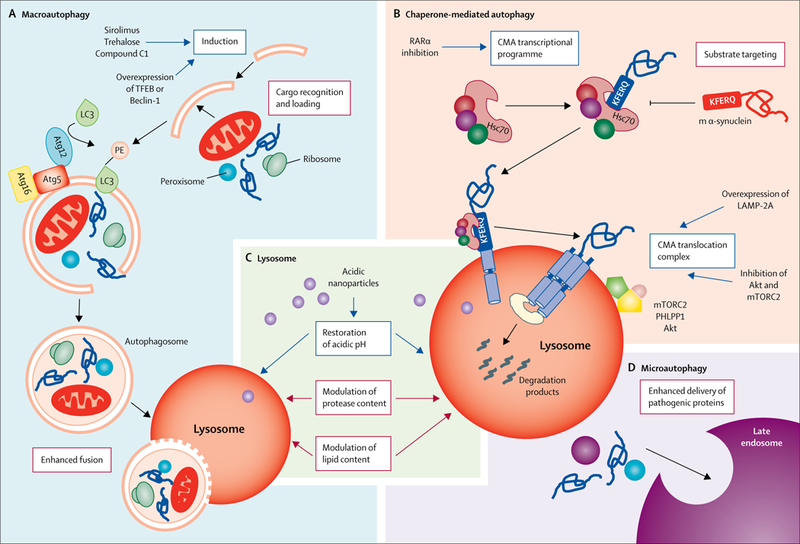
Cells rely on surveillance systems such as autophagy to handle protein alterations and organelle damage. Dysfunctional autophagy, an evolutionarily conserved cellular mechanism for degradation of intracellular components in lysosomes, frequently leads to neurodegeneration. The neuroprotective effect of autophagy stems from its ability to eliminate pathogenic forms of proteins such as α-synuclein or tau. However, the same pathogenic proteins often affect different types and steps of the autophagic process. Furthermore, genetic studies have shown that some proteins related to neurodegeneration, such as huntingtin, participate in autophagy as one of their physiological functions. This complex interplay between autophagy and neurodegeneration suggests that targeting autophagy as a whole might have limited applicability in neurodegenerative diseases, and that future efforts should focus instead on targeting specific types and steps of the autophagic process. This change of strategy in the modulation of autophagy might hold promise for future disease-modifying therapies for patients with neurodegenerative disorders.


Conflict of interest statement

Declaration of interests

None of the authors declares a competing interest.

Figures

Figure 1:
Figure 1:. Autophagic pathways in mammalian cells
The different autophagic processes that commonly coexist in most mammalian cells can be grouped into three types. (A) Macroautophagy: cytosolic cargo is first trapped inside double-membrane vesicles (autophagosomes) whose membranes form through conjugation of the autophagy-related protein LC3 with the lipid PE and other autophagy-related proteins (ie, Atg5, Atg16, and Atg12, depicted here). Formation of these double-membrane vesicles is initiated by the phosphorylation of lipids in the membrane of organelles such as the endoplasmic reticulum, mitochondria, and Golgi apparatus. This phosphorylation is triggered by a kinase complex regulated by Beclin-1. Cargo can be trapped in bulk (ie, several types of cargo in the same autophagosome) or in a selective manner (ie, only one type of cargo is trapped inside the autophagosome). Examples of selective macroautophagy include aggrephagy (degradation of protein aggregates after their recognition by autophagy cargo receptors such as p62 or neighbour of BCRA1), chaperone-assisted selective autophagy (degradation of protein aggregates targeted to autophagosomes, in this case by chaperones such as Hsc70 and Bag3; in contrast to chaperone-mediated autophagy, chaperone-assisted selective autophagy does not require binding of Hsc70 to a KFERQ-like motif and is dependent on the macroautophagy machinery), mitophagy (selective degradation of mitochondria by macroautophagy, often mediated by autophagy receptors such as PTEN-induced putative kinase 1, Parkin, NIX, and BNIP, which link the mitochondria to be degraded with the autophagy machinery), lipophagy (selective sequestration of lipid droplets by autophagosomes for their degradation in lysosomes), and ribophagy (selective sequestration of lysosomes in autophagosomes for degradation in lysosomes). Autophagosomes are targeted to lysosomes and, after fusion of both vesicles, cargo is delivered to lysosomes for complete degradation. (B) Chaperone-mediated autophagy: single cytosolic proteins bearing a KFERQ-like motif in their sequence are recognised by Hsc70 and brought to the lysosomal membrane for translocation across the LAMP-2A multimeric complex. Lysosomal Hsc70 aids translocation of the substrate protein, which is rapidly degraded once inside the lysosomal lumen. (C) Microautophagy: proteins and organelles can be degraded in bulk through invaginations at the lysosomal membrane. Cytosolic proteins are selectively targeted by Hsc70 to late endosomes, using the same KFERQ-like motif as in chaperone-mediated autophagy, resulting in their internalisation and degradation in a process known as endosomal microautophagy. Hsc70=heat shock cognate 70 kDa protein. LC3=microtubule-associated protein 1 light chain 3. Atg=autophagy-related protein. PE=phosphatidyl ethanolamine. LAMP-2A=lysosome-associated membrane protein-2A. Bag3=Bcl2-associated athanogene 3.
Figure 2:
Figure 2:. Autophagy of pathogenic proteins
(A) Examples of pathogenic proteins (red boxes) related to neurodegeneration that are cleared up by two types of selective macroautophagy: aggrephagy or chaperone-assisted selective autophagy. In aggrephagy, cytosolic aggregates of these pathogenic proteins labelled with ubiquitin are recognised by p62, which either brings LC3 directly and initiates formation of the autophagosome around the aggregate, or (in a process mediated by ALFY) assembles with Atg12 and Atg5 prior to LC3 conjugation, to initiate formation of the autophagosome. In chaperone-assisted selective autophagy, polyubiquitinated aggregate proteins are brought to the autophagosome by Hsc70 and Bag3. (B) Examples of proteins (green box) related to neurodegeneration that are degraded by chaperone-mediated autophagy, in which they are identified through their KFERQ-like motif by Hsc70, which brings them to the lysosomal surface for translocation across the lysosomal membrane. However, the pathogenic variants of these proteins (red box) are similarly targeted to lysosomes but fail to cross the lysosomal membrane, resulting in blockage of chaperone-mediated autophagy. Some other pathogenic variants of neurodegeneration-related proteins (blue box) are capable of blocking both chaperone-mediated autophagy (B) and endosomal microautophagy (C), wherein substrate proteins bearing KFERQ-like motifs are normally targeted to endosomes (green box) and internalised for degradation through invaginations in their membrane. m=mutant. K63-polyUb=polyubiquitin tagged to proteins through lysine 63. Atg=autophagy-related protein. ALFY=autophagy-linked FYVE protein. Htt=huntingtin. SOD1=superoxide dismutase 1. LC3=microtubule-associated protein 1 light chain 3. Bag3=Bcl2-associated athanogene 3. polyQ=polyglutamine. LRRK2=leucine-rich repeat kinase 2. UCH-L1=ubiquitin carboxy-terminal hydrolase L1. DJ-1=deglycase J-1. GCase=glucocerebrosidase. TDP-43=TAR DNA-binding protein 43.
Figure 3:
Figure 3:. Toxic effects of pathogenic proteins on autophagy
Several proteins related to neurodegeneration have been shown to interfere with macroautophagy (A) and chaperone-mediated autophagy (B). (A) The inhibitory effect of pathogenic proteins on macroautophagy can take place at each of the steps of this process. For instance, binding of mutant α-synuclein to TFEB has been shown to reduce autophagy induction by preventing TFEB nuclear translocation. Initiation of autophagy is also reduced by the interaction of mutant Htt with Rhes, an activator of autophagy through Beclin-1. Mutant Htt also interferes with cargo recognition by autophagy receptors such as p62. Mutant α-synuclein and mutant Htt can also interfere with the trafficking of the autophagosomes to the lysosome, and mutant α-synuclein can also prevent autophagosome fusion with lysosomes. (B) Inhibition of chaperone-mediated autophagy by pathogenic proteins occurs mainly during the steps of this process that take place at the lysosomal membrane. For example, pathogenic variants of α-synuclein, LRKK2, or UCH-L1 can inhibit assembly of monomers of LAMP-2A into the translocation complex, whereas pathogenic forms of tau block translocation of proteins through the translocation complex. Parkinson’s disease-related mutations in VPS35 or DJ-1 seem to promote accelerated degradation of LAMP-2A in lysosomes through still unknown mechanisms, leading to reduced chaperone-mediated autophagy activity. m=mutant. TFEB=transcription factor EB. Rhes=Ras homolog enriched in striatum. Htt=huntingtin. Atg=autophagy-related protein. SOD1=superoxide dismutase 1. LRRK2=leucine-rich repeat kinase 2. UCH-L1=ubiquitin carboxy-terminal hydrolase L1. LAMP-2A=lysosome-associated membrane protein-2A. VPS35=vacuolar protein sorting-associated protein 35. DJ-1=deglycase J-1. LC3=microtubule-associated protein 1 light chain 3.
Figure 4:
Figure 4:. Therapeutic modulation of autophagy in neurodegenerative diseases
Current strategies (blue boxes) and possible future strategies (red boxes) for enhancing or repairing the autophagic defects described in different neurodegenerative disorders. (A) Macroautophagy: induction of autophagosome biogenesis with chemical compounds (ie, sirolimus or trehalose) or through genetic manipulations (ie, overexpression of TFEB or Beclin-1) has successfully reinstated autophagy function, in cell cultures and in animal models of disease.,,– Interventions targeting the other steps in macroautophagy to either enhance their activity or prevent the inhibitory effect of pathogenic proteins have not been attempted yet. (B) Chaperone-mediated autophagy: chemical activation of the transcriptional programme of chaperone-mediated autophagy through inhibition of the retinoic acid receptor-α signaling has been proven effective in reducing α-synuclein toxicity in vitro. Drugs such as inhibitors of Akt, inhibitors of mTORC2, or activators of PHLPP1 (proteins that modulate LAMP-2A dynamics of assembly and disassembly at the lysosomal membrane) are also available, but are not selective to this autophagic pathway and their effect on proteotoxicity has not been tested in contexts with defective chaperone-mediated autophagy. Genetic upregulation of LAMP-2A expression to enhance chaperone-mediated autophagy has proven effective in preventing α-synuclein-mediated toxicity. Interventions at the level of substrate targeting that could prevent delivery of pathogenic proteins to lysosomes to avoid their toxic effect on chaperone-mediated autophagy have also not been attempted. (C) Lysosome: restoration of lysosomal acidification with acidic nanoparticles has been successful in restoring autophagy function in animal models., Interventions to modulate protease or lipid content have not been attempted yet. (D) Endosomal microautophagy: although no interventions to modulate this form of degradation have been attempted, the existence of this degradation pathway at the neuronal synapsis provides a good opportunity to enhance delivery of pathogenic proteins to alleviate their toxicity. m=mutant. TFEB=transcription factor EB. LC3=microtubule-associated protein 1 light chain 3. Atg=autophagy-related protein. RARα=retinoic acid receptor-α. CMA=chaperone-mediated autophagy. Hsc70=heat shock cognate 70 kDa protein. LAMP-2A=lysosome-associated membrane protein-2A. mTORC2=mTOR complex 2. PHLPP1=PH domain leucine-rich repeat-containing protein 1.

References

    1. Labbadia J, Morimoto RI. The biology of proteostasis in aging and disease. Annu Rev Biochem 2015; 84: 435–64. - PMC - PubMed
    1. Kaushik S, Cuervo AM. Proteostasis and aging. Nat Med 2015; 21: 1406–15. - PubMed
    1. Kaushik S, Cuervo AM. The coming of age of chaperone-mediated autophagy. Nat Rev Mol Cell Biol 2018; 19: 365–81. - PMC - PubMed
    1. Menzies FM, Fleming A, Caricasole A, et al. Autophagy and neurodegeneration: pathogenic mechanisms and therapeutic opportunities. Neuron 2017; 93: 1015–34. - PubMed
    1. Bento CF, Renna M, Ghislat G, et al. Mammalian autophagy: how does it work? Annu Rev Biochem 2016; 85: 685–713. - PubMed

MeSH terms