Review
. 2018 Sep;17(9):816-828.
doi: 10.1016/S1474-4422(18)30254-0.

Classification and management of adult inflammatory myopathies

Affiliations
Review

Classification and management of adult inflammatory myopathies

Albert Selva-O'Callaghan et al. Lancet Neurol. 2018 Sep.

Abstract

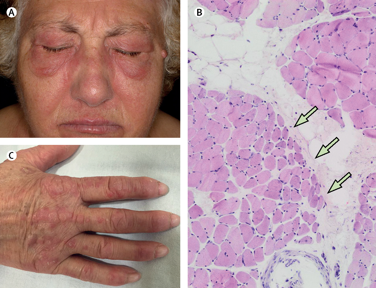
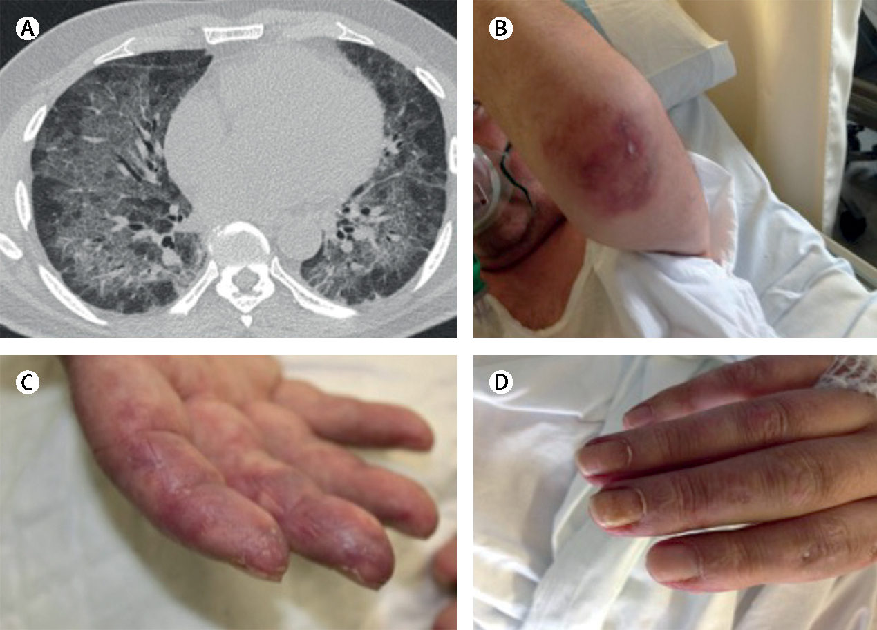
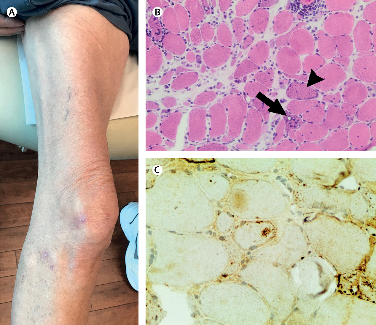
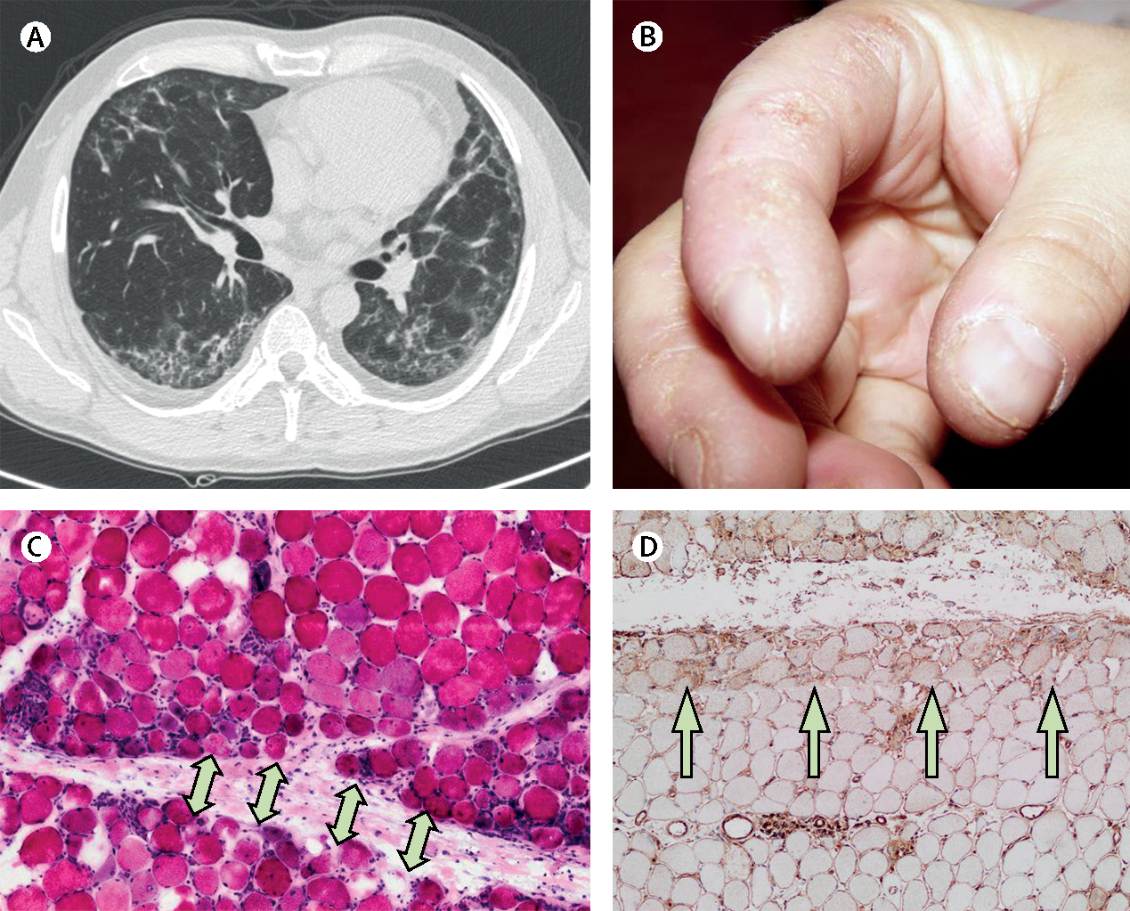
Inflammatory myopathies, collectively known as myositis, are heterogeneous disorders characterised by muscle inflammation, and frequently accompanied by extramuscular manifestations that affect the skin, lung, and joints. Patients with inflammatory myopathies were previously classified as having dermatomyositis if characteristic rashes accompanied the muscle involvement, and as having polymyositis if no rashes were present. Five main types of inflammatory myopathies are now widely recognised: dermatomyositis, immune-mediated necrotising myopathy, sporadic inclusion-body myositis, overlap myositis (including antisynthetase syndrome), and polymyositis. The discovery of autoantibodies that are specifically associated with characteristic clinical phenotypes has been instrumental to the understanding of inflammatory myopathies. Treatment is still largely based on expert opinion, but several studies have shown effectiveness of different therapies in various subsets of inflammatory myopathies. These advances will undoubtedly improve the outcomes of patients with inflammatory myopathies.


Conflict of interest statement

Declaration of interests

ALM has a patent on an anti-HMGCR assay. AS-O’C, IP-F, ET-A, JCM, and JMG-J declare no competing interests.

Figures

Figure 1:
Figure 1:. Clinical features and pathological findings of dermatomyositis
A women aged 67 years presented with muscle weakness. She had (A) a heliotrope rash and (C) Gottron’s papules, and a muscle biopsy revealed (B) perifascicular atrophy. The patient was positive for anti-Mi2 autoantibodies and was diagnosed with dermatomyositis. Arrows indicate perifascicular atrophy.
Figure 2:
Figure 2:. Clinical features and radiological findings of anti-melanoma differentiation-associated gene 5 syndrome
A man aged 52 years presented with progressive dyspnoea that had been present for 2 months. At the emergency room, he was hypoxemic with a ratio of arterial oxygen partial pressure to fractional inspired oxygen of less than 300 (normal >500). The patient was positive for high concentrations of anti-melanoma differentiation-associated gene-5 autoantibodies . (A) A high-resolution chest CT scan showed alveolar infiltrate in both lungs. Infectious and neoplastic causes were ruled out. (B) Characteristic skin lesion ulcer on elbow, (C) fingers suggesting vasculopathy , and (D) erythematous skin changes due to dermatomyositis. The patient was intubated and mechanic ventilation was started at the intensive care unit. After treatment with polymyxin-B haemoperfusion, glucocorticoids, tacrolimus, plasmapheresis, and intravenous immunoglobulin, the patient’s general condition improved to the extent that the orotracheal tube was withdrawn and he was discharged to a conventional ward.
Figure 3:
Figure 3:. Clinical features and pathological findings of sporadic inclusion-body myositis
A man aged 65 years presented with slowly progressing muscle weakness that included weakness of distal muscle groups. (A) Quadriceps were markedly atrophied and the muscle biopsy showed (B) fibres with rimmed vacuoles (black arrow), ragged-red fibres (black arrowhead), and (C) p62 positivity. Quadriceps image courtesy of Dr Tom Lloyd (Department of Neurology, Johns Hopkins University School of Medicine, Baltimore, MD, USA).
Figure 4:
Figure 4:. Clinical features and radiological and pathological findings of antisynthetase syndrome
A woman aged 45 years presented with muscle weakness and dyspnoea. (A) A high-resolution chest CT scan showed interstitial lung disease. She had crackles in both lung bases and (B) mechanic’s hands. Muscle biopsy showed (C) necrotic and regenerating muscle fibres in the perifascicular area (arrows) and (D) prominent class-1 major histocompatibility complex positivity predominantly in the perifascicular area (arrows). Serum was positive for anti-Jo1 antibodies. Jo1=histidyl tRNA synthetase.

References

    1. Senecal JL, Raynauld JP, Troyanov Y. Editorial: a new classification of adult autoimmune myositis. Arthritis Rheumatol 2017; 69: 878–84. - PubMed
    1. Lundberg IE, Tjarnlund A, Bottai M, et al. 2017 European League Against Rheumatism/American College of Rheumatology classification criteria for adult and juvenile idiopathic inflammatory myopathies and their major subgroups. Ann Rheum Dis 2017; 76: 1955–64. - PMC - PubMed
    1. Dalakas MC. Inflammatory muscle diseases. N Engl J Med 2015; 373: 393–94. - PubMed
    1. Betteridge Z, McHugh N. Myositis-specific autoantibodies: an important tool to support diagnosis of myositis. J Intern Med 2016; 280: 8–23. - PubMed
    1. Paganoni S, Amato A. Electrodiagnostic evaluation of myopathies. Phys Med Rehabil Clin N Am 2013; 24: 193–207. - PMC - PubMed