. 2019 Jan;60(1):41-48.
doi: 10.1165/rcmb.2018-0033OC.

Mitogen-activated Protein Kinase-activated Protein Kinase 2 Inhibition Attenuates Fibroblast Invasion and Severe Lung Fibrosis

Affiliations

Mitogen-activated Protein Kinase-activated Protein Kinase 2 Inhibition Attenuates Fibroblast Invasion and Severe Lung Fibrosis

Jiurong Liang et al. Am J Respir Cell Mol Biol. 2019 Jan.

Abstract

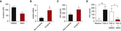
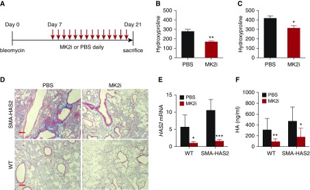
Severe pulmonary fibrosis such as idiopathic pulmonary fibrosis (IPF) is characterized by the accumulation of extracellular matrix and fibroblast activation. Targeting fibroblast activation has contributed to the development of antifibrotic therapeutics for patients with IPF. Mitogen-activated protein kinase-activated protein kinase 2 (MK2), downstream in the transforming growth factor-β/p38 mitogen-activated protein kinase pathway, has been implicated in inflammatory and fibrosing diseases. Increased concentrations of activated MK2 were expressed in IPF lung and in the mouse bleomycin model of lung fibrosis. The aim of the present study was to determine the role and the mechanisms of MK2 in fibroblast invasion and lung fibrosis. Our results showed that an MK2 inhibitor (MMI-0100) was able to inhibit the invasive capacity of lung fibroblasts isolated from patients with IPF, as well as fibroblasts isolated from both wild-type mice and mice with overexpressing hyaluronan synthase 2 (HAS2) in the myofibroblast compartment. We previously showed that hyaluronan and HAS2 regulate fibroblast invasion and lung fibrosis in vivo. The results of the present study showed that MMI-0100 reduced transforming growth factor-β-induced hyaluronan production in human and mouse fibroblasts in vitro and that HAS2 mediated MK2 activation, suggesting a feed-forward loop in fibroblast activation. More importantly, MK2 inhibition attenuated hyaluronan accumulation and reduced collagen content in bleomycin-injured mouse lungs in vivo. Conditional deletion of MK2 in fibroblasts attenuated bleomycin-induced lung fibrosis. These data provide evidence that MK2 has a role in fibroblast invasion and fibrosis and may be a novel therapeutic target in pulmonary fibrosis.

Keywords: fibroblast invasion; hyaluronan synthase 2; idiopathic pulmonary fibrosis; lung fibrosis; mitogen-activated protein kinase.


Figures

Figure 1.
Figure 1.
Mitogen-activated protein kinase–activated protein kinase 2 inhibitor (MK2i, MMI-0100) inhibition reduced hyaluronan (HA) production and invasion of idiopathic pulmonary fibrosis (IPF) fibroblasts. Fibroblasts were isolated from lung tissues of patients with IPF and healthy donors. (A) IPF fibroblasts were subjected to invasion assay with and without MMI-0100 treatment. The number of invasive cells per view was counted (n = 3 wells per treatment). *P < 0.05. (B) HAS2 (hyaluronan synthase 2) mRNA was determined with RT-PCR in invasive and noninvasive fibroblasts from 11 patients. *P < 0.05. (C) Invasive and noninvasive IPF fibroblasts were plated into multiple well plates. HA in conditioned media was measured (n = 4). *P < 0.05. (D) Equal numbers of IPF fibroblasts were plated into multiple-well plates and serum starved overnight. Cells were then treated with transforming growth factor (TGF)-β and with or without MMI-0100. HA concentrations were measured in the medium collected 24 hours after treatment (n = 3 or 4 wells per treatment). *P < 0.05; **P < 0.01.
Figure 2.
Figure 2.
Knockdown of HAS2 reduced MK2 activation of IPF fibroblasts. IPF fibroblasts were serum starved after cultured to 80% confluence overnight. The cells were then transfected with HAS2 siRNA and control (Ctl) siRNA. (A) HAS2 siRNA interference reduced HAS2 expression and (B) HA production. (C) Total cell lysates were harvested 48 hours after transfection and subjected to Western blotting for phospho-MK2 (pMK2) and total MK2. (D) Densitometric analyses of individual band intensities of expression of activated Thr334-MK2 when normalized to β-actin (n = 3). **P < 0.01. hHAS2 = human HAS2. Complete images of uncropped gels are shown in Figure E4 in the data supplement.
Figure 3.
Figure 3.
MK2 activation was correlated with HA production and fibroblast invasion. Lung tissues were harvested from mice overexpressing HAS2 in the myofibroblast compartment (SMA-HAS2–transgenic mice) and littermate controls 10 days after bleomycin treatment, and lung fibroblasts were isolated. (A) HA concentration in 24-hour culture medium of fibroblasts treated with TGF-β for 24 hours (n = 3). ***P < 0.001. (B) Invasive capacity of fibroblasts from SMA-HAS2–transgenic mice and littermate controls (n = 3 or 4). ***P < 0.001. (C) Total lung tissue proteins were subjected to Western blot analysis for p-MK2 and total MK2. (D) Densitometric analyses of individual band intensities of activated Thr334-MK2 expression when normalized to GAPDH (n = 4). **P < 0.01. Complete images of uncropped gels are shown in Figure E5 in the data supplement. (E) Total proteins of lung fibroblasts treated with and without MK2i were subjected to Western blot analysis for p-MK2 and total MK2. (F) Densitometric analyses of individual band intensities of activated Thr334-MK2 expression when normalized to GAPDH (n = 4). ***P < 0.001; ****P < 0.0001. Complete images of uncropped gels are shown in Figure E6 in the data supplement. WT = wild type.
Figure 4.
Figure 4.
MK2i reduced HA production and fibroblast invasion. Mouse lung fibroblasts were isolated from mice overexpressing HAS2 in the myofibroblast compartment (SMA-HAS2 mice) and WT mice 10 days after bleomycin treatment. Equal numbers of cells were plated into 24-well plates. The cells were starved overnight before being treated with TGF-β, and with or without MK2 inhibition with MMI-0100. HA concentrations were measured in 24-hour culture media of fibroblasts from (A) WT mice and (B) SMA-HAS2 mice (n = 4). **P < 0.01; ****P < 0.0001. (C and D) Fibroblasts from bleomycin-treated SMA-HAS2 and control mice with and without MMI-0100 treatment at either (C) 4 μM or (D) 6 μM were subjected to invasion assay. Invasive capacity (invaded cells per view) of primary lung fibroblasts from (C) control mice and (D) SMA-HAS2 mice was compared (n = 3). *P < 0.05; **P < 0.01.
Figure 5.
Figure 5.
MK2i reduced HA accumulation in bleomycin-injured mouse lung in vivo. WT C57BL/6J mice were subjected to bleomycin-induced lung injury. From Day 3, mice started to receive intraperitoneal injections of MMI-0100 37.5 μg/kg or PBS daily. (A) Mice were killed on Day 10, and their lung tissues were harvested. (B) HA contents in lung tissues were compared between the MMI-0100 treatment group and the PBS control group (n = 4). **P < 0.01.
Figure 6.
Figure 6.
MK2i attenuated mouse lung fibrosis in vivo. Mice overexpressing HAS2 in the myofibroblast compartment (SMA-HAS2–transgenic mice) and littermates were subjected to bleomycin-induced lung injury. From Day 7, the mice started to receive MMI-0100 at 37.5 μg/kg or PBS daily. The mice were killed on Day 21, and BAL fluid and lung tissues were harvested (A). Hydroxyproline contents in lung tissues were compared between the MMI-0100 treatment group and the PBS control group in (B) WT mice and (C) SMA-HAS2–transgenic mice, respectively (n = 7 or 8). *P < 0.05. (D) Trichrome staining of lung slides from bleomycin Day 21 WT and SMA-HAS2–transgenic mice with and without MMI-0100 treatment (scale bars: 100 μm). (E) Has2 mRNA expression (n = 4) and (F) hyaluronan concentrations in BAL fluid in four groups of mice were compared (n = 5–9). *P < 0.05; **P < 0.01; ***P < 0.001.
Figure 7.
Figure 7.
Deletion of MK2 in collagen, type I, α2 chain (Col1a2)-expressing fibroblasts attenuated lung fibrosis. Mouse lung fibroblasts were isolated from Col1a2-CreER;Rosa-Tomato;MK2flox/flox mice (MK2△Col) and littermate Col1a2-CreER;Rosa-Tomato mice 10 days after bleomycin treatment. (A) Fibroblast total cell protein lysis was subjected to Western blot analysis for p-MK2 and total MK2. (B) Densitometric analyses of individual band intensities of activated Thr334-MK2 expression when normalized to GAPDH knockdown (n = 4). ***P < 0.001. (C) Murine Has2 (mHas2) gene expression in fibroblasts with and without MK2 knockdown (n = 4). **P < 0.01. (D) Invasive cells per view of fibroblasts isolated from MK△Col mice and littermate control mice (n = 3). *P < 0.05. (EG) Col1a2-CreER;Rosa-Tomato;MK2flox/flox mice (MK2△Col) and littermate MK2flox/flox mice were subjected to bleomycin for 21 days. (E) Hydroxyproline contents (n = 7–10; *P < 0.05) and (F) trichrome staining (scale bars: 100 μm), as well as (G) HA concentrations in BAL fluid (n = 4–6; *P < 0.05) were determined. Complete images of uncropped gels in A are shown in Figure E7 in the data supplement.

References

    1. American Thoracic Society; European Respiratory Society. Idiopathic pulmonary fibrosis: diagnosis and treatment. International consensus statement. Am J Respir Crit Care Med. 2000;161:646–664. - PubMed
    1. Martinez FJ, Collard HR, Pardo A, Raghu G, Richeldi L, Selman M, et al. Idiopathic pulmonary fibrosis. Nat Rev Dis Primers. 2017;3:17074. - PubMed
    1. Olson AL, Swigris JJ, Lezotte DC, Norris JM, Wilson CG, Brown KK. Mortality from pulmonary fibrosis increased in the United States from 1992 to 2003. Am J Respir Crit Care Med. 2007;176:277–284. - PubMed
    1. Lederer DJ, Martinez FJ. Idiopathic pulmonary fibrosis. N Engl J Med. 2018;378:1811–1823. - PubMed
    1. Noble PW, Barkauskas CE, Jiang D. Pulmonary fibrosis: patterns and perpetrators. J Clin Invest. 2012;122:2756–2762. - PMC - PubMed

Publication types

MeSH terms