. 2018 Aug 21:38:197-217.
doi: 10.1146/annurev-nutr-071816-064642.

Mechanisms Underlying Muscle Protein Imbalance Induced by Alcohol

Affiliations

Mechanisms Underlying Muscle Protein Imbalance Induced by Alcohol

Scot R Kimball et al. Annu Rev Nutr. .

Abstract

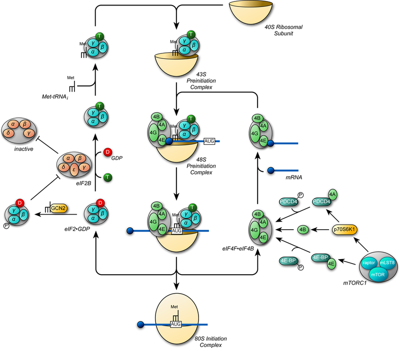
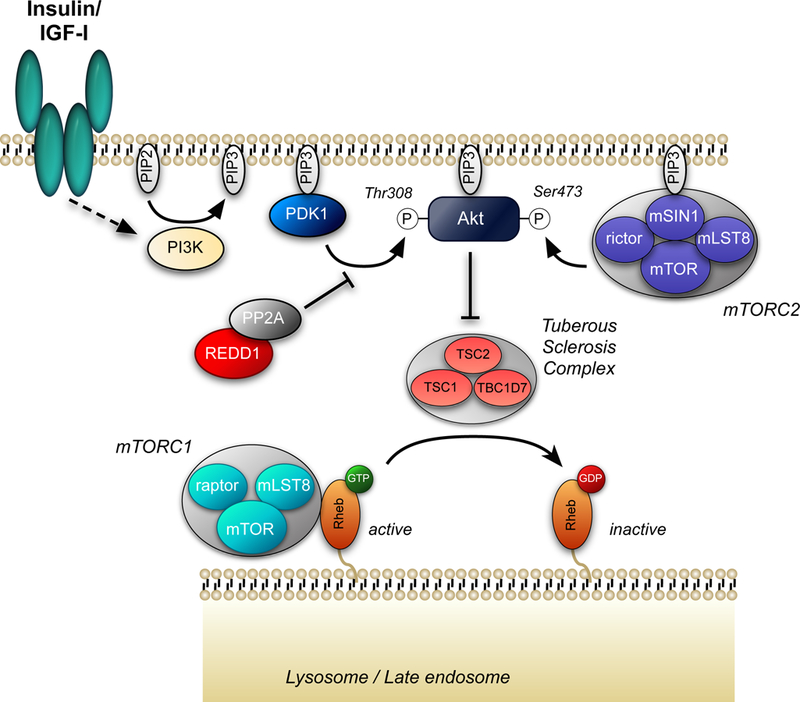
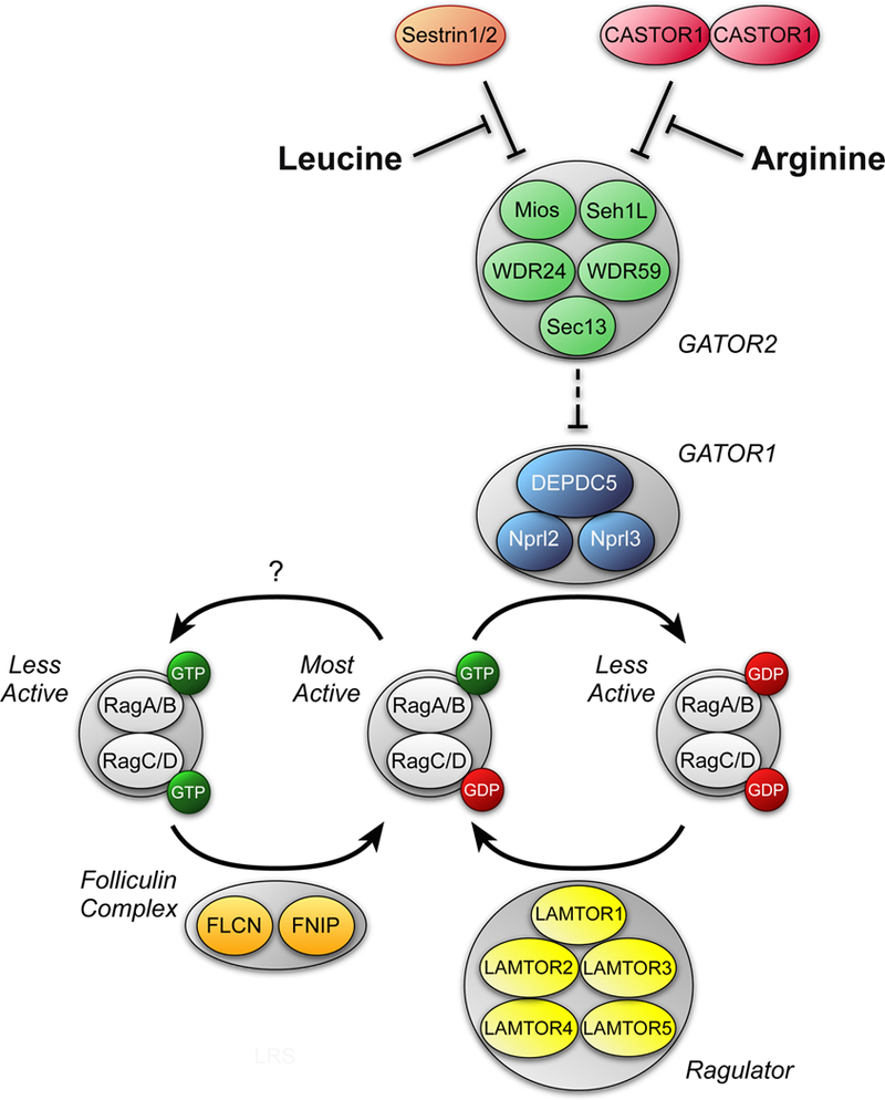
Both acute intoxication and longer-term cumulative ingestion of alcohol negatively impact the metabolic phenotype of both skeletal and cardiac muscle, independent of overt protein calorie malnutrition, resulting in loss of skeletal muscle strength and cardiac contractility. In large part, these alcohol-induced changes are mediated by a decrease in protein synthesis that in turn is governed by impaired activity of a protein kinase, the mechanistic target of rapamycin (mTOR). Herein, we summarize recent advances in understanding mTOR signal transduction, similarities and differences between the effects of alcohol on this central metabolic controller in skeletal muscle and in the heart, and the effects of acute versus chronic alcohol intake. While alcohol-induced alterations in global proteolysis via activation of the ubiquitin-proteasome pathway are equivocal, emerging data suggest alcohol increases autophagy in muscle. Further studies are necessary to define the relative contributions of these bidirectional changes in protein synthesis and autophagy in the etiology of alcoholic myopathy in skeletal muscle and the heart.

Keywords: alcoholic myopathy; amino acids; autophagy; mTOR; protein synthesis; translational control; ubiquitin-proteasome pathway.


Figures

Figure 1.
Figure 1.
Key steps in the initiation phase of mRNA translation. The initiation step in translation involves the binding of the ternary complex, consisting of eukaryotic initiation factor 2 (eIF2) associated with GTP and the initiator form of methionyl-tRNA (met-tRNAi), to the 40S ribosomal subunit to form the 43S preinitiation complex. The eIF4F•eIF4B complex in association with mRNA subsequently binds to form the 48S preinitiation complex. The 40S ribosomal subunit then scans along the 5’-untranslated region of the mRNA and stops at an AUG start codon, triggering hydrolysis of GTP bound to eIF2 to GDP leading to release of the eIF2•GDP and eIF4F•eIF4B complexes. The GDP bound to eIF2 is exchanged for GTP by eIF2B and the eIF4F•eIF4B complex binds to another mRNA to re-start the process. GCN2 phosphorylates eIF2 on its α-subunit, converting it from a substrate of eIF2B into a competitive inhibitor, leading to decreased ternary complex formation. mTORC1 promotes assembly of the eIF4F complex through phosphorylation of the eIF4E binding proteins (4E-BPs) and programmed cell death 4 (PDCD4). Phosphorylation of 4E-BP by mTORC1 releases it from the eIF4E•4E-BP complex, allowing eIF4E to bind to eIF4G. Similarly, phosphorylation of PDCD4 by p70S6K1 (which is also activated by mTORC1) frees eIF4A from the eIF4A•PDCD4 complex, allowing it to bind to eIF4G. In addition, p70S6K1 phosphorylates eIF4B, thereby enhancing its stimulatory activity toward eIF4A.
Figure 2.
Figure 2.
Insulin/IGF-I signaling to mTORC1. Insulin and IGF-I both activate phosphatidylinositol 3-kinase (PI3K) leading to production of phosphatidylinositol-3,4,5-trisphosphate (PIP3) on the cytoplasmic face of the plasma membrane. PDK1, Akt, and mSIN1 have pleckstrin homology domains that interact with PIP3 resulting in their localization at the plasma membrane (67, 80). In addition, binding of mSIN1 to PIP3 results in activation of mTORC2 (67). Phosphorylation of Akt on Thr308 is sufficient for phosphorylation, and thus inactivation of TSC2 (23, 37). Phosphorylation of TSC2 by Akt leads to dissociation of the TSC complex from the late endosomal/lysosomal membrane, allowing Rheb to accumulate in the active GTP-bound form (6). Whether or not phosphorylation of TSC2 by either ERK- or p90RSK promotes TSC dissociation from the LEL membrane is unknown.
Figure 3.
Figure 3.
Regulation of mTORC1 by amino acids. Leucine and arginine activate mTORC1 in part by modulating the GTP loading status of Rags A and B. Leucine binds to Sestrins 1 and 2 and promotes their dissociation from GATOR2 (120), which consists of the proteins meiosis regulator for oocyte development (Mios), WD repeat-domain-containing proteins 24 and 59 (WDR24 and WDR59, respectively, Sec13-like protein (Sec13), and Sec13. Similarly, arginine binds to a protein referred to as cellular arginine sensor for mTORC1 (CASTOR) 1 and promotes its dissociation from GATOR2 (8). Binding of either Sestrin1/2 or CASTOR1 to GATOR2 represses the inhibitory effect of GATOR1 (consisting of DEP domain containing 5 (DEPDC5) and nitrogen permease regulator-like 2 and 3 (Nprl2 and Nprl3)) GAP activity toward Rags A and B. Ragulator acts to oppose the GAP activity of GATOR1 (comprised of LEL adaptor and MAPK and mTORC1 activators (LAMTOR) 1–5), and instead acts to promote exchange of GDP for GTP on Rags A and B. However, the mechanism through which amino acids act to regulate Ragulator GEF activity is unknown. The folliculin complex, composed of folliculin (FLCN) and the folliculin-interacting protein (FNIP), has have been reported to act as GAPs for Rags C and/or D. Neither the mechanism through which folliculin is regulated by amino acids nor its amino acid selectivity are known. Note that the RagA/B-GDP•RagC/D-GTP complex is not shown for simplicity, but would be expected to be less active than the RagA/G-GTP•RagC/D-GDP complex in activating mTORC1.

References

    1. Atherton PJ, Greenhaff PL, Phillips SM, Bodine SC, Adams CM, Lang CH. 2016. Control of skeletal muscle atrophy in response to disuse: clinical/preclinical contentions and fallacies of evidence. Am J Physiol Endocrinol Metab 311: E594–604 - PMC - PubMed
    1. Bento CF, Renna M, Ghislat G, Puri C, Ashkenazi A, et al. 2016. Mammalian autophagy: How does It work? Annu Rev Biochem 85: 685–713 - PubMed
    1. Bernal CA, Vazquez JA, Adibi SA. 1993. Leucine metabolism during chronic ethanol consumption. Metabolism: clinical and experimental 42: 1084–6 - PubMed
    1. Broer S, Broer A. 2017. Amino acid homeostasis and signalling in mammalian cells and organisms. Biochem J 474: 1935–63 - PMC - PubMed
    1. Buerger C, DeVries B, Stambolic V. 2006. Localization of Rheb to the endomembrane is critical for its signaling function. Biochemical and Biophysical Research Communications 344: 869–80 - PubMed

Publication types