Review
. 2018 Aug 21:38:97-125.
doi: 10.1146/annurev-nutr-082117-051732.

Iron and Cancer

Affiliations
Review

Iron and Cancer

Suzy V Torti et al. Annu Rev Nutr. .

Abstract

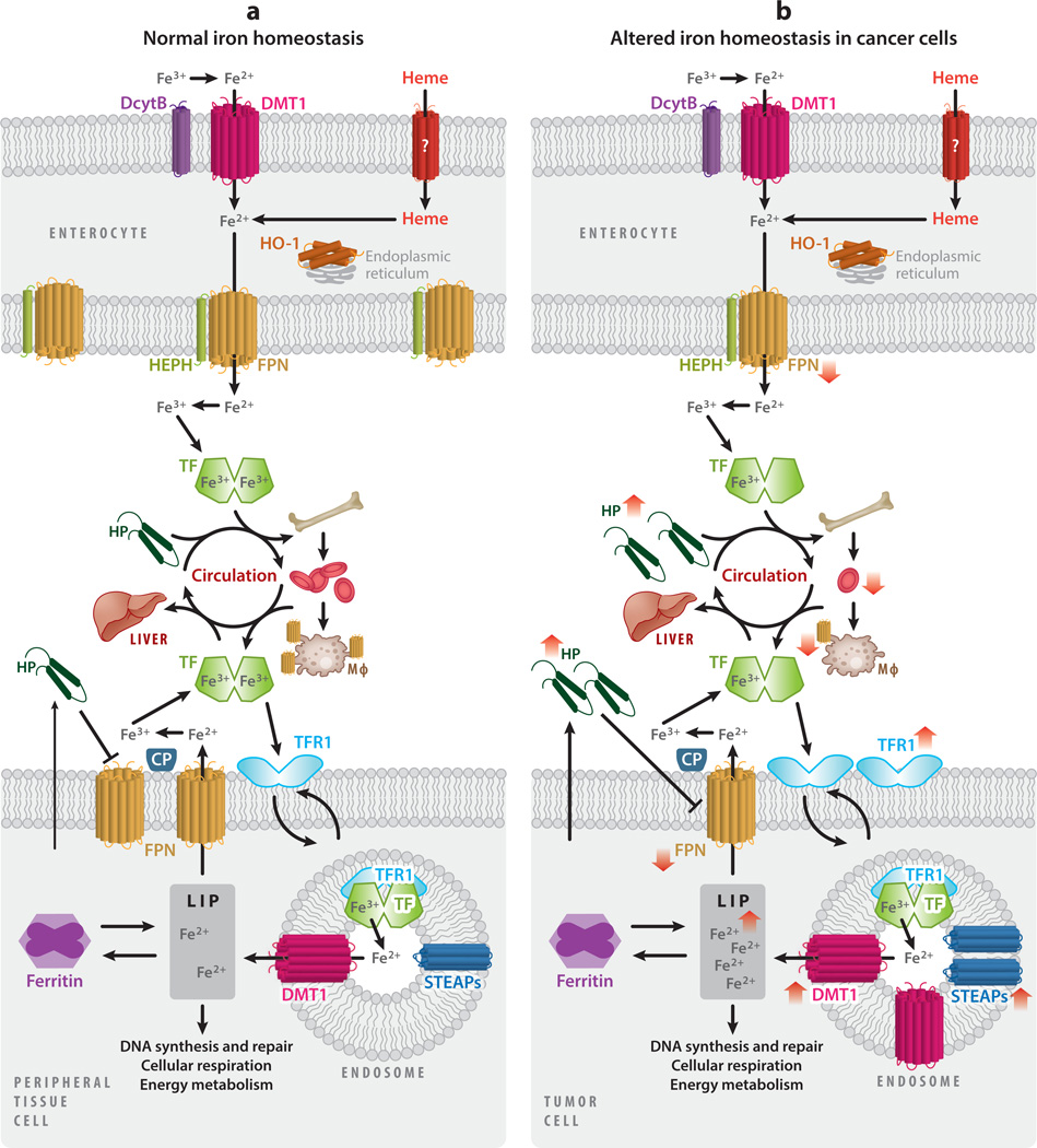
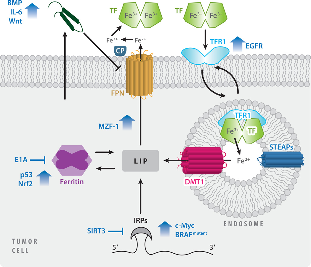
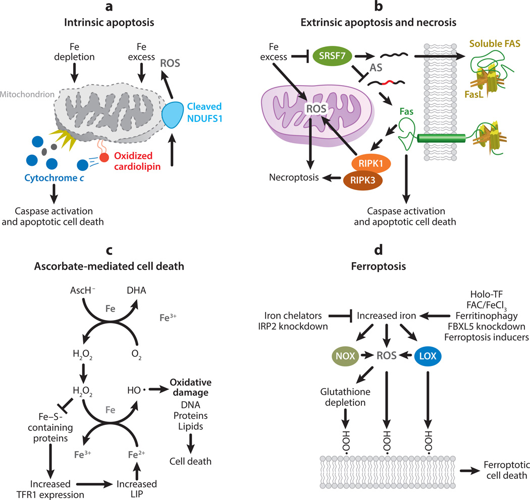
This review explores the multifaceted role that iron has in cancer biology. Epidemiological studies have demonstrated an association between excess iron and increased cancer incidence and risk, while experimental studies have implicated iron in cancer initiation, tumor growth, and metastasis. The roles of iron in proliferation, metabolism, and metastasis underpin the association of iron with tumor growth and progression. Cancer cells exhibit an iron-seeking phenotype achieved through dysregulation of iron metabolic proteins. These changes are mediated, at least in part, by oncogenes and tumor suppressors. The dependence of cancer cells on iron has implications in a number of cell death pathways, including ferroptosis, an iron-dependent form of cell death. Uniquely, both iron excess and iron depletion can be utilized in anticancer therapies. Investigating the efficacy of these therapeutic approaches is an area of active research that promises substantial clinical impact.

Keywords: cancer; carcinogenesis; cell death; iron.


Figures

Figure 1
Figure 1
Iron homeostasis and cancer. (a) Dietary iron is absorbed as inorganic iron or heme by enterocytes of the intestine. Dietary ferric iron (Fe3+) is reduced to ferrous iron (Fe2+) by duodenal cytochrome B (DcytB) and imported into the enterocyte by divalent metal transporter 1 (DMT1). Heme is absorbed by an unidentified heme transporter and subsequently degraded by heme oxygenase 1 (HO-1), present on the endoplasmic reticulum, liberating iron. Ferrous iron is transported out of the enterocyte by ferroportin (FPN), where it is oxidized by hephaestin (HEPH) and loaded onto transferrin (TF) for systemic circulation. A large percentage of iron is delivered to the bone marrow for the production of hemoglobin and red blood cells. Iron is recycled from senescent red blood cells through endocytosis by macrophages, reentering circulation via FPN. Alternatively, iron can be transported to the liver for storage. The liver acts as an iron-sensing organ and regulates the absorption, recycling, and tissue levels of iron by secreting hepcidin (HP), a negative regulator of FPN. Iron is imported into the cells of peripheral tissues by binding of TF to its receptor, transferrin receptor 1 (TFR1). The complex is endocytosed, and within the endosome ferric iron is released from TF, reduced by six-transmembrane epithelial antigen of prostate (STEAP) proteins, and exported to the cytosol by DMT1. Newly imported iron enters the metabolically active labile iron pool (LIP), where it can be utilized in processes including DNA synthesis and repair, cellular respiration, and energy metabolism. Iron can also be stored in ferritin or exported back into circulation by FPN, oxidized by ceruloplasmin (CP), and loaded onto TF for further rounds of iron delivery. Peripheral tissue cells also secrete HP, which, unlike HP secreted by the liver, is thought to primarily act locally. (b) As a consequence of inflammation, HP secretion by the liver is increased, thereby reducing FPN expression throughout the body. Cancer-induced HP secretion impairs iron absorption and iron recycling, restricting circulating iron levels and leading to reduced erythropoiesis and anemia. Cancer cells generally exhibit an iron-seeking phenotype, with increased levels of TFR1, STEAPs, DMT1, and HP, and decreased FPN as compared with normal cells of the same tissue. The change in direction of iron metabolic proteins in cancer is shown by red arrows.
Figure 2
Figure 2
Regulation of cellular iron metabolism in cancer. Dysregulated iron metabolism in cancer cells is driven by oncogenes and tumor suppressors. The localization of TFR1 to the cell surface, and therefore its ability to bind TF and import iron, is promoted by EGF. Iron storage is altered by the suppression of ferritin by E1A and its induction by p53 and nuclear factor Nrf2. Iron export through FPN is promoted by MZF-1, but inhibited by increased hepcidin expression, driven by BMPs, IL-6, and Wnt signaling. Some factors (c-Myc, mutated BRAF, and SIRT3) dysregulate multiple phases of cellular iron metabolism (import, storage, and export) by targeting IRPs. Abbreviations: BMPs, bone morphogenetic proteins; CP, ceruloplasmin; EGF, epidermal growth factor; EGFR, epidermal growth factor receptor; FPN, ferroportin; IL-6, interleukin-6; IRPs, iron regulatory proteins; LIP, labile iron pool; MZF-1, myeloid zinc finger 1; Nrf2, erythroid 2-related factor 2; STEAP, six-transmembrane epithelial antigen of prostate; TF, transferrin; TFR1, transferrin receptor 1.
Figure 3
Figure 3
Role of iron in cell death. (a) Iron depletion disrupts cellular processes, while iron excess causes cellular damage through the generation of reactive oxygen species (ROS). If excessive, these processes initiate the intrinsic apoptosis pathway and trigger permeabilization of the outer mitochondrial membrane and the release of cytochrome c. Through the generation of ROS, excess iron may promote the release of cytochrome c through the oxidation of cardiolipin. Cytochrome c release leads to the activation of caspases and apoptotic cell death. Caspase activation further contributes to mitochondrial ROS generation through cleavage of the iron–sulfur cluster-containing protein NADH:ubiquinone oxidoreductase core subunit S1 (NDUFS1). (b) Excess iron can promote extrinsic apoptosis or necrosis, or both, through regulating the death receptor FAS. Alternative splicing (AS) results in a soluble, decoy FAS and a membrane-bound Fas capable of initiating cell death. Excess iron promotes the production of membrane-bound FAS by interfering with messenger RNA binding of serine- and arginine-rich splicing factor 7 (SRSF7). The binding of FAS ligand (FasL) to membrane-bound FAS can initiate either caspase activation and apoptosis or necroptosis in a receptor-interacting protein kinase-1 and -3 (RIPK1 and RIPK3)-dependent manner. RIPK1 and RIPK3 induce necroptosis, in part, by initiating the generation of ROS in the mitochondria. (c) Ascorbate (AscH) is oxidized to dehydroascorbic acid (DHA) in the presence of iron (Fe) and oxygen (O2) to generate hydrogen peroxide (H2O2). Through Fenton chemistry, H2O2 generates hydroxyl radicals (HO·), which can cause oxidative damage to cellular components (including DNA, protein, and lipids) and induce cell death. H2O2 can also inhibit the activity of iron–sulfur (Fe–S)-containing proteins, including cytosolic aconitase, increasing transferrin receptor 1 (TFR1) expression and the labile iron pool (LIP). As the LIP is predominately ferrous iron (Fe2+), increased LIP further promotes HO· production via Fenton chemistry. (d) Ferroptosis is a form of cell death caused by the iron-mediated generation of lipid peroxides. The reduction of intracellular iron, through iron chelation and knockdown of iron-responsive element-binding protein 2 (IRP2), protects against ferroptosis. Conversely, several methods to increase intracellular iron promote ferroptotic cell death: iron treatment with holo-transferrin (holo-TF), ferric ammonium citrate (FAC), and iron chloride hexahydrate (FeCl3); ferritinophagy; and knockdown of FBXL5, an inhibitor of IRP2. Furthermore, ferroptosis inducers (such as erastin and RSL3) have been reported to directly increase LIP. Increased iron is thought to promote ferroptosis through an increase in ROS, particularly lipid ROS, and a reduction in glutathione, an antioxidant important in reducing lipid peroxides. Fenton chemistry, nicotinamide adenine dinucleotide phosphate (NADPH) oxidases (NOX), and lipoxygenases (LOX) contribute to ROS generation, lipid peroxidation, and ferroptotic cell death.

References

    1. Abbaspour N, Hurrell R, Kelishadi R. 2014. Review on iron and its importance for human health. J. Res. Med. Sci. 19:164–74 - PMC - PubMed
    1. Abeysinghe RD, Greene BT, Haynes R, Willingham MC, Turner J, et al. 2001. p53-independent apoptosis mediated by tachpyridine, an anti-cancer iron chelator. Carcinogenesis 22:1607–14 - PubMed
    1. Adams PC. 2015. Epidemiology and diagnostic testing for hemochromatosis and iron overload. Int. J. Lab. Hematol. 37(Suppl. 1):25–30 - PubMed
    1. Aisen P, Enns C, Wessling-Resnick M. 2001. Chemistry and biology of eukaryotic iron metabolism. Int. J. Biochem. Cell Biol. 33:940–59 - PubMed
    1. Akatsuka S, Yamashita Y, Ohara H, Liu YT, Izumiya M, et al. 2012. Fenton reaction induced cancer in wild type rats recapitulates genomic alterations observed in human cancer. PLOS ONE 7:e43403 - PMC - PubMed

Publication types

LinkOut - more resources