. 2018 Aug 21;17(1):125.
doi: 10.1186/s12943-018-0871-4.

The chromosome 11q13.3 amplification associated lymph node metastasis is driven by miR-548k through modulating tumor microenvironment

Affiliations

The chromosome 11q13.3 amplification associated lymph node metastasis is driven by miR-548k through modulating tumor microenvironment

Weimin Zhang et al. Mol Cancer. .

Abstract

Background: The prognosis for esophageal squamous cell carcinoma (ESCC) patients with lymph node metastasis (LNM) is still dismal. Elucidation of the LNM associated genomic alteration and underlying molecular mechanisms may provide clinical therapeutic strategies for ESCC treatment.

Methods: Joint analysis of ESCC sequencing data were conducted to comprehensively survey SCNAs and identify driver genes which significantly associated with LNM. The roles of miR-548k in lymphangiogensis and lymphatic metastasis were validated both in vitro and in vivo. ESCC tissue and blood samples were analyzed for association between miR-548k expression and patient clinicopathological features and prognosis and diagnosis.

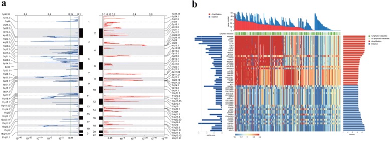
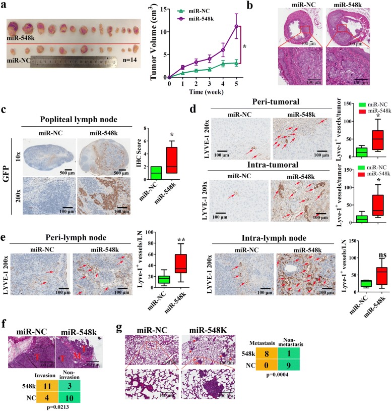
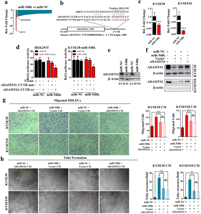
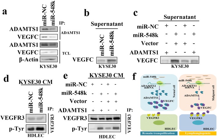
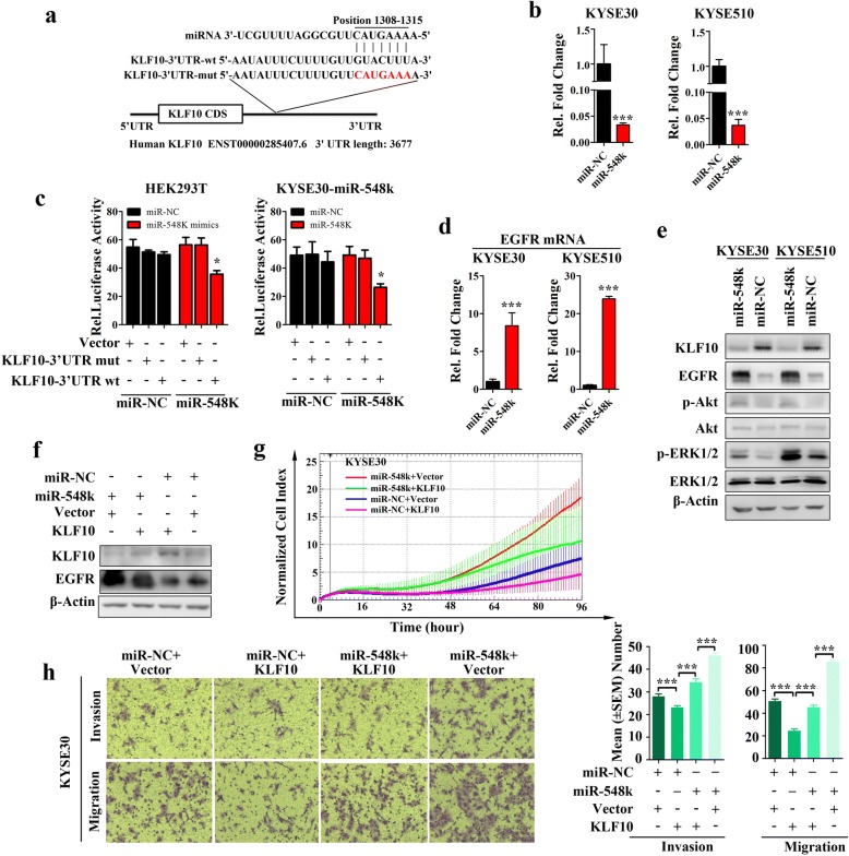
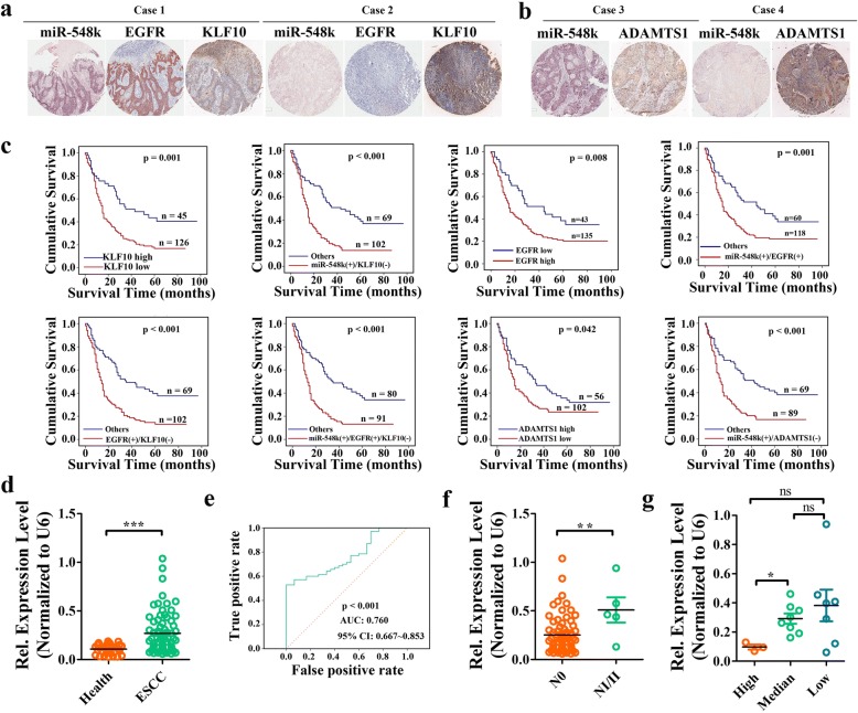
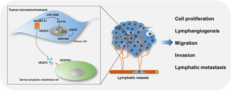
Results: In the pooled cohort of 314 ESCC patients, we found 76 significant focused regions including 43 amplifications and 33 deletions. Clinical implication analysis revealed a panel of genes associated with LNM with the most frequently amplified gene being MIR548K harbored in the 11q13.3 amplicon. Overexpression of miR-548k remarkably promotes lymphangiogenesis and lymphatic metastasis in vitro and in vivo. Furthermore, we demonstrated that miR-548k modulating the tumor microenvironment by promoting VEGFC secretion and stimulating lymphangiogenesis through ADAMTS1/VEGFC/VEGFR3 pathways, while promoting metastasis by regulating KLF10/EGFR axis. Importantly, we found that serum miR-548k and VEGFC of early stage ESCC patients were significantly higher than that in healthy donators, suggesting a promising application of miR-548k and VEGFC as biomarkers in early diagnosis of ESCC.

Conclusions: Our study comprehensively characterized SCNAs in ESCC and highlighted the crucial role of miR-548k in promoting lymphatic metastasis, which might be employed as a new diagnostic and prognostic marker for ESCC.

Keywords: Esophageal squamous cell carcinoma; Lymphangiogenesis. Tumor microenvironment miR-548k; Lymphatic metastasis.


Conflict of interest statement

Ethics approval and consent to participate

Tissue microarrays (TMA) of ESCC specimens were obtained from Shanghai Outdo Biotech Co., Ltd. (SOBC), with the approval of the Institutional Review Board. The ESCC tissues and matched adjacent normal tissues and the serum samples of ESCC patients and health persons used for real time PCR assay were histopathologically and clinically diagnosed at Beijing Cancer Hospital and the Cancer Institute and Hospital, Chinese Academic of Medical Sciences & Peking Union Medical College (Additional file 1: Table S17). Written informed consent was obtained from all patients prior to the study. The use of the clinical specimens for research purposes was approved by the Institutional Research Ethics Committee.

All animal care and procedures were in accordance with national and institutional policies for animal health and well-being. Mouse experimentations were approved by Cancer Institute and Hospital, Chinese Academic of Medical Sciences & Peking Union Medical College Animal Care and Use Committee. All mouse surgery was performed under anesthesia, and all efforts were made to minimize suffering of animals.

Consent for publication

Not applicable

Competing interests

The authors declare that they have no competing interests.

Publisher’s Note

Springer Nature remains neutral with regard to jurisdictional claims in published maps and institutional affiliations.

Figures

Fig. 1
Fig. 1
Somatic copy number alterations analysis in pooled ESCC cohort. a, Significance of SCNAs recurrences across chromosomes. b, The frequency and lymph node metastasis implication of SCNAs harbored genes. Genes of alteration frequency more than 30% were shown here. The upper panel shown the number of alteration genes in each sample. The middle panel shown the lymph node metastasis (LNM) status of each sample. The bottom heatmap shown the copy number of the indicated genes in each sample. The left panel shown the log10 (p value) of LNM association of each gene. The right panel shown the alteration frequency of each gene
Fig. 2
Fig. 2
Clinical implication of miR-548k. a, Kaplan-Meier survival analysis of pooled ESCC cohort stratified by miR-548k amplification (n = 314; p = 0.038, log-rank test). b, The association of miR-548k RNA expression versus DNA copy number in TCGA ESCC cohort (p < 0.001, r = 0.51, Spearman’s Rank Correlation Coefficient analysis). c, Relative expression levels of miR-548k in 23 pairs of human ESCC tissues and matched adjacent normal tissues. d, Left, representative miRNA ISH photos of miR-548k expression in ESCC tissues and matched adjacent normal tissues. Scale bar: 100 μm. Right, MiRNA in situ hybridization (ISH) score in ESCC tissues and matched adjacent normal tissues (n = 185). e, Kaplan-Meier survival analysis of all ESCC patients stratified by miR-548k expression level (n = 178; p = 0.002, log-rank test). f, Multivariate analysis of the hazard ratios (HR) showed that the upregulation of miR-548k may be an independent prognostic factor for the overall survival rate (by the Cox multivariate proportional hazard regression model). The HR is presented as the means (95% confidence interval, 95% CI)
Fig. 3
Fig. 3
In vivo mouse model studies reveal miR-548k promoted xenograft tumor formation, lymphangiogenesis, lymphatic metastasis and distant metastases. a, Stable overexpression of miR-548k in KYSE30 cells enhanced subcutaneous xenograft tumors formation in BALB/c nude mice (n = 14). Left, representative picture. Right, growth curves of xenograft tumors. b, Stable overexpression of miR-548k in KYSE30 cells promoted subserosa tumor growth in esophageal abdominal portion (n = 6 in miR-548k overexpression group and n = 4 in control group). Representative HE photos of esophageal subserosa tumors. Scale bar: 500 μm or 100 μm. c, Representative images and quantitative analysis of the popliteal lymph nodes immunostained with anti-GFP antibody (*p < 0.05, t-test). Scale bar: 500 μm or 100 μm. d, Representative images of peri-tumoral and intra-tumoral sections immunostained with anti-LYVE-l antibody (left), and quantification (right), indicates the microlymphatic vessel density (*p < 0.05, t-test). Scale bar: 100 μm. e, Representative images of peri-lymph node and intra-lymph node sections immunostained with anti-LYVE-l antibody (left), and quantification (right), indicates the microlymphatic vessel density (**p < 0.01, ns, no significance, t-test). Scale bar: 100 μm. f, MiR-548k promoted local invasion in subcutaneous xenograft tumors formation model (p = 0.0213, Fisher’s Exact-test). Representative pictures (up) and quantitative data (bottom), T, tumor; M, muscle. Scale bar: 500 μm. g, Stable overexpression of miR-548k in KYSE30 cells promoted the lung metastasis (p = 0.0004, Fisher’s Exact-test). Representative pictures (up) and quantitative data (bottom). Scale bar: 500 μm or 100 μm
Fig. 4
Fig. 4
ADAMTS1 is a direct target of miR-548k in ESCC. a, Candidate targets of miR-548k. b, The predicted binding sequence of human hsa-miR-548k and its binding site in the 3′-untranslated region (3’-UTR) of ADAMTS1 were presented for alignment. ADAMTS1–3’-UTR-mut was the mutated sequences of 3’-UTR of ADAMTS1 without the binding sites of miR-548k. c, Quantitative real time PCR results show that ADAMTS1 could be downregulated KYSE30 cells (left) and KYSE510 cells (right) (p < 0.001, t-test). Data were expressed as mean ± S.E.M of three independent experiments. d, Luciferase assay was used to confirm the interaction of miR-548k with ADAMTS1. 3’-UTR of ADAMTS1 containing the target binding site (ADAMTS1–3’-UTR-wt) and the mutated sequences of 3’-UTR of ADAMTS1 (ADAMTS1–3’-UTR-mut) were cloned into downstream of a firefly luciferase gene. The plasmids were transfected into empty vector and miR-548k stably expressing cells (KYSE30-lenti-miR-548k). Renilla luciferase plasmid was co-transfected for normalisation. GV126-Control vector was co-transfected as positive control (*p < 0.05). e, Western blot analysis the expression level of ADAMTS1 in miR-548k overexpression cells and control cells. f, Ectopic expressed ADAMTS1 open reading frame plasmid without 3’-UTR (cannot be targeted by miR-548k) in miR-548k overexpression cells (KYSE30-Lenti-548 k and KYSE510-Lenti-548 k) and control cells. g and h, Representative images (left) and quantitative data (right) of HDLECs cultured with conditioned medium derived from miR-548k overexpressing cells and control cells with or without ectopic expression of ADAMTS1. g, Transwell migration assays, left representative images; right, quantitative data. h Matrigel tube formation assay, left representative images; right, quantitative data. CM, conditioned medium. Error bars represent the mean ± S.E.M from three independent experiments, ns, no significance
Fig. 5
Fig. 5
The effect of miR-548k on VEGFR3 phosphorylation. a, Immunoprecipitation and Western blot assays were used to examine the interaction of ADAMTS1 and VEGFC in miR-548k overexpression cells (KYSE30-Lenti-miR-548k) and control cells. Total cell lysates (TCL) of miR-548k overexpression cells and control cells were immunoprecipitated with the antibody against the indicated proteins. Immunocomplexes were then immunoblotted using antibodies against the indicated proteins. TCL were also immunoblotted using antibodies against the indicated proteins. b, The secretory VEGFC was examined in supernatants of miR-548k overexpression cells and control cells. c, The secretory VEGFC was examined in supernatants of ADAMTS1 and or miR-548k overexpression cells and control cells. d, The phosphorylation level of VEGFR3 in HDLEC cells. HDLEC cells were cultured in conditioned media of miR-548k overexpression cells and control cells. Cells were immunoprecipitated with the antibody against VEGFR3. Immunocomplexes were then immunoblotted using antibodies against the indicated proteins. e, The phosphorylation level of VEGFR3 in HDLEC cells. HDLEC cells were cultured in conditioned media of ADAMTS1 and/or miR-548k overexpression cells and control cells. Cells were immunoprecipitated with the antibody against VEGFR3. Immunocomplexes were then immunoblotted using antibodies against the indicated proteins. f, Illustrative model showing the proposed mechanism by which miR-548k promotes cell lymphangiogenesis and lymphatic metastasis in ESCC via suppressing ADAMTS1 expression
Fig. 6
Fig. 6
MiR-548k modulates EGFR pathways by directly targeting KLF10 in ESCC. a, The predicted binding sequence of human hsa-miR-548k and its binding site in the 3′-untranslated region (3’-UTR) of KLF10 were presented for alignment. KLF10–3’-UTR-mut was the mutated sequences of 3’-UTR of KLF10 without the binding sites of miR-548k. b, Quantitative real time PCR results show that KLF10 could be downregulated by miR-548k in KYSE30 cells and KYSE510 cells (p < 0.001, t-test). Data were expressed as mean ± S.E.M of three independent experiments. c, Luciferase assay was used to confirm the interaction of miR-548k with KLF10. 3’-UTR of KLF10 containing the target binding site (KLF10–3’-UTR-wt) and the mutated sequences of 3’-UTR of KLF10 (KLF10–3’-UTR-mut) were cloned into downstream of a firefly luciferase gene. The plasmids were transfected into empty vector and miR-548k stably expressing cells (KYSE30-lenti-miR-548k). Renilla luciferase plasmid was co-transfected for normalisation. GV126-Control vector was co-transfected as positive control (*p < 0.05). d, Real time PCR examined the mRNA level of EGFR in miR-548k overexpression cells (KYSE30-Lenti-548 k and KYSE510-Lenti-548 k) and control cells. e, Western blot analysis the expression level of KLF10, EGFR and the phosphorylation protein level and total protein level of ERK1/2, Akt in miR-548k overexpression cells and control cells. f, Ectopic expressed KLF10 open reading frame plasmid without 3’-UTR (cannot be targeted by miR-548k) in miR-548k overexpression cells (KYSE30-Lenti-548 k and KYSE510-Lenti-548 k) and control cells. g, Growth curves of miR-548k overexpressing cells (KYSE30-Lenti-miR-548k) and control cells (KYSE30-Lenti-miR-NC) with or without ectopic expression KLF10 open reading frame. h, Transwell assays evaluated the migration and invasion capacities of miR-548k overexpressing cells (KYSE30-Lenti-miR-548k) and control cells (KYSE30-Lenti-miR-NC) with or without ectopic expression KLF10 open reading frame. Left, representative images; right, quantitative data. Error bars represent the mean ± S.E.M from three independent experiments, ns, no significance
Fig. 7
Fig. 7
Up-regulated miR-548k expression levels predict aggressive clinicopathological characteristics and a poor prognosis in ESCC patients. a, Representative miRNA ISH photos of miR-548k expression and EGFR, KLF10 IHC images in the same ESCC samples. Scale bar: 100 μm. b, Representative miRNA ISH photos of miR-548k expression and ADAMTS1 IHC images in the same ESCC samples. Scale bar: 100 μm. c, Kaplan–Meier survival analysis (log-rank test) and multivariate Cox analysis of all ESCC patients stratified by different clasifiers as indicated. d, The abundance of miR-548k in serum of early ESCC patients and health people were detected by real time PCR. Relative expression level were normalized to U6. e, ROC curve evaluated the diagnostic accuracy of miR-548k abundance. ROC, receiver operator characteristic. AUC, area under the curve. f, MiR-548k abundance in the serum of ESCC patients with or without lymph node metastasis. g, MiR-548k abundance in the serum of ESCC patients with different grade of differentiation. ***p < 0.001. **p < 0.01. ns, no significant
Fig. 8
Fig. 8
Schematic representation of the miR-548k proposed mode of action in modulating ESCC tumor responses. miR-548k functions as an oncogene in ESCC cells by directly dampening KLF10, a transcriptional repressor of EGFR, thus leading to transcriptional activation of EGFR and subsequently activation of its downstream effectors ERK and Akt, which promote cell proliferation, migration and invasion. In addition, miR-548k functions as a mediator of intercellular communication within the tumor microenvironment by targeting ADAMTS1, resulting in more amount of VEGFC releasing from ADAMTS1 sequestration. Secretory VEGFC stimulates VEGFR3 activation in infiltrated dermal lymphatic endothelial cells and leads to lymphangiogensis and promotes lymphatic metastasis

Similar articles

Cited by

References

    1. Song Y, Li L, Ou Y, Gao Z, Li E, Li X, Zhang W, Wang J, Xu L, Zhou Y, Ma X, Liu L, Zhao Z, Huang X, Fan J, Dong L, Chen G, Ma L, Yang J, Chen L, He M, Li M, Zhuang X, Huang K, Qiu K, Yin G, Guo G, Feng Q, Chen P, Wu Z, Wu J, Zhao J, Luo L, Fu M, Xu B, Chen B, Li Y, Tong T, Wang M, Liu Z, Lin D, Zhang X, Yang H, Zhan Q. Identification of genomic alterations in oesophageal squamous cell cancer. Nature. 2014;509(7498):91–95. doi: 10.1038/nature13176. - DOI - PubMed
    1. Lin DC, Hao JJ, Nagata Y, Xu L, Shang L, Meng X, Sato Y, Okuno Y, Varela AM, Ding LW, Garg M, Liu LZ, Yang H, Yin D, Shi ZZ, Jiang YY, Gu WY, Gong T, Zhang Y, Xu X, Kalid O, Shacham S, Ogawa S, Wang MR, Koeffler HP. Genomic and molecular characterization of esophageal squamous cell carcinoma. Nat Genet. 2014;46(5):467–473. doi: 10.1038/ng.2935. - DOI - PMC - PubMed
    1. Alitalo K, Tammela T, Petrova TV. Lymphangiogenesis in development and human disease. Nature. 2005;438(7070):946–953. doi: 10.1038/nature04480. - DOI - PubMed
    1. Achen MG, McColl BK, Stacker SA. Focus on lymphangiogenesis in tumor metastasis. Cancer Cell. 2005;7(2):121–127. doi: 10.1016/j.ccr.2005.01.017. - DOI - PubMed
    1. Tammela T, Alitalo K. Lymphangiogenesis: molecular mechanisms and future promise. Cell. 2010;140(4):460–476. doi: 10.1016/j.cell.2010.01.045. - DOI - PubMed

Publication types

MeSH terms