

The chromosome 11q13.3 amplification associated lymph node metastasis is driven by miR-548k through modulating tumor microenvironment
- PMID: 30131072
- PMCID: PMC6103855
- DOI: 10.1186/s12943-018-0871-4
The chromosome 11q13.3 amplification associated lymph node metastasis is driven by miR-548k through modulating tumor microenvironment
Erratum in
-
Correction: The chromosome 11q13.3 amplification associated lymph node metastasis is driven by miR-548k through modulating tumor microenvironment.Mol Cancer. 2026 Feb 9;25(1):27. doi: 10.1186/s12943-026-02586-w. Mol Cancer. 2026. PMID: 41656277 No abstract available.
Abstract
Background: The prognosis for esophageal squamous cell carcinoma (ESCC) patients with lymph node metastasis (LNM) is still dismal. Elucidation of the LNM associated genomic alteration and underlying molecular mechanisms may provide clinical therapeutic strategies for ESCC treatment.
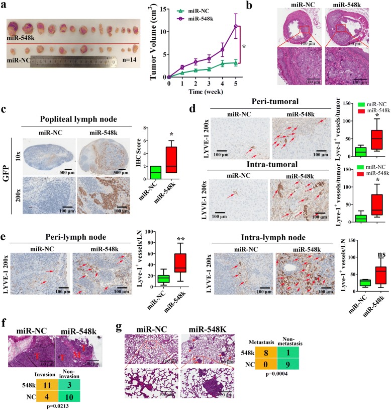
Methods: Joint analysis of ESCC sequencing data were conducted to comprehensively survey SCNAs and identify driver genes which significantly associated with LNM. The roles of miR-548k in lymphangiogensis and lymphatic metastasis were validated both in vitro and in vivo. ESCC tissue and blood samples were analyzed for association between miR-548k expression and patient clinicopathological features and prognosis and diagnosis.
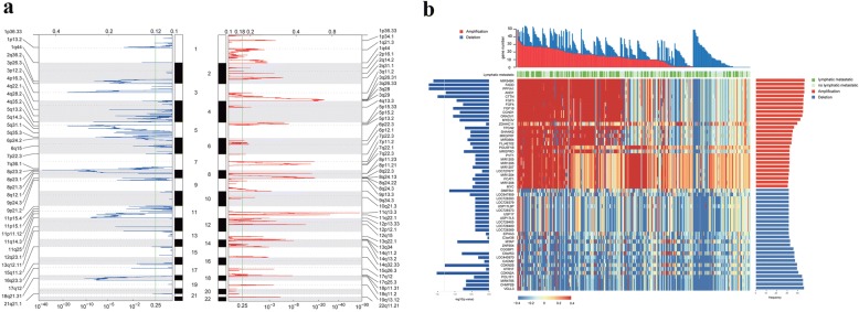
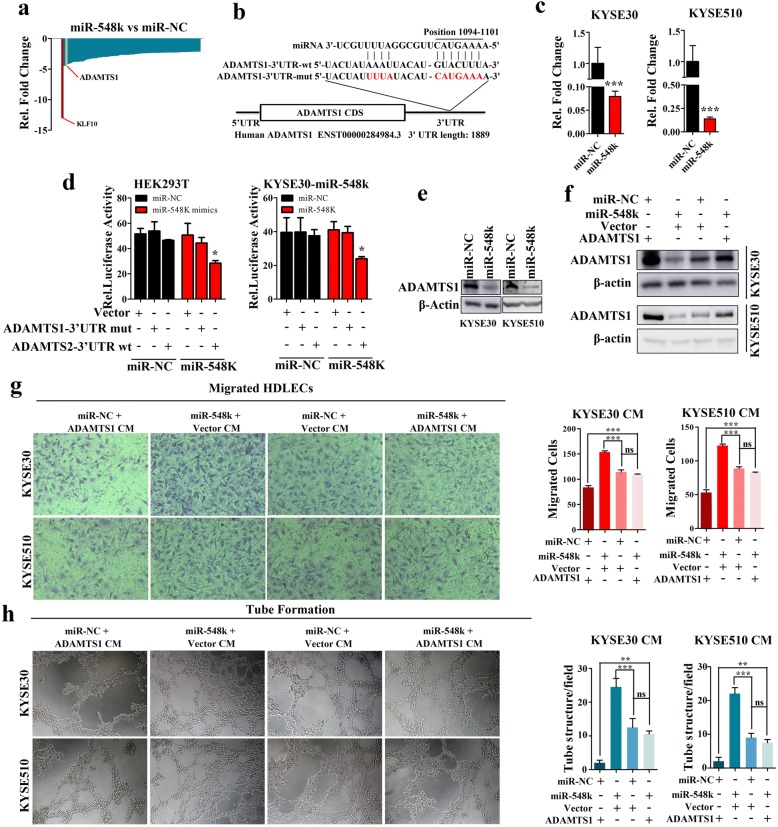
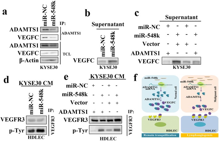
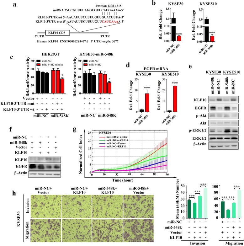
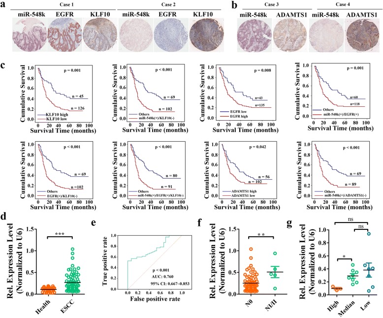
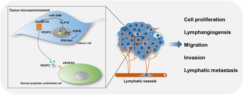
Results: In the pooled cohort of 314 ESCC patients, we found 76 significant focused regions including 43 amplifications and 33 deletions. Clinical implication analysis revealed a panel of genes associated with LNM with the most frequently amplified gene being MIR548K harbored in the 11q13.3 amplicon. Overexpression of miR-548k remarkably promotes lymphangiogenesis and lymphatic metastasis in vitro and in vivo. Furthermore, we demonstrated that miR-548k modulating the tumor microenvironment by promoting VEGFC secretion and stimulating lymphangiogenesis through ADAMTS1/VEGFC/VEGFR3 pathways, while promoting metastasis by regulating KLF10/EGFR axis. Importantly, we found that serum miR-548k and VEGFC of early stage ESCC patients were significantly higher than that in healthy donators, suggesting a promising application of miR-548k and VEGFC as biomarkers in early diagnosis of ESCC.
Conclusions: Our study comprehensively characterized SCNAs in ESCC and highlighted the crucial role of miR-548k in promoting lymphatic metastasis, which might be employed as a new diagnostic and prognostic marker for ESCC.
Keywords: Esophageal squamous cell carcinoma; Lymphangiogenesis. Tumor microenvironment miR-548k; Lymphatic metastasis.
Conflict of interest statement
Ethics approval and consent to participate
Tissue microarrays (TMA) of ESCC specimens were obtained from Shanghai Outdo Biotech Co., Ltd. (SOBC), with the approval of the Institutional Review Board. The ESCC tissues and matched adjacent normal tissues and the serum samples of ESCC patients and health persons used for real time PCR assay were histopathologically and clinically diagnosed at Beijing Cancer Hospital and the Cancer Institute and Hospital, Chinese Academic of Medical Sciences & Peking Union Medical College (Additional file 1: Table S17). Written informed consent was obtained from all patients prior to the study. The use of the clinical specimens for research purposes was approved by the Institutional Research Ethics Committee.
All animal care and procedures were in accordance with national and institutional policies for animal health and well-being. Mouse experimentations were approved by Cancer Institute and Hospital, Chinese Academic of Medical Sciences & Peking Union Medical College Animal Care and Use Committee. All mouse surgery was performed under anesthesia, and all efforts were made to minimize suffering of animals.
Consent for publication
Not applicable
Competing interests
The authors declare that they have no competing interests.
Publisher’s Note
Springer Nature remains neutral with regard to jurisdictional claims in published maps and institutional affiliations.
Figures

References
-
- Song Y, Li L, Ou Y, Gao Z, Li E, Li X, Zhang W, Wang J, Xu L, Zhou Y, Ma X, Liu L, Zhao Z, Huang X, Fan J, Dong L, Chen G, Ma L, Yang J, Chen L, He M, Li M, Zhuang X, Huang K, Qiu K, Yin G, Guo G, Feng Q, Chen P, Wu Z, Wu J, Zhao J, Luo L, Fu M, Xu B, Chen B, Li Y, Tong T, Wang M, Liu Z, Lin D, Zhang X, Yang H, Zhan Q. Identification of genomic alterations in oesophageal squamous cell cancer. Nature. 2014;509(7498):91–95. doi: 10.1038/nature13176. - DOI - PubMed
-
- Lin DC, Hao JJ, Nagata Y, Xu L, Shang L, Meng X, Sato Y, Okuno Y, Varela AM, Ding LW, Garg M, Liu LZ, Yang H, Yin D, Shi ZZ, Jiang YY, Gu WY, Gong T, Zhang Y, Xu X, Kalid O, Shacham S, Ogawa S, Wang MR, Koeffler HP. Genomic and molecular characterization of esophageal squamous cell carcinoma. Nat Genet. 2014;46(5):467–473. doi: 10.1038/ng.2935. - DOI - PMC - PubMed
Publication types
MeSH terms
Substances
LinkOut - more resources
Full Text Sources
Other Literature Sources
Medical
Research Materials
Miscellaneous
