. 2018 Aug:34:46-60.
doi: 10.1016/j.ebiom.2018.07.040. Epub 2018 Aug 18.

Systematic Analysis of Survival-Associated Alternative Splicing Signatures in Gastrointestinal Pan-Adenocarcinomas

Affiliations

Systematic Analysis of Survival-Associated Alternative Splicing Signatures in Gastrointestinal Pan-Adenocarcinomas

Peng Lin et al. EBioMedicine. 2018 Aug.

Abstract

Background: Gastrointestinal pan-adenocarcinomas, which mainly include adenocarcinomas of the esophagus, stomach, colon, and rectum, place a heavy burden on society owing to their poor prognoses. Since aberrant alternative splicing (AS) are starting to be considered as efficacious signatures for tumor prognosis predicting and therapeutic targets, systematic analysis of AS events is urgent.

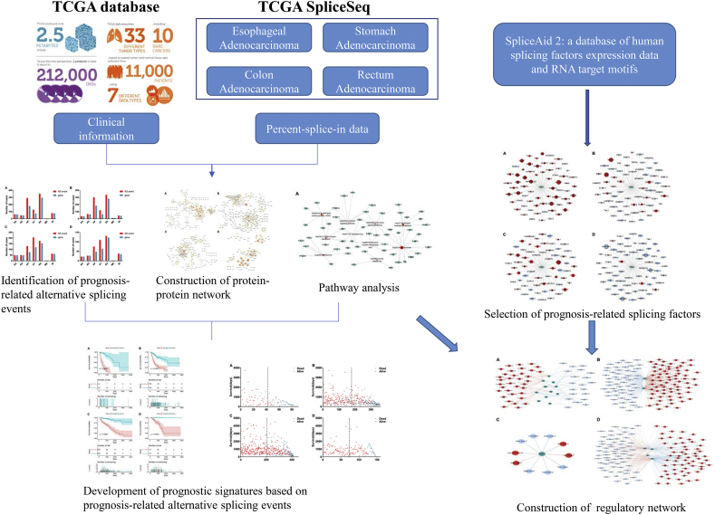
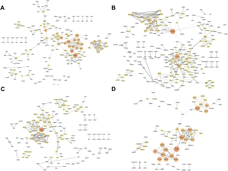
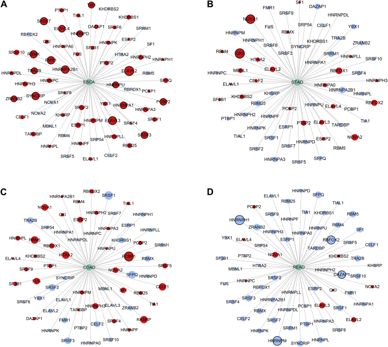
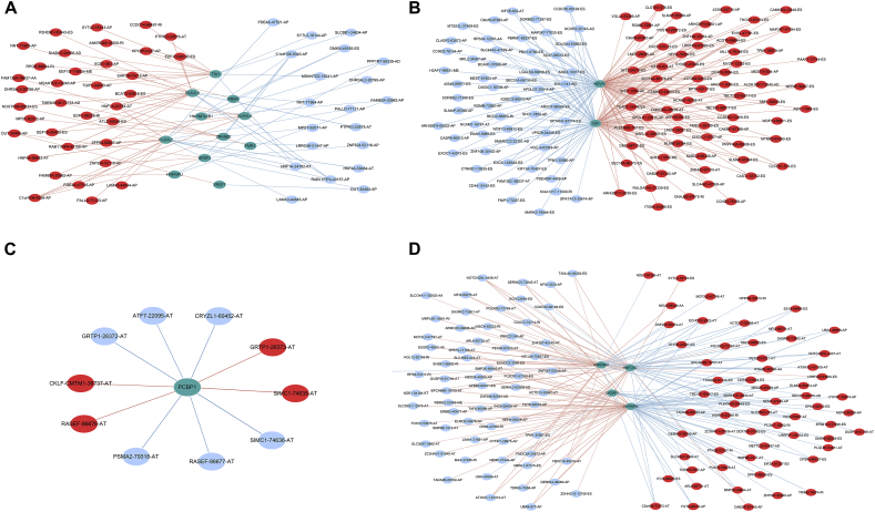
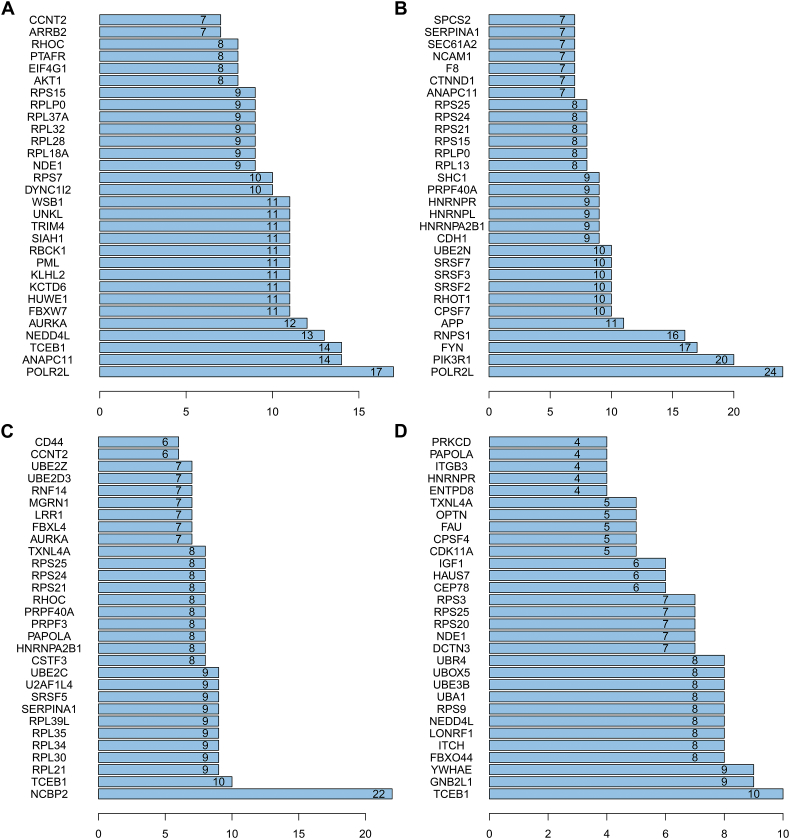
Methods: Prognosis-related AS events were selected by using univariate COX regression analysis. Gene functional enrichment analysis revealed the pathways enriched by prognosis-related AS. Then, prognostic signatures based on AS events were developed for prognosis prediction. Potential mechanism to regulate splicing events by splicing factors was analyzed via Pearson correlation and regulatory networks were constructed.

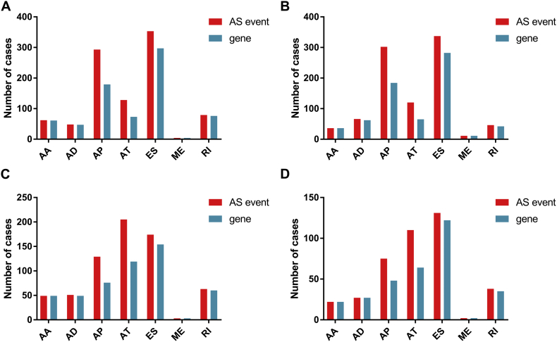
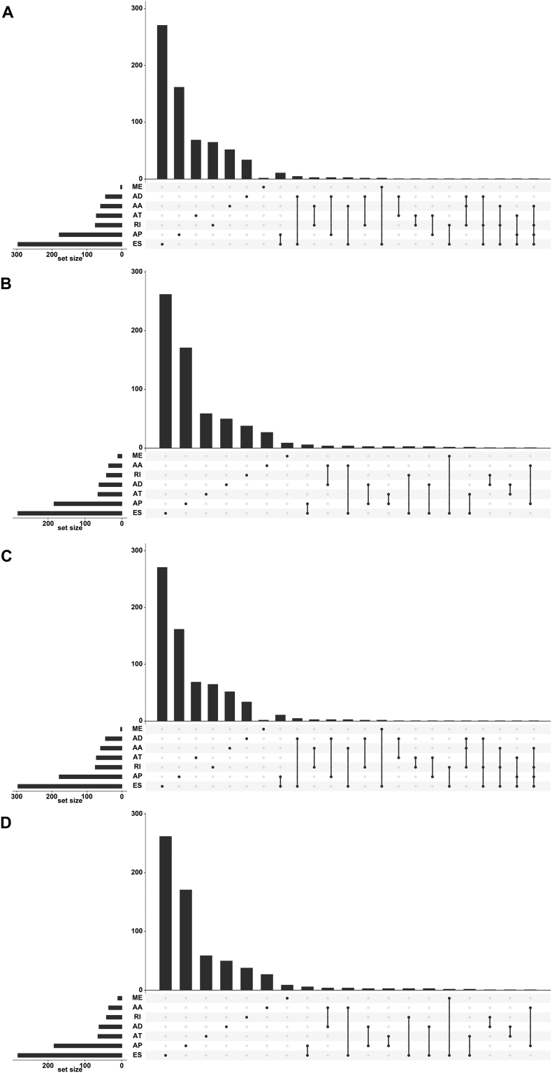
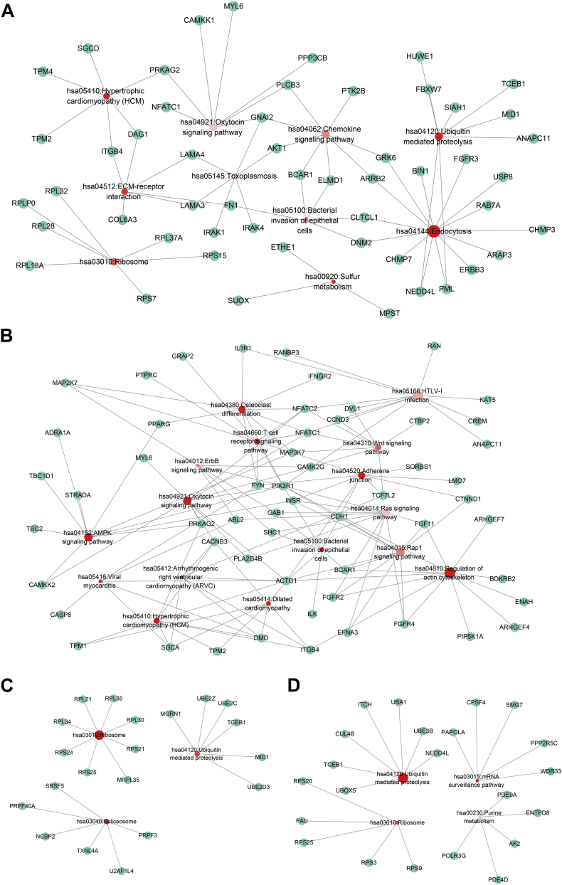
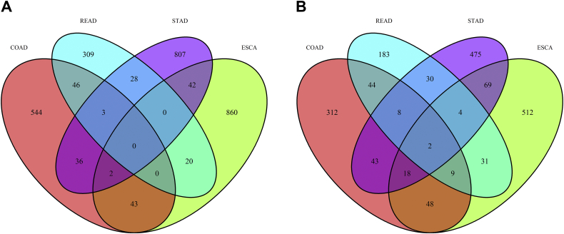
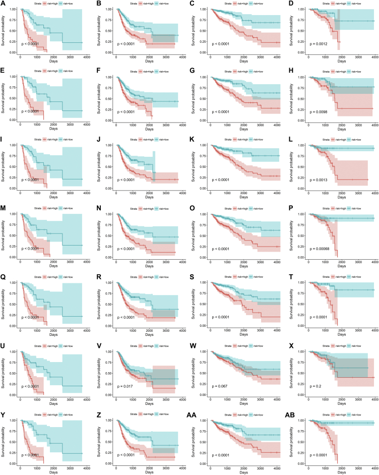
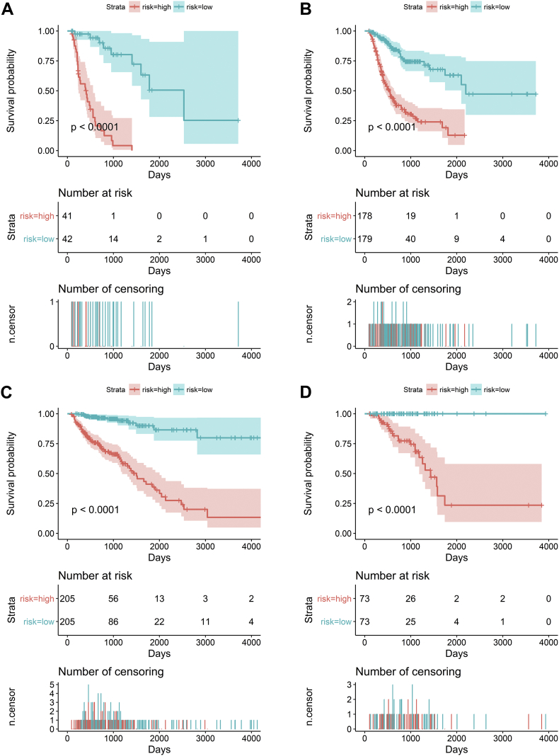
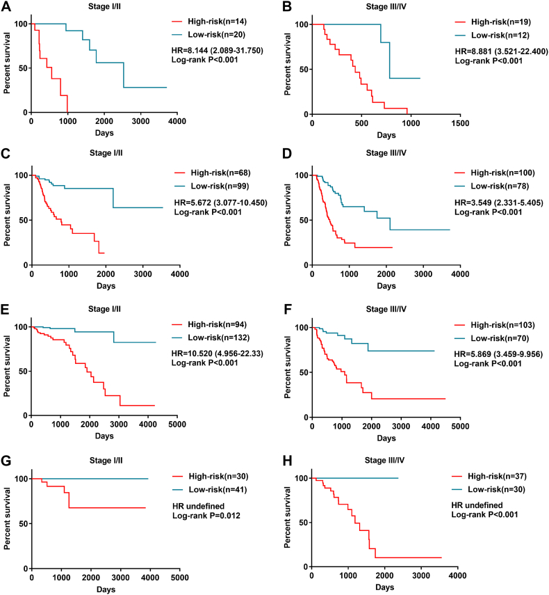
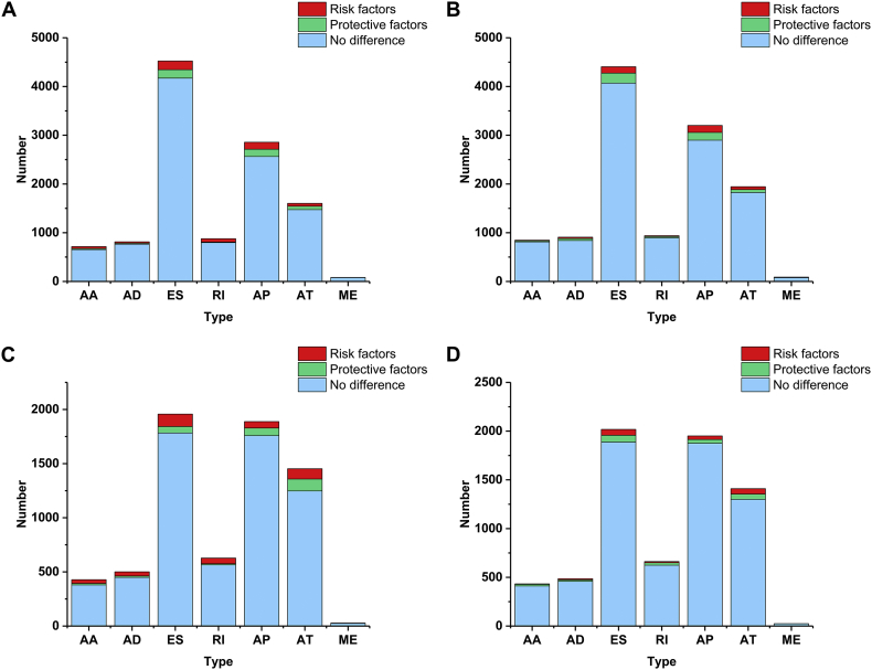
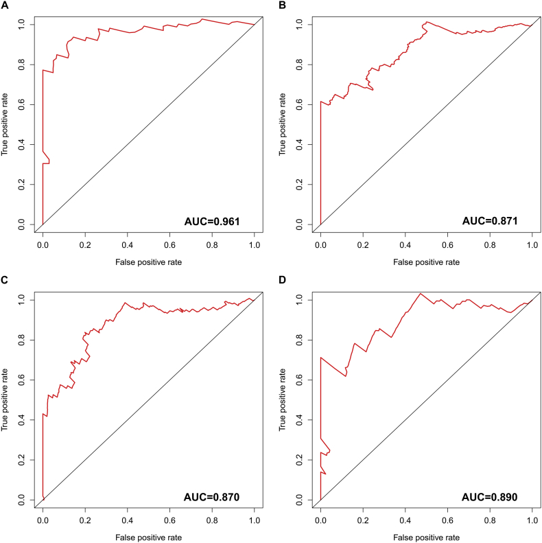
Findings: A total of 967, 918, 674, and 406 AS events were identified as prognosis-related AS events in esophagus, stomach, colon, and rectum adenocarcinomas, respectively. Survival-associated AS events were distinguishing in the four subtypes of adenocarcinoma. Furthermore, computational algorithm results indicated that perturbation of ribosome and ubiquitin-mediated proteolysis pathways were the potential molecular mechanisms corresponding to inferior prognoses. Most notably, several prognostic signatures based on AS events displayed moderate performance in prognosis predicting. The area under curve values of the time-dependent receiver operating characteristic were 0.961, 0.871, 0.870, and 0.890 in esophagus, stomach, colon, and rectum adenocarcinomas. Survival-associated splicing factors were submitted to construct the AS regulatory network, which could be an underlying mechanism of AS events.

Interpretation: AS may could be ideal indiactors in the prognosis of gastrointestinal pan-adenocarcinomas. Exploring interesting splicing regulatory networks is conducive to solve the puzzles of AS.

Keywords: Alternative splicing; Gastrointestinal pan-adenocarcinomas; Prognosis; Splicing factors.


Figures

Fig. 1
Fig. 1
Flowchart of the present study.
Fig. 2
Fig. 2
The number of prognosis-related alternative splicing events and involved genes. Red indicates the number of prognosis-related alternative splicing events and green indicates the number of genes with prognosis-related alternative splicing events. Green columns are equally high or higher than the red columns owing to one gene may have up to one or more types of prognosis-related alternative splicing events. (A) Esophageal adenocarcinoma; (B) stomach adenocarcinoma; (C) colon adenocarcinoma; and (D) rectum adenocarcinoma.
Fig. 3
Fig. 3
UpSet plot of alternative splicing events. One gene may have several types of alternative splicing to be associated with patient survival. UpSet plot of interactions between the seven types of prognosis-related alternative splicing events. One gene may have up to four types of prognosis-related alternative splicing events. (A) Esophageal adenocarcinoma; (B) stomach adenocarcinoma; (C) colon adenocarcinoma; and (D) rectum adenocarcinoma.
Fig. 4
Fig. 4
Protein-protein interaction networks of genes of alternative splicing events. Information of interactions were extracted from online database STRING (http://string-db.org/). The larger, the brighter circles are more important in the network. The thicker lines between two nodules represents the higher combined score. (A) Esophageal adenocarcinoma; (B) stomach adenocarcinoma; (C) colon adenocarcinoma; and (D) rectum adenocarcinoma.
Fig. 5
Fig. 5
Kyoto Encyclopedia of Genes and Genomes analysis. Red circles represent the enriched pathways. The size of the circles represents the number of the gene enriched in the pathway. A greater size indicates a larger number. The colour depth displays P value of pathways. A darker colour indicates a smaller P value. Green circles represent genes. (A) Esophageal adenocarcinoma; (B) stomach adenocarcinoma; (C) colon adenocarcinoma; and (D) rectum adenocarcinoma.
Fig. 6
Fig. 6
Venn plot of alternative splicing events. Red stands for colon adenocarcinoma (COAD), light blue means rectum adenocarcinoma (READ), purple means stomach adenocarcinoma (STAD), and green means studious and green represents esophageal carcinoma (ESCA). (A) A Venn diagram shows the overlap of prognosis-related alternative splicing events in COAD, READ, STAD and ESCA; and (B) A Venn diagram shows the overlap of genes with prognosis-related alternative splicing events in COAD, READ, STAD and ESCA.
Fig. 7
Fig. 7
Kaplan-Meier survival analysis of prognostic signatures based on seven types of alternative splicing events in gastrointestinal tract adenocarcinomas. Prognostic signatures based on Alternate Acceptor site for esophageal adenocarcinoma (A), stomach adenocarcinoma (B), colon adenocarcinoma (C), and rectum adenocarcinoma (D). Prognostic signatures based on Alternate Donor site for esophageal adenocarcinoma (E), stomach adenocarcinoma (F), colon adenocarcinoma (G), and rectum adenocarcinoma (H). Prognostic signatures based on Alternate Promoter for esophageal adenocarcinoma (I), stomach adenocarcinoma (J), colon adenocarcinoma (K), and rectum adenocarcinoma (L). Prognostic signatures based on Alternate Terminator for esophageal adenocarcinoma (M), stomach adenocarcinoma (N), colon adenocarcinoma (O), and rectum adenocarcinoma (P). Prognostic signatures based on Exon Skip for esophageal adenocarcinoma (Q), stomach adenocarcinoma (R), colon adenocarcinoma (S), and rectum adenocarcinoma (T). Prognostic signatures based on Mutually Exclusive Exons for esophageal adenocarcinoma (U), stomach adenocarcinoma (V), colon adenocarcinoma (W), and rectum adenocarcinoma (X). Prognostic signatures based on Retained Intron for esophageal adenocarcinoma (Y), stomach adenocarcinoma (Z), colon adenocarcinoma (AA), and rectum adenocarcinoma (AB).
Fig. 8
Fig. 8
Prognostic signatures based on all types of alternative splicing events in gastrointestinal tract adenocarcinomas. Patients were divided into high- and low-risk groups according to prognostic signatures. Each panel contains three parts: [1] survival differences were estimated by Kaplan-Meier survival curve; [2] number of patients in different groups; and [3] number of censoring at different times. (A) Esophageal adenocarcinoma; (B) stomach adenocarcinoma; (C) colon adenocarcinoma; and (D) rectum adenocarcinoma.
Fig. 9
Fig. 9
The four prognosis-relevant splicing event groups influencing the survival of gastrointestinal tract adenocarcinoma patients. X-axis represents the orders of patients based on prognostic signatures and Y-axis represents the survival days of patients. Dotted lines were used to distinguish patients in high-risk group and low-risk group. (A) Esophageal adenocarcinoma; (B) stomach adenocarcinoma; (C) colon adenocarcinoma; and (D) rectum adenocarcinoma.
Fig. 10
Fig. 10
Kaplan-Meier curves of overall survival (OS) among patients with low stage and advanced tumor stage. Esophageal adenocarcinoma (A) early stage, (B) advanced stage. Stomach adenocarcinoma (C) early stage, (D) advanced stage. Colon adenocarcinoma (E) early stage, (F) advanced stage. Rectum adenocarcinoma (G) early stage, (H) advanced stage. Patients in high-risk group suffer poor OS whether with early or advanced tumor stage.
Fig. 11
Fig. 11
The prognostic analysis of splicing factors in four subtypes of gastrointestinal pan-adenocarcinoma. Red circles represent splicing factors with hazard ratio >1 and blue circles represent splicing factors with hazard ratio <1. Circles with black edge indicate significant (P < 0.05). (A) Esophageal adenocarcinoma; (B) stomach adenocarcinoma; (C) colon adenocarcinoma; and (D) rectum adenocarcinoma.
Fig. 12
Fig. 12
Survival-associated splicing factors and the splicing correlation network in gastrointestinal tract adenocarcinomas. Expression of prognosis-related splicing factors (green dots) were positively (red line) or negatively (blue line) correlated with the PSI values of prognosis-related alternative splicing events. (A) Esophageal adenocarcinoma; (B) stomach adenocarcinoma; (C) colon adenocarcinoma; and (D) rectum adenocarcinoma.
Supplementary Fig. 1
The proportion of prognosis-related alternative splicing events. Red represents the number of risk factors while green represents the number of protective factors. And blue was alternative splicing events that have not prognostic value. (A) Esophageal adenocarcinoma; (B) stomach adenocarcinoma; (C) colon adenocarcinoma; and (D) rectum adenocarcinoma.
Supplementary Fig. 2
Hub genes of the protein-protein interaction network. The number represents the connected nodes of the correspondence genes. (A) Esophageal adenocarcinoma; (B) stomach adenocarcinoma; (C) colon adenocarcinoma; and (D) rectum adenocarcinoma.
Supplementary Fig. 3
Time-dependent receiver operating characteristic curves of four prognostic signatures in gastrointestinal tract adenocarcinomas. (A) Esophageal adenocarcinoma; (B) stomach adenocarcinoma; (C) colon adenocarcinoma; and (D) rectum adenocarcinoma.

Similar articles

Cited by

References

    1. Black D.L. Mechanisms of alternative pre-messenger RNA splicing. Annu. Rev. Biochem. 2003;72:291–336. - PubMed
    1. Jin Y., Dong H., Shi Y., Bian L. Mutually exclusive alternative splicing of pre-mRNAs. Wiley Interdiscip. Rev. RNA. 2018;9(3) - PubMed
    1. Yang X., Coulombe-Huntington J., Kang S. Widespread expansion of protein interaction capabilities by alternative splicing. Cell. 2016;164(4):805–817. - PMC - PubMed
    1. Baralle F.E., Giudice J. Alternative splicing as a regulator of development and tissue identity. Nat. Rev. Mol. Cell Biol. 2017;18(7):437–451. - PMC - PubMed
    1. Park E., Pan Z., Zhang Z., Lin L., Xing Y. The expanding landscape of alternative splicing variation in human populations. Am. J. Hum. Genet. 2018;102(1):11–26. - PMC - PubMed