. 2018 Dec;77(12):1825-1833.
doi: 10.1136/annrheumdis-2018-213746. Epub 2018 Aug 21.

Dysregulated neutrophil responses and neutrophil extracellular trap formation and degradation in PAPA syndrome

Affiliations

Dysregulated neutrophil responses and neutrophil extracellular trap formation and degradation in PAPA syndrome

Pragnesh Mistry et al. Ann Rheum Dis. 2018 Dec.

Abstract

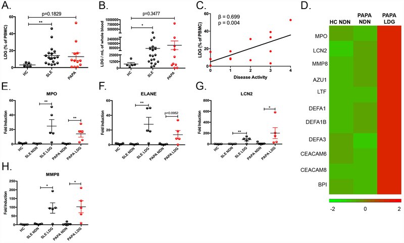
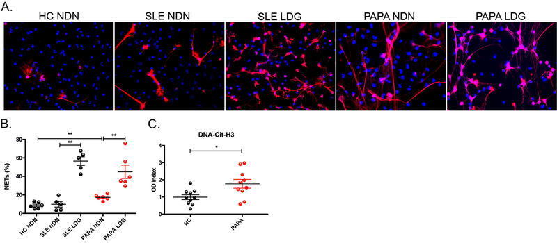
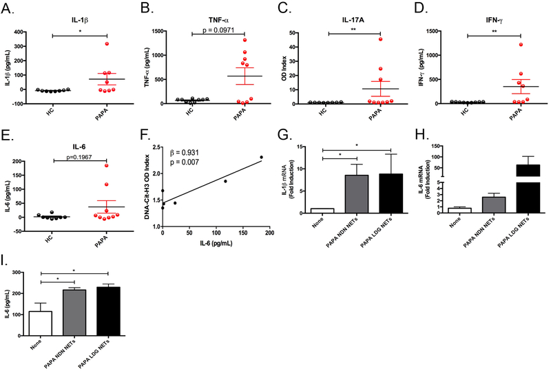
Objectives: Pyogenic arthritis, pyoderma gangrenosum and acne (PAPA) syndrome is characterised by flares of sterile arthritis with neutrophil infiltrate and the overproduction of interleukin (IL)-1β. The purpose of this study was to elucidate the potential role of neutrophil subsets and neutrophil extracellular traps (NET) in the pathogenesis of PAPA.

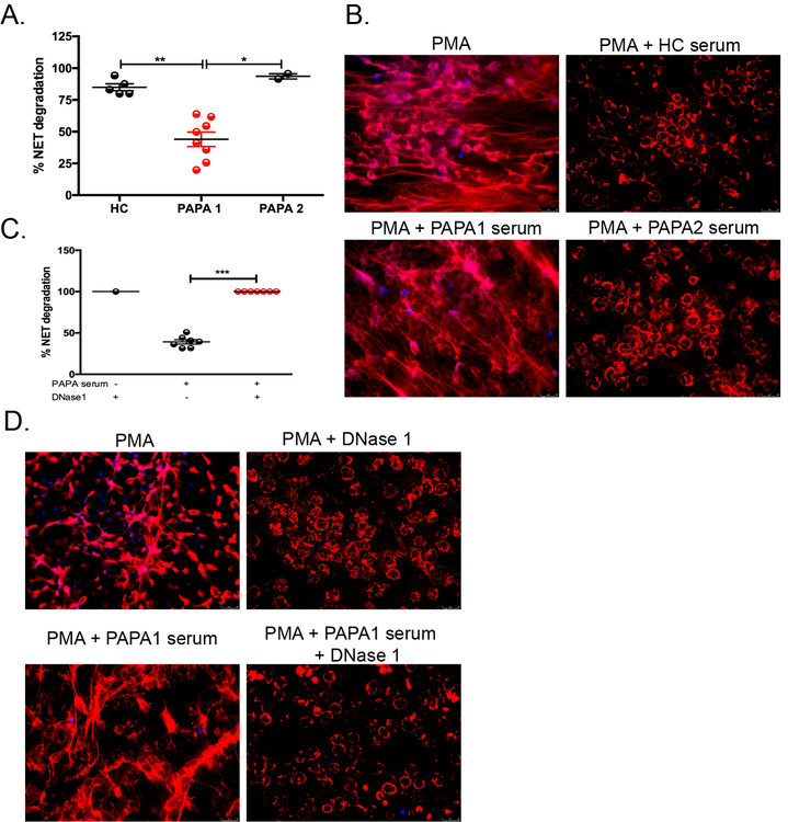
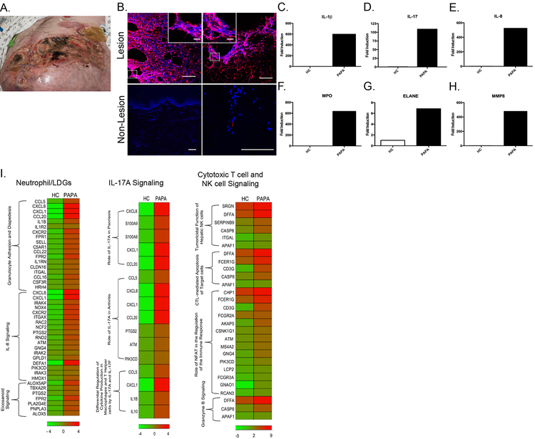
Methods: Neutrophils and low-density granulocytes (LDG) were quantified by flow cytometry. Circulating NETs were measured by ELISA and PAPA serum was tested for the ability to degrade NETs. The capacity of NETs from PAPA neutrophils to activate macrophages was assessed. Skin biopsies were analysed for NETs and neutrophil gene signatures.

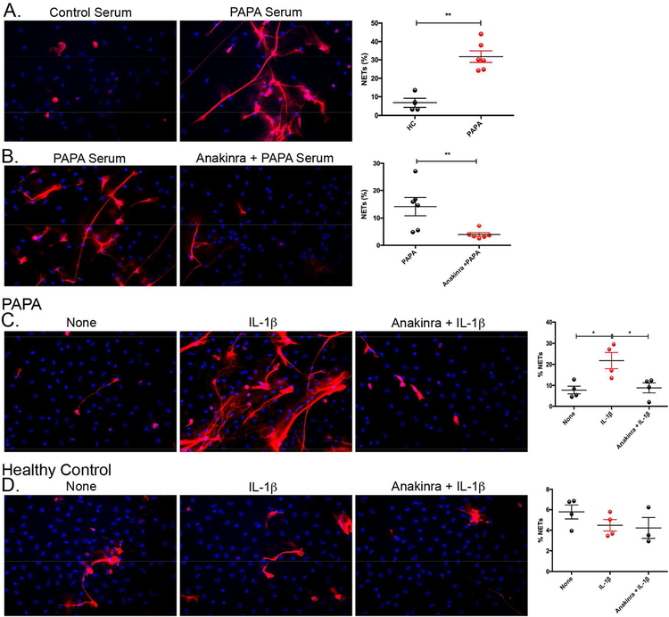
Results: Circulating LDGs are elevated in PAPA subjects. PAPA neutrophils and LDGs display enhanced NET formation compared with control neutrophils. PAPA sera exhibit impaired NET degradation and this is corrected with exogenous DNase1. Recombinant human IL-1β induces NET formation in PAPA neutrophils but not healthy control neutrophils. NET formation in healthy control neutrophils is induced by PAPA serum and this effect is inhibited by the IL-1 receptor antagonist, anakinra. NETs from PAPA neutrophils and LDGs stimulate IL-6 release in healthy control macrophages. NETs are detected in skin biopsies of patients with PAPA syndrome in association with increased tissue IL-1β, IL-8 and IL-17. Furthermore, LDG gene signatures are detected in PAPA skin.

Conclusions: PAPA syndrome is characterised by an imbalance of NET formation and degradation that may enhance the half-life of these structures in vivo, promoting inflammation. Anakinra ameliorates NET formation in PAPA and this finding supports a role for IL-1 signalling in exacerbated neutrophil responses in this disease. The study also highlights other inflammatory pathways potentially pathogenic in PAPA, including IL-17 and IL-6, and these results may help guide new therapeutic approaches in this severe and often treatment-refractory condition.

Keywords: cytokines; fever syndromes; inflammation.


Conflict of interest statement

Competing interests: None declared.

Figures

Fig. 1:
Fig. 1:. Identification and characterization of LDGs in PAPA syndrome.
(A) Percentage of LDGs in the PBMC fraction of healthy controls (HC) (n=5), SLE patients (n=18), and PAPA patients (n=12). (B) Total LDG numbers per mL of whole blood in HC (n=5), SLE (n=18), and PAPA patients (n=10). (C) A linear regression analysis of the percentage of LDGs in PBMC fraction and disease activity (n=15). (D) RNASeq analysis of HC normal-dense neutrophils (NDN), PAPA NDN, and PAPA LDG (n=1/each group) identified upregulated expression of neutrophil granule genes in PAPA LDGs. (EH) Total RNA was extracted from unstimulated HC NDN, SLE NDN and autologous LDGs, and PAPA NDN and autologous LDGs (n=5/group) and neutrophil granule gene expression was measured by qRT-PCR. Expression values were normalized to the expression of the glyceraldehyde 3-phosphate dehydrogenase (GAPDH) housekeeping gene and fold induction over HC NDN was calculated. The results shown in A-H represent the mean ± SEM (*P ≤ 0.05; **P ≤ 0.01).
Fig. 2.
Fig. 2.. PAPA neutrophils and LDGs undergo enhanced NET formation.
(A) Representative images of unstimulated HC NDN, SLE NDN, SLE LDGs, PAPA NDN, and PAPA LDGs isolated from peripheral blood after 1 h (LDG) or 2 h (NDN) of incubation at 37°C. NETs were identified by colocalization of extracellular MPO (red) and DNA labeled with Hoechst33342 (blue). Original magnification x40. (B) Quantification of the percentage of cells undergoing NET formation are plotted as mean ± SEM (n=6 for HC and PAPA, n=5 for SLE, **P ≤ 0.01). (C) Circulating NET remnants in HC and PAPA subjects quantified (n=10 subjects/group) plotted as mean ± SEM (*P ≤ 0.05). The OD index was calculated by dividing the individual OD450 values by the average of HC OD450 values.
Fig. 3.
Fig. 3.. Impaired NET degradation in a subset of PAPA subjects.
(A and B) HC NDN were stimulated with PMA (2.5 μM) for 4 h to induce NETs and subsequently incubated with 1% serum from either HC (n=5) or PAPA subjects (n=10) for 16 h. “PAPA1” represents PAPA subjects who did not degrade NETs efficiently and “PAPA2” represents PAPA subjects that were efficient degraders of NETs. (C and D) HC NDN were stimulated with PMA (2.5 μM) for 4 h to induce NETs, followed by incubation with or without 1% serum from PAPA1 subjects (n=7) in the presence or absence of 1U/mL DNase1for 16 h. (A) Represents the mean ± SEM from three independent experiments (*P ≤ 0.05; **P ≤ 0.01; ***P < 0.001).
Fig. 4.
Fig. 4.. PAPA serum induces NET formation in healthy control neutrophils in an IL-1-dependent manner.
(A) HC NDN were stimulated with 10% serum from either HC (n=4) or PAPA subjects (n=6) for 2.5 h and NET formation was quantified and reported as mean ± SEM (**P ≤ 0.01). (B) HC NDN were pretreated with/without Anakinra (149 μg/mL) for 15 min prior to stimulation with 10% PAPA serum (n=6) for 2.5 h and NET formation was quantified and plotted as mean ± SEM (**P ≤ 0.01). (C and D) PAPA or HC NDN were left unstimulated or stimulated with recombinant human IL-1β (100 ng/mL) for 2.5 h in the absence/presence of anakinra (149 μg/mL) pretreatment, respectively. C and D are the mean ± SEM from four different samples for each group (*P ≤ 0.05).
Fig. 5.
Fig. 5.. Proinflammatory cytokines are elevated in PAPA serum.
(A-E) Serum concentrations of IL-1β, TNF-α, IFN-γ, and IL-6 (A, B, D, and E) and OD Index for IL-17A (C) in HC and PAPA subjects (n=9/group). Results represent mean ± SEM (*P ≤ 0.05; **P ≤ 0.01). (F) A linear regression analysis of the OD Index from citrullinated histone (H3)-DNA complexes in the plasma from PAPA subjects and IL-6 protein in PAPA sera (n=6). (G-I) HC macrophages were stimulated with spontaneously formed NETs from PAPA NDN or LDGs for 2 h (G and H) or 24 h (I). (G and H) Total RNA was isolated and qRT-PCR was performed to measure IL-1β and IL-6 gene expression; results represent mean ± SEM from four independent experiments (*P ≤ 0.05). (I) Culture supernatants were analyzed by ELISA for IL-6 protein and results represent the mean ± SEM from four independent experiments (*P ≤ 0.05).
Fig. 6.
Fig. 6.. NETs are present in PAPA skin lesion.
(A) Pyoderma gangrenosum lesion that developed in a PAPA patient with the A230T mutation. The lesion developed while receiving anakinra, infliximab and prednisone at doses up to 60 mg/daily. A skin biopsy taken from this lesion was obtained and used to perform experiments depicted in B-I. (B) Immunohistochemistry was performed on skin lesions from the PAPA patient described in A and in non-lesional skin sample from another PAPA subject. NETs are identified by colocalization of citrullinated histone H4 (cit-H4) (red) and DNA labeled with Hoechst33342 (blue). Main figure scale bar 100 μM, inset scale bar 20 μM. (C-H) Total RNA was extracted from HC skin biopsy and from lesional skin from the PAPA patient described in A. Proinflammatory cytokine and neutrophil granule gene expression were measured by qRT-PCR, normalized to the expression of GAPDH, and fold induction was calculated to HC skin. (I) RNASeq analysis of HC skin (n=1) and PAPA lesional skin (n=1) identified upregulated expression of neutrophil/LDG, IL-17A signaling, and cytotoxic T cell and NK cell signaling genes.

References

    1. Smith EJ, Allantaz F, Bennett L, Zhang D, Gao X, Wood G, et al. Clinical, Molecular, and Genetic Characteristics of PAPA Syndrome: A Review. Curr Genomics. 2010. November; 11(7):519–527. - PMC - PubMed
    1. Masters SL, Simon A, Aksentijevich I, Kastner DL. Horror autoinflammaticus: the molecular pathophysiology of autoinflammatory disease (*). Annu Rev Immunol. 2009; 27:621–668. - PMC - PubMed
    1. Wise CA, Gillum JD, Seidman CE, Lindor NM, Veile R, Bashiardes S, et al. Mutations in CD2BP1 disrupt binding to PTP PEST and are responsible for PAPA syndrome, an autoinflammatory disorder. Hum Mol Genet. 2002. April 15; 11(8):961–969. - PubMed
    1. Spencer S, Dowbenko D, Cheng J, Li W, Brush J, Utzig S, et al. PSTPIP: a tyrosine phosphorylated cleavage furrow-associated protein that is a substrate for a PEST tyrosine phosphatase. J Cell Biol. 1997. August 25; 138(4):845–860. - PMC - PubMed
    1. Badour K, Zhang J, Shi F, McGavin MK, Rampersad V, Hardy LA, et al. The Wiskott-Aldrich syndrome protein acts downstream of CD2 and the CD2AP and PSTPIP1 adaptors to promote formation of the immunological synapse. Immunity. 2003. January; 18(1):141–154. - PubMed

Publication types

Supplementary concepts