

Systematic Dissection of the Evolutionarily Conserved WetA Developmental Regulator across a Genus of Filamentous Fungi
- PMID: 30131357
- PMCID: PMC6106085
- DOI: 10.1128/mBio.01130-18
Systematic Dissection of the Evolutionarily Conserved WetA Developmental Regulator across a Genus of Filamentous Fungi
Abstract
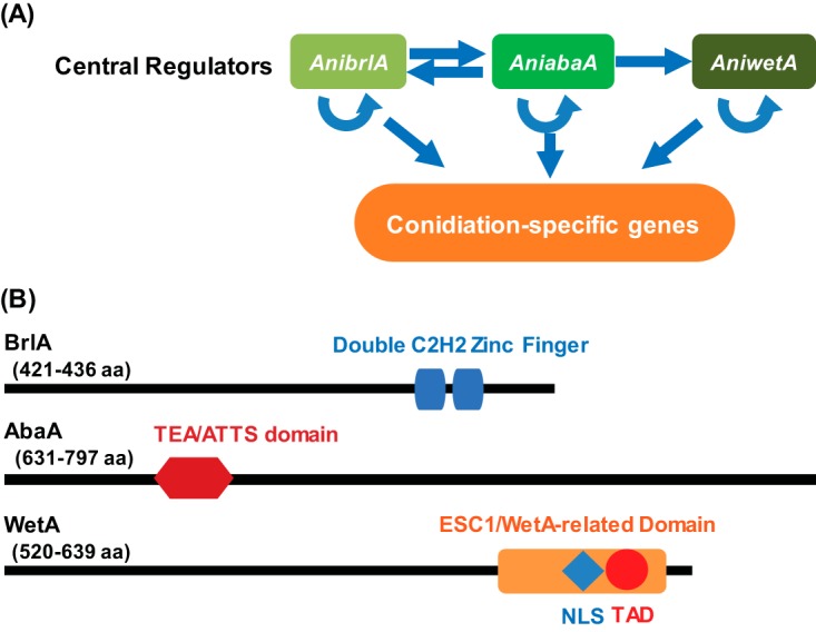
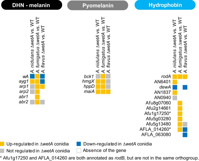
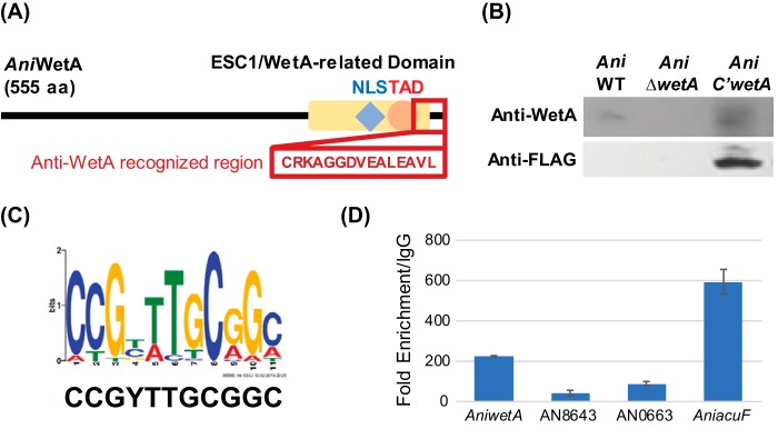
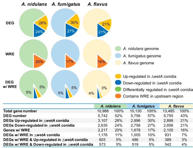
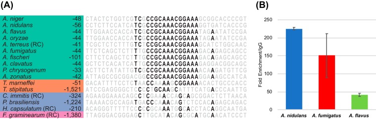
Asexual sporulation is fundamental to the ecology and lifestyle of filamentous fungi and can facilitate both plant and human infection. In Aspergillus, the production of asexual spores is primarily governed by the BrlA→AbaA→WetA regulatory cascade. The final step in this cascade is controlled by the WetA protein and governs not only the morphological differentiation of spores but also the production and deposition of diverse metabolites into spores. While WetA is conserved across the genus Aspergillus, the structure and degree of conservation of the wetA gene regulatory network (GRN) remain largely unknown. We carried out comparative transcriptome analyses of comparisons between wetA null mutant and wild-type asexual spores in three representative species spanning the diversity of the genus Aspergillus: A. nidulans, A. flavus, and A. fumigatus We discovered that WetA regulates asexual sporulation in all three species via a negative-feedback loop that represses BrlA, the cascade's first step. Furthermore, data from chromatin immunoprecipitation sequencing (ChIP-seq) experiments in A. nidulans asexual spores suggest that WetA is a DNA-binding protein that interacts with a novel regulatory motif. Several global regulators known to bridge spore production and the production of secondary metabolites show species-specific regulatory patterns in our data. These results suggest that the BrlA→AbaA→WetA cascade's regulatory role in cellular and chemical asexual spore development is functionally conserved but that the wetA-associated GRN has diverged during Aspergillus evolution.IMPORTANCE The formation of resilient spores is a key factor contributing to the survival and fitness of many microorganisms, including fungi. In the fungal genus Aspergillus, spore formation is controlled by a complex gene regulatory network that also impacts a variety of other processes, including secondary metabolism. To gain mechanistic insights into how fungal spore formation is controlled across Aspergillus, we dissected the gene regulatory network downstream of a major regulator of spore maturation (WetA) in three species that span the diversity of the genus: the genetic model A. nidulans, the human pathogen A. fumigatus, and the aflatoxin producer A. flavus Our data show that WetA regulates asexual sporulation in all three species via a negative-feedback loop and likely binds a novel regulatory element that we term the WetA response element (WRE). These results shed light on how gene regulatory networks in microorganisms control important biological processes and evolve across diverse species.
Keywords: Aspergillus; WetA; asexual development; gene regulatory network; sporulation.
Copyright © 2018 Wu et al.
Figures
Similar articles
-
Transcriptomic, Protein-DNA Interaction, and Metabolomic Studies of VosA, VelB, and WetA in Aspergillus nidulans Asexual Spores.mBio. 2021 Feb 9;12(1):e03128-20. doi: 10.1128/mBio.03128-20. mBio. 2021. PMID: 33563821 Free PMC article.
-
Species-specific gene regulatory network rewiring mediated by the GATA-type regulator NsdD in Aspergillus.mBio. 2025 Aug 13;16(8):e0118125. doi: 10.1128/mbio.01181-25. Epub 2025 Jul 3. mBio. 2025. PMID: 40607808 Free PMC article.
-
Recurrent Loss of abaA, a Master Regulator of Asexual Development in Filamentous Fungi, Correlates with Changes in Genomic and Morphological Traits.Genome Biol Evol. 2020 Jul 1;12(7):1119-1130. doi: 10.1093/gbe/evaa107. Genome Biol Evol. 2020. PMID: 32442273 Free PMC article.
-
Regulation of Conidiogenesis in Aspergillus flavus.Cells. 2022 Sep 7;11(18):2796. doi: 10.3390/cells11182796. Cells. 2022. PMID: 36139369 Free PMC article. Review.
-
Regulators of the Asexual Life Cycle of Aspergillus nidulans.Cells. 2023 Jun 4;12(11):1544. doi: 10.3390/cells12111544. Cells. 2023. PMID: 37296664 Free PMC article. Review.
Cited by
-
A methanolic extract of Zanthoxylum bungeanum modulates secondary metabolism regulator genes in Aspergillus flavus and shuts down aflatoxin production.Sci Rep. 2022 Apr 9;12(1):5995. doi: 10.1038/s41598-022-09913-3. Sci Rep. 2022. PMID: 35397670 Free PMC article.
-
Transcription in fungal conidia before dormancy produces phenotypically variable conidia that maximize survival in different environments.Nat Microbiol. 2021 Aug;6(8):1066-1081. doi: 10.1038/s41564-021-00922-y. Epub 2021 Jun 28. Nat Microbiol. 2021. PMID: 34183813
-
The Tudor Domain-Containing Protein BbTdp1 Contributes to Fungal Cell Development, the Cell Cycle, Virulence, and Transcriptional Regulation in the Insect Pathogenic Fungus Beauveria bassiana.Microbiol Spectr. 2021 Sep 3;9(1):e0056421. doi: 10.1128/Spectrum.00564-21. Epub 2021 Aug 11. Microbiol Spectr. 2021. PMID: 34378960 Free PMC article.
-
Molecular Mechanisms of Conidial Germination in Aspergillus spp.Microbiol Mol Biol Rev. 2019 Dec 4;84(1):e00049-19. doi: 10.1128/MMBR.00049-19. Print 2020 Feb 19. Microbiol Mol Biol Rev. 2019. PMID: 31801804 Free PMC article. Review.
-
Transcriptomic, Protein-DNA Interaction, and Metabolomic Studies of VosA, VelB, and WetA in Aspergillus nidulans Asexual Spores.mBio. 2021 Feb 9;12(1):e03128-20. doi: 10.1128/mBio.03128-20. mBio. 2021. PMID: 33563821 Free PMC article.
References
-
- Ebbole DJ. 2010. The conidium, p 577–590. In Cellular and molecular biology of filamentous fungi. American Society of Microbiology, Washington, DC.
-
- Bennett JW, Klich MA. 1992, Aspergillus: biology and industrial applications. Butterworth-Heinemann, Boston, MA.
MeSH terms
Substances
LinkOut - more resources
Full Text Sources
Other Literature Sources
Molecular Biology Databases
Miscellaneous
