. 2018 Aug 21;9(4):e01581-18.
doi: 10.1128/mBio.01581-18.

High-Throughput Screening Identifies Genes Required for Candida albicans Induction of Macrophage Pyroptosis

Affiliations

High-Throughput Screening Identifies Genes Required for Candida albicans Induction of Macrophage Pyroptosis

Teresa R O'Meara et al. mBio. .

Abstract

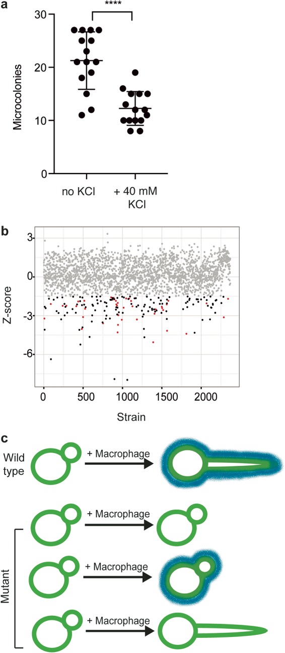
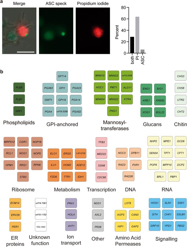
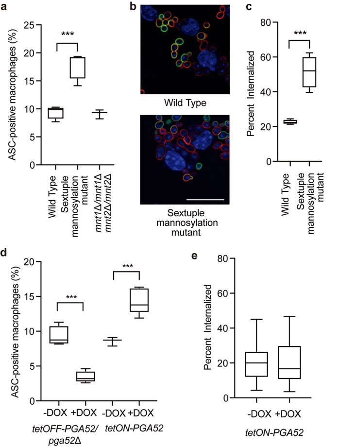
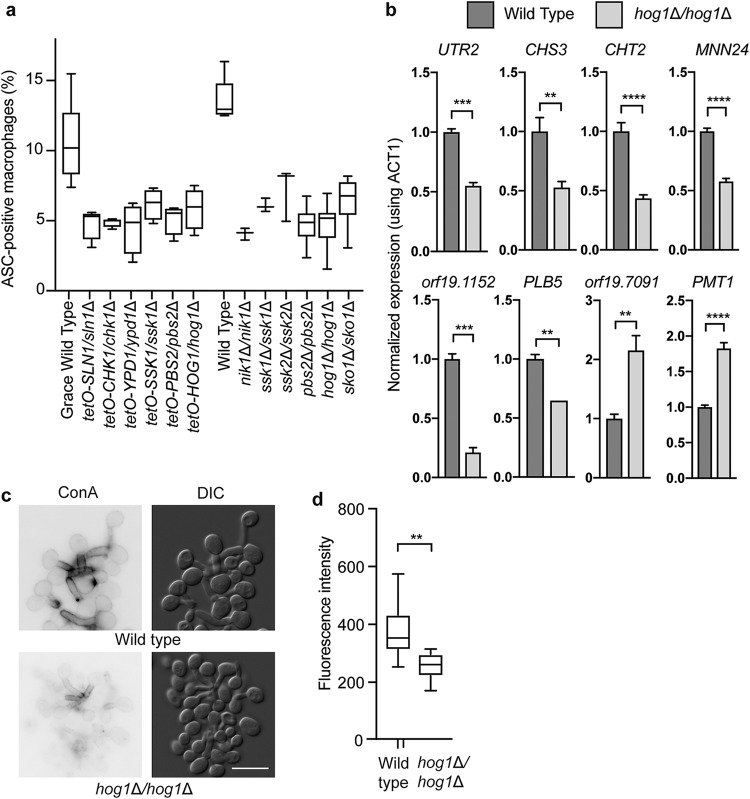
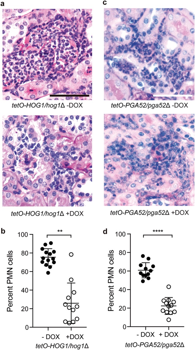
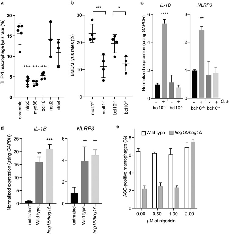
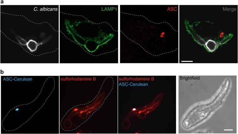
The innate immune system is the first line of defense against invasive fungal infections. As a consequence, many successful fungal pathogens have evolved elegant strategies to interact with host immune cells. For example, Candida albicans undergoes a morphogenetic switch coupled to cell wall remodeling upon phagocytosis by macrophages and then induces macrophage pyroptosis, an inflammatory cell death program. To elucidate the genetic circuitry through which C. albicans orchestrates this host response, we performed the first large-scale analysis of C. albicans interactions with mammalian immune cells. We identified 98 C. albicans genes that enable macrophage pyroptosis without influencing fungal cell morphology in the macrophage, including specific determinants of cell wall biogenesis and the Hog1 signaling cascade. Using these mutated genes, we discovered that defects in the activation of pyroptosis affect immune cell recruitment during infection. Examining host circuitry required for pyroptosis in response to C. albicans infection, we discovered that inflammasome priming and activation can be decoupled. Finally, we observed that apoptosis-associated speck-like protein containing a CARD (ASC) oligomerization can occur prior to phagolysosomal rupture by C. albicans hyphae, demonstrating that phagolysosomal rupture is not the inflammasome activating signal. Taking the data together, this work defines genes that enable fungal cell wall remodeling and activation of macrophage pyroptosis independently of effects on morphogenesis and identifies macrophage signaling components that are required for pyroptosis in response to C. albicans infection.IMPORTANCECandida albicans is a natural member of the human mucosal microbiota that can also cause superficial infections and life-threatening systemic infections, both of which are characterized by inflammation. Host defense relies mainly on the ingestion and destruction of C. albicans by innate immune cells, such as macrophages and neutrophils. Although some C. albicans cells are killed by macrophages, most undergo a morphological change and escape by inducing macrophage pyroptosis. Here, we investigated the C. albicans genes and host factors that promote macrophage pyroptosis in response to intracellular fungi. This work provides a foundation for understanding how host immune cells interact with C. albicans and may lead to effective strategies to modulate inflammation induced by fungal infections.

Keywords: Candida; cell wall remodelling; functional genomics; fungal morphogenesis; fungal pathogenesis; host-pathogen interaction; pyroptosis.


Figures

FIG 1
FIG 1
High-throughput screening to examine C. albicans-macrophage interactions. (a) Macrophage pyroptosis increases fungal survival. Macrophages were incubated with diluted C. albicans cells in the presence or absence of 40 mM KCl for 24 h. Microcolonies were counted from two biological replicates, with 8 technical replicates. ****, P < 0.0001 (unpaired t test). Error bars represent standard deviations. (b) Screening approach. J774A.1 macrophages were coincubated for 3 h with GRACE strains in the presence of 0.05 µg/ml DOX to repress target gene expression. Lysis events were determined by counting the number of propidium iodide foci. To detect whether a mutant has a significant defect in macrophage pyroptosis, we developed a model for host cell lysis in response to the wild-type strain using the locally weighted scatterplot smoothing method (LOESS) and evaluated each mutant relative to the model. Each dot represents an individual GRACE strain, with the calculated Z score shown. The grey dots represent strains with no significant difference from the wild-type results. Red dots represent strains with a significant decrease in induction of host cell lysis rates and a defect in filamentation. Black dots represent strains with a significant decrease in host cell lysis rates and no defect in filamentation. (c) Model for C. albicans interactions with macrophages. Blue lines represent cell surface moieties that are required for pyroptosis.
FIG 2
FIG 2
High-throughput screening identifies filamentation-competent mutants that are defective in activation of macrophage pyroptosis. (a) Images showing ASC-cerulean macrophages infected with C. albicans and stained with propidium iodide. Bar, 20 µm. The bar chart shows the percentage of cells positive for ASC specks only, propidium iodide staining only, or both. (b) We identified 98 genes that are required for wild-type levels of pyroptosis but that have no effect on filamentation. Functional clusters were defined by Gene Ontology (GO) term annotation. ER, endoplasmic reticulum.
FIG 3
FIG 3
Defects in cell wall affect pyroptosis and phagocytosis. (a) Levels of ASC speck formation in response to mannosylation mutants. The sextuple mannosylation mutant has deletions in the MNN2, MNN24, MNN22, MNN23, MNN26, and MNN21 genes. Macrophages were infected at an MOI of 1:3. ASC specks were quantified in ASC-mCherry macrophages after 4 h of infection in at least two biological replicates. ***, P < 0.0005 (1-way ANOVA). Error bars represent standard deviations. (b) The sextuple mannosylation mutant has increased phagocytosis. Cells were visualized by staining for DAPI (blue channel). Extracellular fungi are stained in green, and total fungi are stained in red. Bar, 20 µm. (c) The sextuple mannosylation mutant has increased rates of internalization. Internalization was measured after 30 min of coincubation. The numbers of internalized cells were determined from two biological replicates and plotted as the average rate of internalization from at least 4 fields of view. ***, P < 0.0005 (1-way ANOVA). Error bars represent standard deviations. (d) Levels of ASC speck formation for PGA52 mutants. GRACE tetracycline-repressible strains were incubated with or without 0.5 µg/ml DOX overnight and during infection. Tetracycline-inducible strains were incubated overnight in 50 µg/ml DOX and 10 µg/ml DOX during infection. Macrophages were infected at an MOI of 1:3. ASC specks were calculated in ASC-mCherry macrophages after 4 h of infection in at least two biological replicates. ***, P < 0.0005 (unpaired t tests). Error bars represent standard deviations. (e) Overexpression of PGA52 does not influence phagocytosis rates. The PGA52 tetracycline-inducible strain was incubated overnight in 50 µg/ml DOX and 10 µg/ml DOX during infection. Internalization was measured after 30 min of coincubation. The numbers of internalized cells were determined from two biological replicates and plotted as the average rate of internalization from at least 10 fields of view. Error bars represent standard deviations. Significance was determined by 1-way ANOVAs.
FIG 4
FIG 4
Hog1 signaling is required for induction of macrophage pyroptosis via transcriptional regulation of cell wall genes. (a) Levels of ASC speck formation for mutants of genes in the Hog1 cascade. GRACE strains were incubated with 0.5 µg/ml DOX overnight and during infection to repress target gene expression. Macrophages were infected at an MOI of 1:3. ASC specks were quantified in ASC-mCherry macrophages after 4 h of infection from the average results from two technical replicates from at least two biological replicates. All mutants were significantly (P < 0.0001) different from the parent strains, as determined by 1-way ANOVA with Dunnett’s multiple-comparison test. Error bars represent standard deviations. (b) Hog1 is required for transcription of C. albicans genes encoding pyroptosis activators in macrophages. RNA was collected from the indicated strains after incubation with macrophages for 1 h. Significance was determined using 1-way ANOVA. ****, P < 0.0001; ***, P < 0.005; *, P < 0.05. Error bars represent standard errors of the means. (c) Hog1 is required for cell wall remodeling. Cells were incubated with macrophages for 1 h before collection and staining with concanavalin A for mannose. Bar, 10 µm. (d) Quantification of concanavalin A staining after incubation in macrophages for 1 h. Fluorescence levels were quantified using ImageJ from two biological replicates. Significance was determined by unpaired t tests. **, P < 0.01.
FIG 5
FIG 5
Neutrophil recruitment is influenced by induction of pyroptosis by C. albicans. (a) PAS-treated kidneys from mice infected with the tetO-HOG1/hog1Δ strain and treated with or without DOX. Bar, 50 µm. (b) Hog1 induces PMN-dominated inflammatory lesions. For each lesion from the PAS-treated kidneys, the percentage of PMN cells was determined by nuclear morphology. n = 14 lesions for kidneys without DOX; n = 12 lesions for kidneys with DOX. Significance was determined by t tests. **, P < 0.005. Error bars represent standard deviations. (c) PAS-treated kidneys from mice infected with the tetO-PGA52/pga52Δ strain and treated with or without DOX. Bar, 50 µm. (d) Pga52 induces PMN-dominated inflammatory lesions. For each lesion from the PAS-treated kidneys, the percentage of PMN cells was determined by nuclear morphology. n = 12 lesions for kidneys without DOX; n = 12 lesions for kidneys with DOX. Significance was determined by t tests. ****, P < 0.0001. Error bars represent standard deviations.
FIG 6
FIG 6
Host cells actively respond to C. albicans infection. (a) Nlrp3, Myd88, and Bcl10 are required for lysis in response to C. albicans. Lentiviral shRNA knockdowns of the indicated genes were performed in THP-1 macrophages, which were then infected with wild-type C. albicans at an MOI of 1:3 for 4 h before imaging for propidium iodide staining was performed. ****, P < 0.0001 (one-way ANOVA). Data are combined from two biological replicates. Error bars represent standard deviations. (b) Bcl10 and Malt1 are required for pyroptosis. Bone marrow-derived macrophages were obtained from Bcl10−/− and Malt1−/− knockout mice and their heterozygote littermates. Macrophages were incubated for 4 h with wild-type C. albicans at an MOI of 1:3 before imaging. ***, P < 0.005; *, P < 0.05 (one-way ANOVAs). Data are combined from two biological replicates. Error bars represent standard deviations. (c) Bcl10 is required for priming the inflammasome. Bone marrow-derived macrophages were obtained from Bcl10−/− knockout mice and their heterozygote littermates. Macrophages were incubated for 3 h with or without C. albicans at an MOI of 1:3 before RNA extraction. ****, P < 0.001; **, P < 0.01 (one-way ANOVAs). Error bars represent standard errors of the means. (d) IL-1β and NLRP3 transcriptional inductions are not impaired in the hog1Δ/hog1Δ mutant. RNA was collected from macrophages infected for 3 h at an MOI of 1:3 with the indicated strains. Significance was determined using one-way ANOVA. Data are representative of two biological replicates. Error bars represent standard errors of the means. (e) The hog1Δ/hog1Δ mutant has a specific defect in inflammasome activation. ASC-mCherry macrophages were incubated with wild-type cells or hog1Δ/Δ cells at an MOI of 1:3 for 3 h before the indicated doses of nigericin were added. ASC speck formation was determined after a further 30 min. Error bars represent standard deviations.
FIG 7
FIG 7
ASC oligomerization does not depend on phagolysosomal rupture during C. albicans infection. (a) Bone marrow-derived macrophages from C57/BL6 mice were infected with wild-type C. albicans for 2.5 h at an MOI of 1:2, fixed with 4% PFA, and immunostained with an anti-Lamp1 antibody to mark the late phagolysosomes, anti-ASC antibody to mark inflammasomes, and calcofluor white to mark C. albicans cells. The images represent one macrophage from two biological replicates. Bar, 10 µm. (b) ASC-cerulean cells were loaded overnight with sulforhodamine B, chased for 1 h with fresh RPMI medium, infected with wild-type C. albicans for 2.5 h at an MOI of 1:2, and then imaged. The images represent one macrophage from two biological replicates. Bar, 10 µm.

Comment in

Similar articles

Cited by

References

    1. Drummond RA, Gaffen SL, Hise AG, Brown GD. 2014. Innate defense against fungal pathogens. Cold Spring Harb Perspect Med 5:a019620. doi:10.1101/cshperspect.a019620. - DOI - PMC - PubMed
    1. Netea MG, Brown GD, Kullberg BJ, Gow NAR. 2008. An integrated model of the recognition of Candida albicans by the innate immune system. Nat Rev Microbiol 6:67–78. doi:10.1038/nrmicro1815. - DOI - PubMed
    1. Noble SM, French S, Kohn LA, Chen V, Johnson AD. 2010. Systematic screens of a Candida albicans homozygous deletion library decouple morphogenetic switching and pathogenicity. Nat Genet 42:590–598. doi:10.1038/ng.605. - DOI - PMC - PubMed
    1. Rooney PJ, Klein BS. 2002. Linking fungal morphogenesis with virulence. Cell Microbiol 4:127–137. doi:10.1046/j.1462-5822.2002.00179.x. - DOI - PubMed
    1. Lo HJ, Köhler JR, DiDomenico B, Loebenberg D, Cacciapuoti A, Fink GR. 1997. Nonfilamentous C. albicans mutants are avirulent. Cell 90:939–949. doi:10.1016/S0092-8674(00)80358-X. - DOI - PubMed

Publication types