

Chemoreception of Mouthparts: Sensilla Morphology and Discovery of Chemosensory Genes in Proboscis and Labial Palps of Adult Helicoverpa armigera (Lepidoptera: Noctuidae)
- PMID: 30131703
- PMCID: PMC6091246
- DOI: 10.3389/fphys.2018.00970
Chemoreception of Mouthparts: Sensilla Morphology and Discovery of Chemosensory Genes in Proboscis and Labial Palps of Adult Helicoverpa armigera (Lepidoptera: Noctuidae)
Abstract
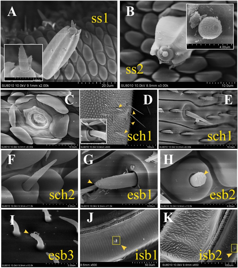
Siphoning mouthparts, consisting of proboscis and labial palps, are the exclusive feeding organs and important chemosensory organs in most adult Lepidoptera. In this study, the general morphology of the mouthpart organs and precision architecture of the proboscis was described in adult Helicoverpa armigera. Three major sensilla types with nine subtypes including three novel subtypes were identified. The novel sensilla styloconica subtype 2 was the only one having a multiporous structure, which may play olfactory roles. For further understanding of the chemosensory functions of mouthpart organs, we conducted transcriptome analysis on labial palps and proboscises. A total of 84 chemosensory genes belonging to six different families including 4 odorant receptors (ORs), 6 ionotropic receptors (IRs), 7 gustatory receptors (GRs), 39 odorant binding proteins (OBPs), 26 chemosensory proteins (CSPs), and 2 sensory neuron membrane proteins (SNMPs) were identified. Furthermore, eight OBPs and six CSPs were identified as the novel genes. The expression level of candidate chemosensory genes in the proboscis and labial palps was evaluated by the differentially expressed gene (DEG) analysis, and the expression of candidate chemosensory receptor genes in different tissues was further investigated by quantitative real-time PCR (qRT-PCR). All the candidate receptors were detected by DEG analysis and qRT-PCR, but only a small part of the OR or IR genes was specifically or partially expressed in proboscis or labial palps, such as HarmOR58 and HarmIR75p.1, however, most of the GRs were abundantly expressed in proboscis or labial palps. The reported CO2 receptors such as HarmGR1, GR2, and GR3 were mainly expressed in labial palps. HarmGR5, GR6, and GR8, belonging to the "sugar receptor" clade, were mainly expressed in proboscis or antenna and were therefore suggested to perceive saccharide. The results suggest that the mouthparts are mutually cooperative but functionally concentrated system. These works contribute to the understanding of chemical signal recognition in mouthpart organs and provide the foundation for further functional studies.
Keywords: Helicoverpa armigera; chemosensory genes; mouthparts; sensilla; transcriptome.
Figures


Similar articles
-
A mouthpart transcriptome for Spodoptera frugiperda adults: identification of candidate chemoreceptors and investigation of expression patterns.Front Physiol. 2023 Apr 25;14:1193085. doi: 10.3389/fphys.2023.1193085. eCollection 2023. Front Physiol. 2023. PMID: 37179830 Free PMC article.
-
Molecule characterization of chemosensory and metabolism-related genes in the proboscis of Athetis lepigone.Front Physiol. 2023 Dec 22;14:1287353. doi: 10.3389/fphys.2023.1287353. eCollection 2023. Front Physiol. 2023. PMID: 38187138 Free PMC article.
-
Identification and expression profiling of candidate chemosensory membrane proteins in the band-winged grasshopper, Oedaleus asiaticus.Comp Biochem Physiol Part D Genomics Proteomics. 2019 Jun;30:33-44. doi: 10.1016/j.cbd.2019.02.002. Epub 2019 Feb 10. Comp Biochem Physiol Part D Genomics Proteomics. 2019. PMID: 30771563
-
Beyond chemoreception: diverse tasks of soluble olfactory proteins in insects.Biol Rev Camb Philos Soc. 2018 Feb;93(1):184-200. doi: 10.1111/brv.12339. Epub 2017 May 7. Biol Rev Camb Philos Soc. 2018. PMID: 28480618 Review.
-
Feeding mechanisms of adult Lepidoptera: structure, function, and evolution of the mouthparts.Annu Rev Entomol. 2010;55:307-27. doi: 10.1146/annurev-ento-112408-085338. Annu Rev Entomol. 2010. PMID: 19961330 Free PMC article. Review.
Cited by
-
Odorant Receptors for Detecting Flowering Plant Cues Are Functionally Conserved across Moths and Butterflies.Mol Biol Evol. 2021 Apr 13;38(4):1413-1427. doi: 10.1093/molbev/msaa300. Mol Biol Evol. 2021. PMID: 33231630 Free PMC article.
-
An Odorant Receptor from the Proboscis of the Cotton Bollworm Helicoverpa armigera (Lepidoptera: Noctuidae) Narrowly Tuned to Indole.Insects. 2022 Apr 13;13(4):385. doi: 10.3390/insects13040385. Insects. 2022. PMID: 35447827 Free PMC article.
-
How do moth and butterfly taste?-Molecular basis of gustatory receptors in Lepidoptera.Insect Sci. 2020 Dec;27(6):1148-1157. doi: 10.1111/1744-7917.12718. Epub 2019 Sep 12. Insect Sci. 2020. PMID: 31433559 Free PMC article. Review.
-
Gustatory function of sensilla chaetica on the labial palps and antennae of three tortricid moths (Lepidoptera: Tortricidae).Sci Rep. 2022 Nov 7;12(1):18882. doi: 10.1038/s41598-022-21825-w. Sci Rep. 2022. PMID: 36344566 Free PMC article.
-
Genome-wide analysis of gustatory receptor genes and identification of the fructose gustatory receptor in Arma chinensis.Heliyon. 2024 May 9;10(10):e30795. doi: 10.1016/j.heliyon.2024.e30795. eCollection 2024 May 30. Heliyon. 2024. PMID: 38765039 Free PMC article.
References
LinkOut - more resources
Full Text Sources
Other Literature Sources